0674
Moving MRI (mMRI): imaging a moving body with synchronized magnet movement1Radiology, Massachusetts General Hospital, Charlestown, MA, United States, 2Harvard Medical School, Boston, MA, United States, 3Radiology, Massachusetts General Hospital, Boston, MA, United States, 4The Ohio State University College of Medicine, Columbus, OH, United States
Synopsis
Keywords: Hybrid & Novel Systems Technology, Hybrid & Novel Systems Technology, Artifacts, Brain, Motion Correction
Motivation: MRI is largely limited to scenarios involving small-scale bodily movements to minimize artifacts and field-induced physiological effects.
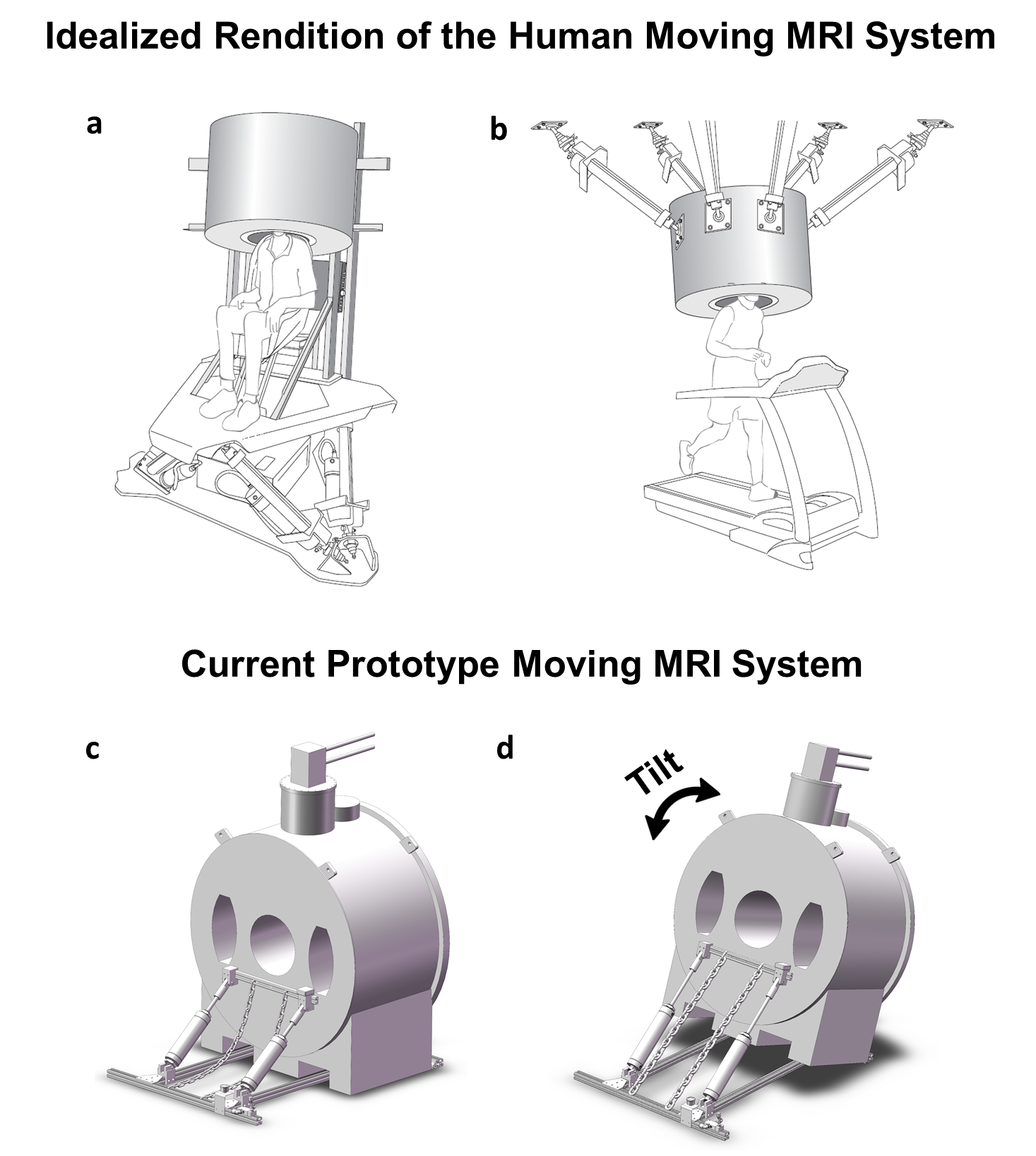
Goal(s): We are developing a moving MRI system where the magnet and subject’s head remain stationary with respect to each other during large-scale motion.
Approach: Utilizing a compact, dry 1.5T magnet, we built an apparatus that tilted the entire magnet assembly, including the cold head, gradient/shim/RF coils, and the subject, up and down during scanning.
Results: We demonstrated the ability to scan phantoms and live animals while the magnet is in motion and to correct for imaging artifacts caused by tilting the magnet.
Impact: Our proof-of-concept prototype moving MRI system supports the future viability of developing a human-scale moving MRI system, which has the potential to advance studies in vestibular research, traumatic brain injury, and brain-behavior interactions, among other areas.
Introduction
MRI generally requires the subject to remain stationary in the magnet to eliminate motion artifacts in the image and to reduce unwanted field-induced brain stimulation1. However, the ability to perform imaging during movement would provide novel insights2. For instance, large-scale head motion stimulates the vestibular system, generating neuronal and perceptual responses that might be gainfully studied with fMRI both in the clinic (e.g., vestibular migraine) and in research lab3. Yet, because of the need to keep the head motionless in the magnet, vestibular fMRI is presently restricted to unnatural vestibular stimulation like galvanic currents4,5, hot or cold water, or loud sounds. An additional application is the study of brain tissue and fluid displacement during head acceleration. We propose a novel method, Moving MRI (mMRI), which enables brain imaging during large-scale body movement by moving the magnet synchronously with the subject, eliminating relative motion between the two (Fig. 1a,b). Here we demonstrate a proof-of-concept mMRI prototype with a simple dynamic tilt motion paradigm.Methods
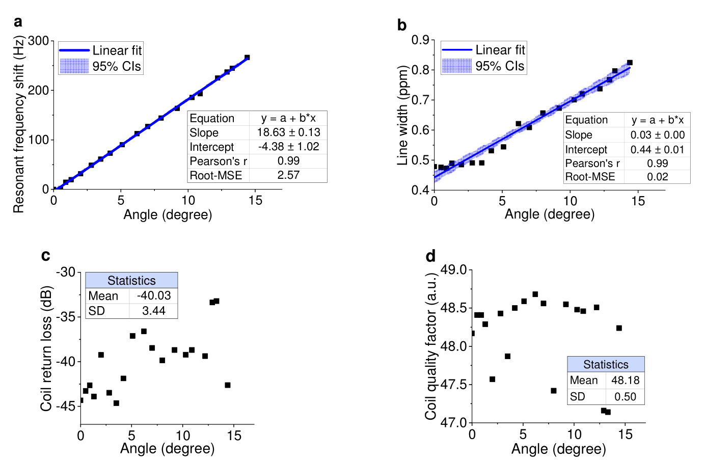
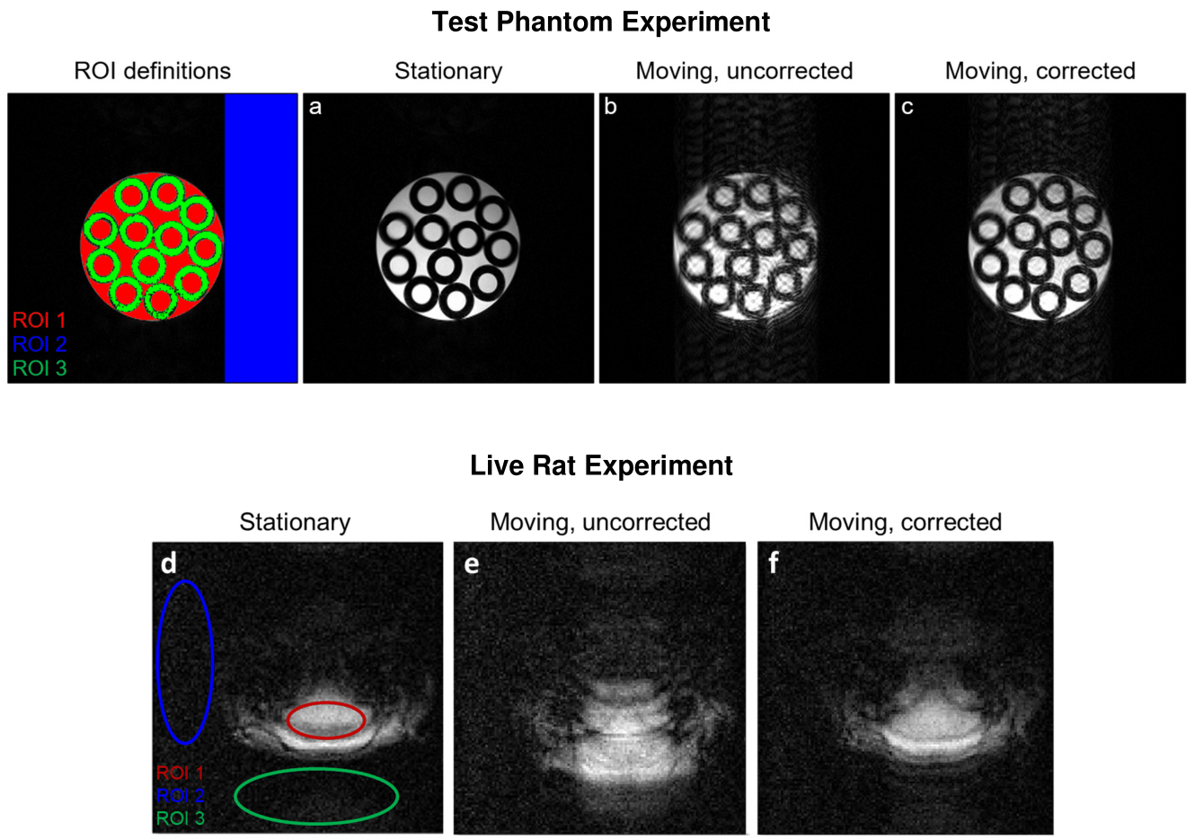
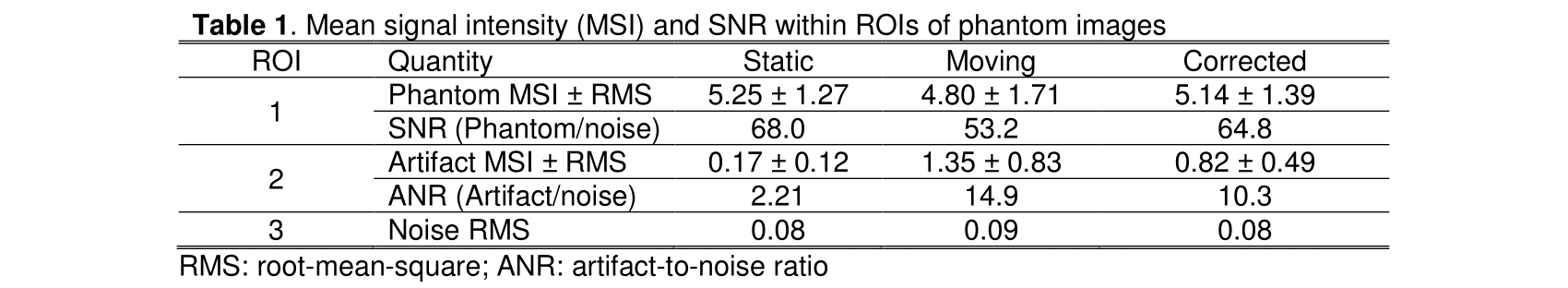
We used a compact cryogen-free 1.5T superconducting magnet capable of withstanding tilting while at field (Fig. 1c,d). Tilting was accomplished with a pair of pneumatic cylinders supplied with compressed air. Airflow was controlled with either manual or computer-programmed valves. The angle was recorded at a rate of about 10 Hz during scanning by a digital 3-axis accelerometer. SE and GE scans of phantoms and anesthetized rats respectively were acquired while the magnet underwent several cycles of tilting up and down.To correct tilt-induced image artifacts, a phase ramp determined by the recorded tilt angle was applied to each raw phase encode line. Image quality was quantitatively characterized in terms of signal-to-noise ratio (SNR) and artifact-to-noise ratio (ANR). For the phantom, the signal region was defined by an ROI chosen with an intensity threshold mask to exclude the noise background and the internal dark rings of the static image. For the rat brain, an elliptical ROI was placed over the brain. The artifact measurement was obtained in a region displaced along the phase encoding direction where there should be only noise and no signal. The noise was measured in a region displaced in the frequency encoding direction where substantial artifacts were not expected.
Results
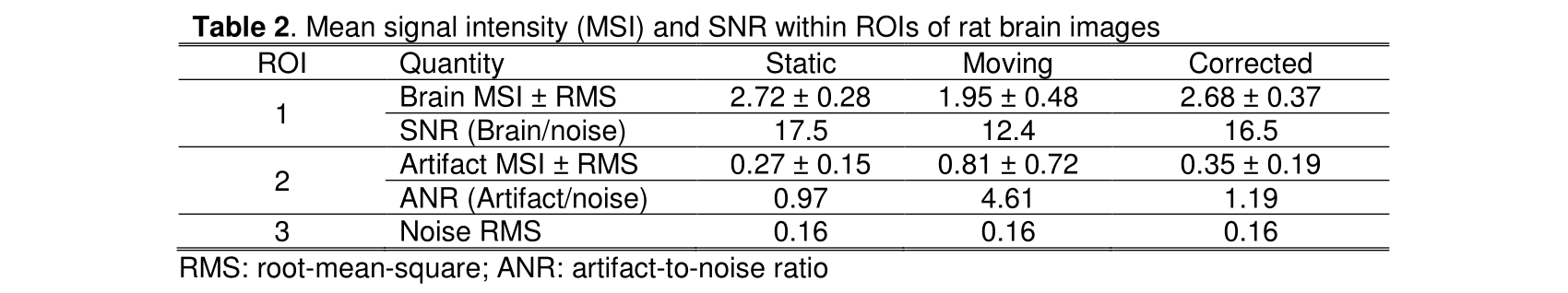
Although scanning during tilting was possible, severe artifacts degraded the images. The primary source of the artifacts was attributed to a magnetic field shift with tilt angle, which was presumed to originate from small mechanical deformations (Fig. 2). At static tilts or modest tilt rates the relationship between angle and field shift was reproducible and linear (275 Hz at 15°), enabling artifact correction with postprocessing. The linewidth was also found to increase with tilt. A small slice displacement during dynamic tilting was observed. No correction for linewidth or slice displacement was applied. Fig. 3a-c show SE images of the static, tilting, and artifact-corrected phantom respectively. Tilting the magnet while scanning reduces the signal intensity and creates artifacts in the phase encoding dimension analogous to those induced by subject motion. Correction of the central frequency of each phase encoding raw data line reduces the artifacts and regains nearly the full original signal intensity (Table 1). The constancy of the noise background among the three images shows that the scanner electronic performance is reasonably stable.Fig. 3d-f show GE images of the static, tilting, and artifact-corrected rat brain respectively. Again, magnet motion degrades the image, but correction of the instantaneous frequency shift at each phase encoding step recovers most of the static signal intensity and substantially reduces artifacts (Table 2).
Discussion
In the context of vestibular testing, the human threshold for sensing tilt is typically less than 1°/s 6, which is well within the capabilities of this motion platform. Although tilting the magnet results in image degradation, the images can be mostly restored with a simple correction procedure.Conclusion
We demonstrated the feasibility of MRI scanning in a moving superconducting magnet. Tilting-induced image artifacts, caused by a field shift, may be largely corrected. Other sources of artifact, such as slice displacement, degradation of field homogeneity, and residual motion between the magnet and subject, were not addressed. Despite the growing development of portable7-10 and head-mounted11,12 MRI where the magnet and head are stationary during imaging, the potential of imaging while the magnet and subject undergo large-scale motion remains untapped.Acknowledgements
This research is funded by NIH Grant R01EB029818. Support for the scanner was provided by R01AR075077.References
- Ward, B. K., Roberts, D. C., Otero-Millan, J. & Zee, D. S. A decade of magnetic vestibular stimulation: from serendipity to physics to the clinic. J Neurophysiol 121, 2013-2019, https://doi.org/10.1152/jn.00873.2018 (2019).
- He, B. et al. Grand challenges in mapping the human brain: NSF workshop report. IEEE Trans Biomed Eng 60, 2983-2992, https://doi.org/10.1109/tbme.2013.2283970 (2013).
- Dieterich, M. & Brandt, T. Functional brain imaging of peripheral and central vestibular disorders. Brain 131, 2538-2552, https://doi.org/10.1093/brain/awn042 (2008).
- Lopez, C., Blanke, O. & Mast, F. W. The human vestibular cortex revealed by coordinate-based activation likelihood estimation meta-analysis. Neuroscience 212, 159-179, https://doi.org/10.1016/j.neuroscience.2012.03.028 (2012).
- Lopez, C. et al. Distorted own-body representations in patients with dizziness and during caloric vestibular stimulation. J Neurol 265, 86-94, https://doi.org/10.1007/s00415-018-8906-8 (2018).
- Bermúdez Rey, M. C. et al. Vestibular Perceptual Thresholds Increase above the Age of 40. Frontiers in Neurology 7, 10.3389/fneur.2016.00162 (2016).
- Yuen, M. M. et al. Portable, low-field magnetic resonance imaging enables highly accessible and dynamic bedside evaluation of ischemic stroke. Science Advances 8, eabm3952, https://doi.org/10.1126/sciadv.abm3952 (2022).
- Deoni, S. C. L. et al. Development of a mobile low-field MRI scanner. Scientific Reports 12, 5690, https://doi.org/10.1038/s41598-022-09760-2 (2022).
- Guallart-Naval, T. et al. Portable magnetic resonance imaging of patients indoors, outdoors and at home. Scientific Reports 12, 13147, https://doi.org/10.1038/s41598-022-17472-w (2022).
- Cooley, C. Z. et al. A portable scanner for magnetic resonance imaging of the brain. Nature Biomedical Engineering 5, 229-239, https://doi.org/10.1038/s41551-020-00641-5 (2021).
- Vaughan, J. T. et al. An RF coil for a head-only MR system. Proceedings of the 29th Annual Meeting of the ISMRM, Virtual Meeting. Abstract 0137 (2021).
- Theilenberg, S. et al. Design and realization of a multi-coil array for B0 field control in a compact 1.5T head-only MRI scanner. Magnetic Resonance in Medicine, 1-14, https://doi.org/10.1002/mrm.29692 (2023).
Figures