0673
Dynamic tracking of cardiac motion fields using multi-frequency scattering parameters of an RF array – an MRI based feasibility study1Computational Imaging Group, UMC Utrecht, Utrecht, Netherlands, 2PrecorDx, Utrecht, Netherlands
Synopsis
Keywords: New Devices, Cardiovascular
Motivation: To enable high frame rate spatial mapping of cardiac mechanical motion with an array of RF antennas.
Goal(s): Investigate the feasibility of estimating 2D motion fields based on measurements of wideband multi-channel RF scattering parameters.
Approach: Paired CINE MRI and multi-channel wideband (55-1300 MHz) RF scattering measurements were acquired in vivo. Motion fields were determined from MRI, a Gaussian process regression model was trained and tested to predict motion fields from the RF scattering parameters.
Results: 2D motion fields in a 4Ch CINE image of the heart can be reconstructed with high precision (RMS error 0.66mm) based on RF scattering measurements.
Impact: We demonstrated feasibility of high precision and high framerate spatial motion mapping with RF antennas. Further validation with real-time MRI is warranted. This method could be applicable for motion tracking during medical imaging or in a low complexity care setting.
Introduction
Scattering parameters of RF antennas on the body are modulated by changing dielectric properties due to motion. This phenomenon can be used in MRI to detect physiological motion [1-4]. The aim of this work is to investigate the feasibility of measurement of cardiovascular motion in 2D based on RF scattering measurements. Motion can be represented by deformation vector fields (motion fields), which consist of a vector per voxel that indicates the magnitude and direction of motion compared to a reference phase. Previous work on MRI motion detection demonstrated that motion fields are highly compressible in space and time [5-7]. RF based imaging will not provide sufficient spatial encoding to make detailed images of the cardiovascular system. However, we hypothesize that is could be feasible to estimate compressed motion fields from multi-channel RF scattering parameters obtained over a wide frequency range (55-1300 MHz). We show here the feasibility of imaging motion fields with RF antennas in an MRI setting. The results were trained and tested with motion fields obtained from ECG triggered CINE MRI.Methods
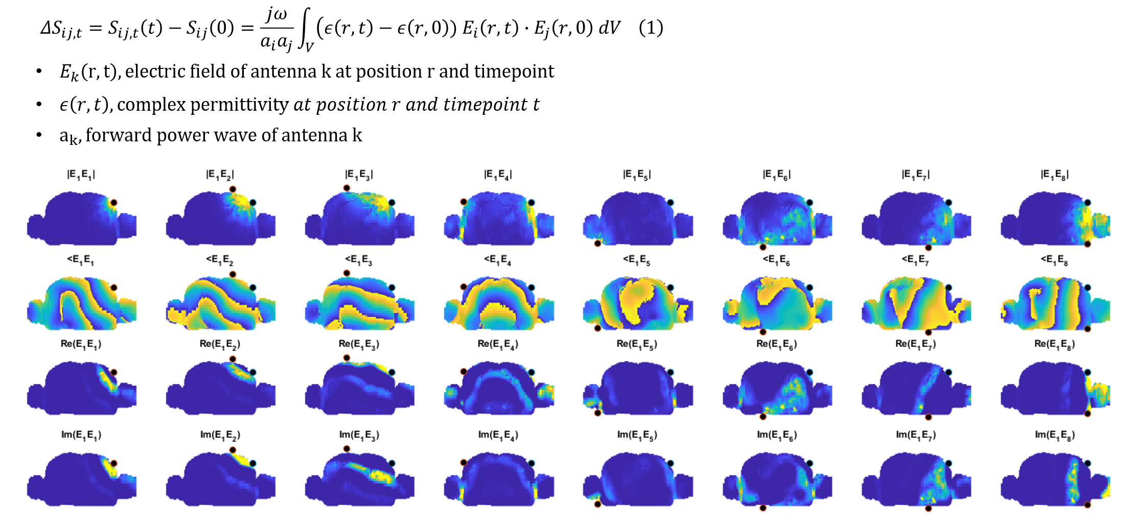
SimulationsTo provide insight on spatial encoding patterns of RF antennas, we performed EM-simulations in Sim4Life (ZMT, Zurich, Switzerland) using the XCAT model [8] and an 8 channel dipole array [9]. An existing signal model [4, 10] was used to visualize the spatial sensitivity of the antennas to motion, described by the multiplied E-fields of two antennas (Figure 1).
MRI Experiments
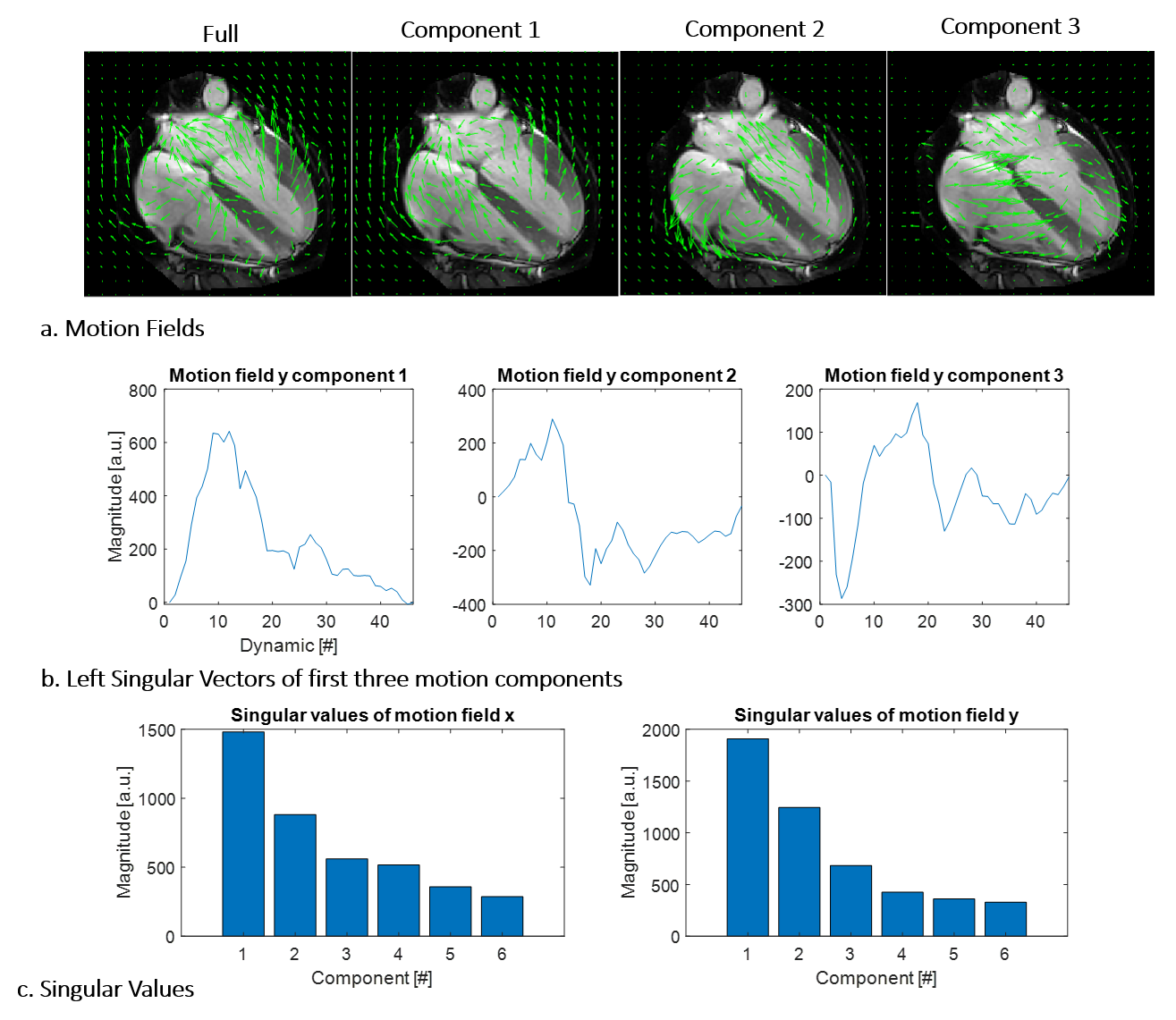
In vivo experiments were performed by acquiring subsequent MRI and multi-frequency scattering parameters. After obtaining IRB approval and informed consent, MRI was acquired in a male subject (23y, 1.83m, 63 kg) at 3T (Ingenia, Philips Healthcare, Best, The Netherlands). A 2D ECG triggered CINE acquisition was acquired in 4Ch view. Motion fields were estimated for every dynamic in Matlab (Mathworks, Natick, USA) and compressed with singular value decomposition (SVD). The MRI data, as well as ground truth and compressed motion fields are shown below.
S-Parameter Measurements
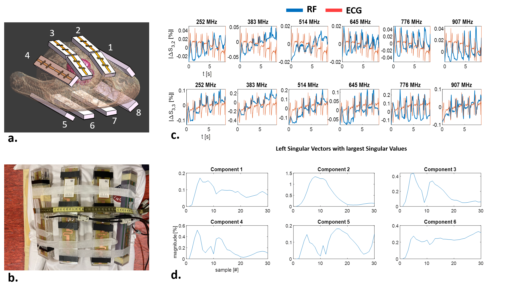
Subsequently, ΔS-parameters were measured on the same a volunteer, using an 8 channel dipole array ([9], Figure 2). The entries of the scattering matrix where measured subsequently. To ensure correct synchronization within a heartbeat between subsequent measurements, all data was acquired with ECG (ECG Shield, Olimex, Plovdiv, Bulgaria).
Regression
The 36 unique S-parameter measurements were compressed with SVD along the frequency and coil axis to obtain a vector of [nsamples ncomponents]. A Gaussian process regression model was trained to map the relation between S-parameters and motion fields. Training was performed on a third of the S-parameter waveform, while testing was done with the full S-parameter measurement.
Results
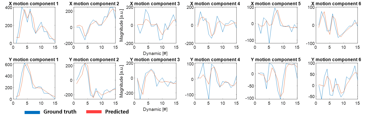
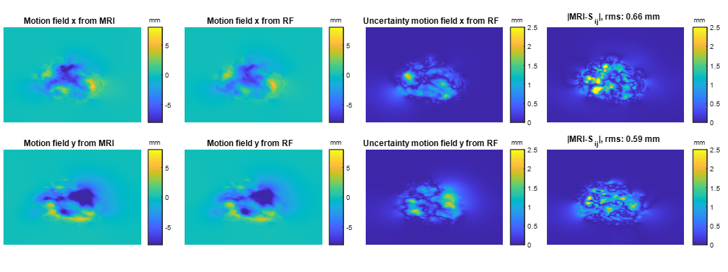
Figure 4 shows the results of the Gaussian process regression. The left singular vectors describing the motion fields can be accurately predicted from the RF measurements. Assuming that a static reference image is available, motion fields can be reconstructed from the RF data.The resulting RF based motion fields, the ground truth motion fields and the error are shown in Figure 5. 2D motion fields can be estimated with a low reconstruction error (rms error 0.66 mm for motion in x direction). The Gaussian process regression method also provides an uncertainty map, which for this example does not show a clear correlation to the error map.
Conclusion and discussion
Preliminary results show that it is feasible to reconstruct 2D motion of the heart from measurements of multi-channel wideband scattering parameters (55-1300 MHz). Results are shown for a 4Ch image, which is relevant since it can be used to determine global longitudinal strain, an important cardiac biomarker. We will further investigate the potential for 3D motion tracking, which requires high frame rate 3D time resolved MRI images. With current hardware, high temporal frame rates (up to 400 Hz) are possible. Potentially, this motion tracking technology could be used as an independent motion tracking technique for image guided interventions with high temporal frame rate, e.g. in MRI guided catheterization. We envision a scenario where dynamic images are acquired at the start of the intervention serve as training, and subsequently RF motion tracking is used for image guidance. Further research is warranted into simultaneous acquisition of scattering parameters and real-time MRI data, as well as validation of RF measurements of global longitudinal strain vs. MRI. In future scenarios, we envision a wearable RF array integrated in a vest to obtain moving images in a low complexity care setting, e.g. at home or in an outpatient clinic. This method could be relevant for monitoring of chronic cardiovascular diseases such as heart failure, where cardiovascular biomarkers need to be tracked in time.Acknowledgements
Funding was obtained from the Dutch Heart Foundation, Dekker Postdoc grant 03-006-2022-002
References
[1] D. Buikman, P. Helzel T Fau - Röschmann, and P. Röschmann, "The rf coil as a sensitive motion detector for magnetic resonance imaging," (in eng), no. 0730-725X (Print).
[2] J. Ludwig, P. Speier, F. Seifert, T. Schaeffter, and C. Kolbitsch, "Pilot tone–based motion correction for prospective respiratory compensated cardiac cine MRI," Magnetic Resonance in Medicine, vol. 85, no. 5, pp. 2403-2416, 2021/05/01 2021, doi: https://doi.org/10.1002/mrm.28580.
[3] R. J. M. Navest et al., "The noise navigator for MRI-guided radiotherapy: an independent method to detect physiological motion," Phys Med Biol, vol. 65, no. 12, p. 12NT01, Jun 18 2020, doi: 10.1088/1361-6560/ab8cd8.
[4] B. R. Steensma, C. A. Louka, A. J. E. Raaijmakers, and C. A. T. van den Berg, "Measuring stroke volume with wearable RF antennas: a validation study with EM simulations and MRI," in Conference of the International Society of Magnetic Resonance in Medicine, London, 2022, 31 ed., p. 3952.
[5] N. R. F. Huttinga, T. Bruijnen, C. A. T. van den Berg, and A. Sbrizzi, "Nonrigid 3D motion estimation at high temporal resolution from prospectively undersampled k-space data using low-rank MR-MOTUS," Magn Reson Med, vol. 85, no. 4, pp. 2309-2326, Apr 2021, doi: 10.1002/mrm.28562.
[6] N. R. F. Huttinga, T. Bruijnen, C. A. T. Van Den Berg, and A. Sbrizzi, "Real-Time Non-Rigid 3D Respiratory Motion Estimation for MR-Guided Radiotherapy Using MR-MOTUS," IEEE Trans Med Imaging, vol. 41, no. 2, pp. 332-346, Feb 2022, doi: 10.1109/TMI.2021.3112818.
[7] N. R. F. Huttinga, C. A. T. van den Berg, P. R. Luijten, and A. Sbrizzi, "MR-MOTUS: model-based non-rigid motion estimation for MR-guided radiotherapy using a reference image and minimal k-space data," Phys Med Biol, vol. 65, no. 1, p. 015004, Jan 10 2020, doi: 10.1088/1361-6560/ab554a.
[8] W. P. Segars, G. Sturgeon, S. Mendonca, J. Grimes, and B. M. Tsui, "4D XCAT phantom for multimodality imaging research," Med Phys, vol. 37, no. 9, pp. 4902-15, Sep 2010, doi: 10.1118/1.3480985.
[9] A. J. Raaijmakers et al., "The fractionated dipole antenna: A new antenna for body imaging at 7 Tesla," Magn Reson Med, vol. 75, no. 3, pp. 1366-74, Mar 2016, doi: 10.1002/mrm.25596.
[10] A. S. Beaverstone, D. S. Shumakov, and N. K. Nikolova, "Frequency-Domain Integral Equations of Scattering for Complex Scalar Responses," IEEE Transactions on Microwave Theory and Techniques, vol. 65, no. 4, pp. 1120-1132, 2017, doi: 10.1109/TMTT.2016.2638428.
[11] N. R. F. Huttinga, T. Bruijnen, C. A. T. van den Berg, and A. Sbrizzi, "Gaussian Processes for real-time 3D motion and uncertainty estimation during MR-guided radiotherapy," Medical Image Analysis, vol. 88, p. 102843, 2023/08/01/ 2023, doi: https://doi.org/10.1016/j.media.2023.102843.
[12] C. E. Rasmussen and
C. K. Williams, Gaussian processes for
machine learning. Springer, 2006.
Figures