0672
Multinuclear MRI Using a Single Adaptable Transmit Hardware1MRIEngT, LFMI,NINDS, NIH, Bethesda, MD, United States
Synopsis
Keywords: Hybrid & Novel Systems Technology, New Devices
Motivation: Routine studies of nuclei other than 1H are constrained by the need of separate fixed tuned hardware.
Goal(s): To increase flexibility of the multinuclear setup by developing an adaptable single transmit hardware to allow excitation of many low-frequency nuclei and 1H.
Approach: The conventional remote broadband amplifier was replaced by an optically controlled dual-tuned on-coil amplifier for 1H and X-nuclei excitation. The amplifier can be automatically tuned to the selected frequency by pulse-width-modulation of an optically transmitted pulse.
Results: Automatic tuning of a first prototype was possible for excitation of 13C, 23Na and 129Xe, while performance of 1H excitation at 7T was unaffected.
Impact: The presented technology combined with new adaptable receive hardware can advance the implementation of routine multinuclear studies to extend research of X-nuclei and their potential clinical use.
Introduction
Multinuclear MRI at high field is a promising tool to study the brain and track diseases like cancer1,2. The implementation of routine multinuclear MRI to extend these studies is constrained by the limited number of dedicated hardware. Normally, only few fixed dual tuned coils for 1H and a target X-nucleus are available in the MRI suite. Recently a multilayer arrangement of dual-tuned coils was presented for the study of 5 nuclei in a scan session3. However, the increase of nuclei to be excited (and detected) using fixed tuned coaxially connected transmit coils is challenged by cable and coil coupling and losses in transmit efficiency. To increase flexibility and performance of the multinuclear hardware, an adaptable optically controlled transmit setup for imaging 1H and multiple X-nuclei without affecting 1H transmit performance is presented.Methods
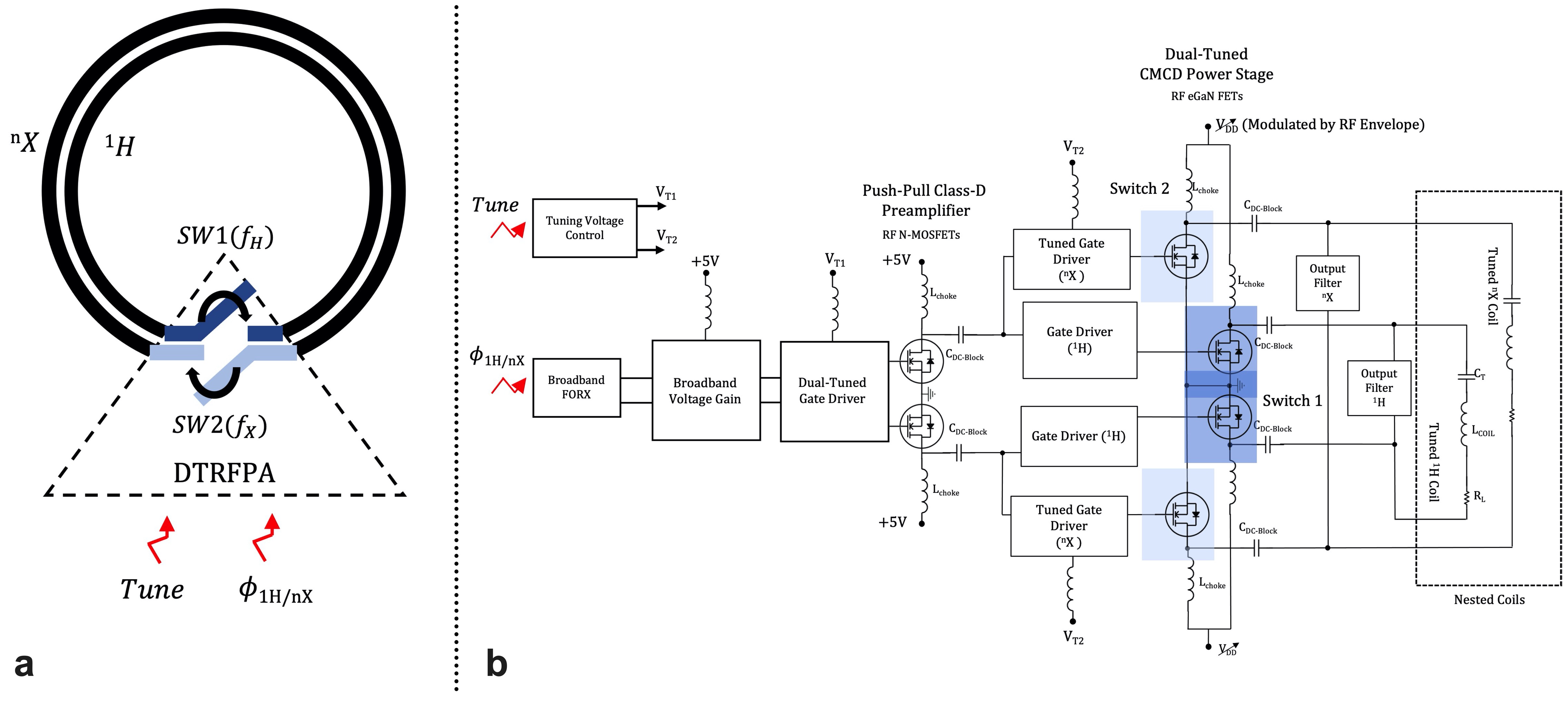
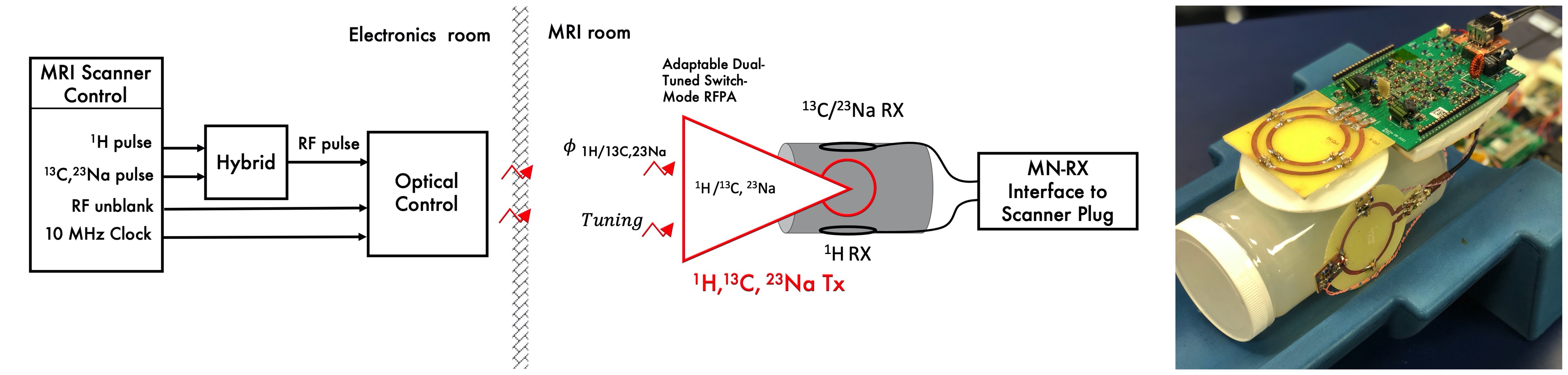
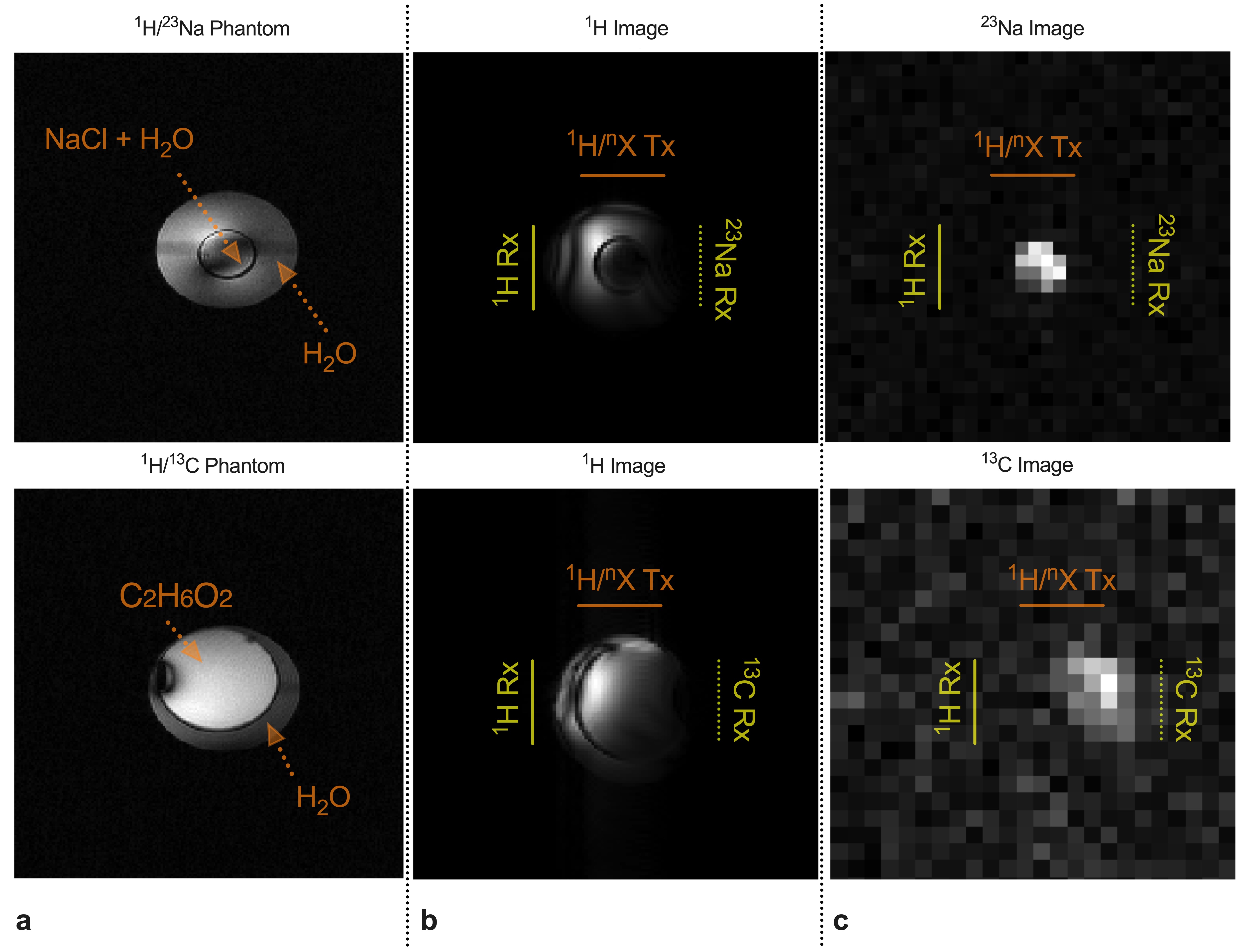
The adaptable optically controlled transmit setup is formed by a novel design of an on-coil switch-mode amplifier4-6 and a nested configuration of two single-tuned coils (Fig. 1a). In this setup, the coil that is not transmitting is intrinsically detuned by the amplifier high output impedance. A simplified diagram of the amplifier is shown in Fig. 1b. The preamplifier drives a separate 1H Current Mode Class-D (CMCD) output stage and a X-nucleus CMCD output stage through a fixed and tunable gate driver respectively7. This last stage has a tunable inductance in the X-nucleus CMCD gate circuit, as similarly implemented for the dual-tuned gate driver in the preamplification stage, to optimize the switching of the power FETs at the selected X-nucleus frequency. The output of the high frequency and the low frequency stages are connected to the corresponding loop in the nested configuration. The new amplifier was tested on the bench for multiple X-nuclei frequencies (13C, 23 Na and 129Xe) and 1H. In a first implementation of the technology at 7 T MRI, 13C and 23Na were selected to simplify implementation of the multinuclear receive hardware using a single low-noise amplifier (LNA) optimized for these nuclei frequencies. The X-nuclei coil was automatically tuned through the total drain-source capacitance of an array of eGaN FETs8. Receive coils were connected to an in-house built interface that contained the LNAs and control signals for connection to the MRI system (Fig. 2). Two-compartment cylindrical phantoms were built, with the inner compartment solution rich in the targeted X-nuclei, and imaged with a volume transmit coil (Fig 4a). 1H and X-nuclei images were acquired using a 3D gradient echo sequence with a non-selective RF pulse, 5 ms TE and 200 x 200 mm2 FOV. For 1H imaging 50 ms TR, 256 base resolution, and no average were selected, while base resolution, TR and number of averages were adjusted for the low-frequency nuclei to maximize the signal considering relaxation constants of the different solutions and acquisition time.Results
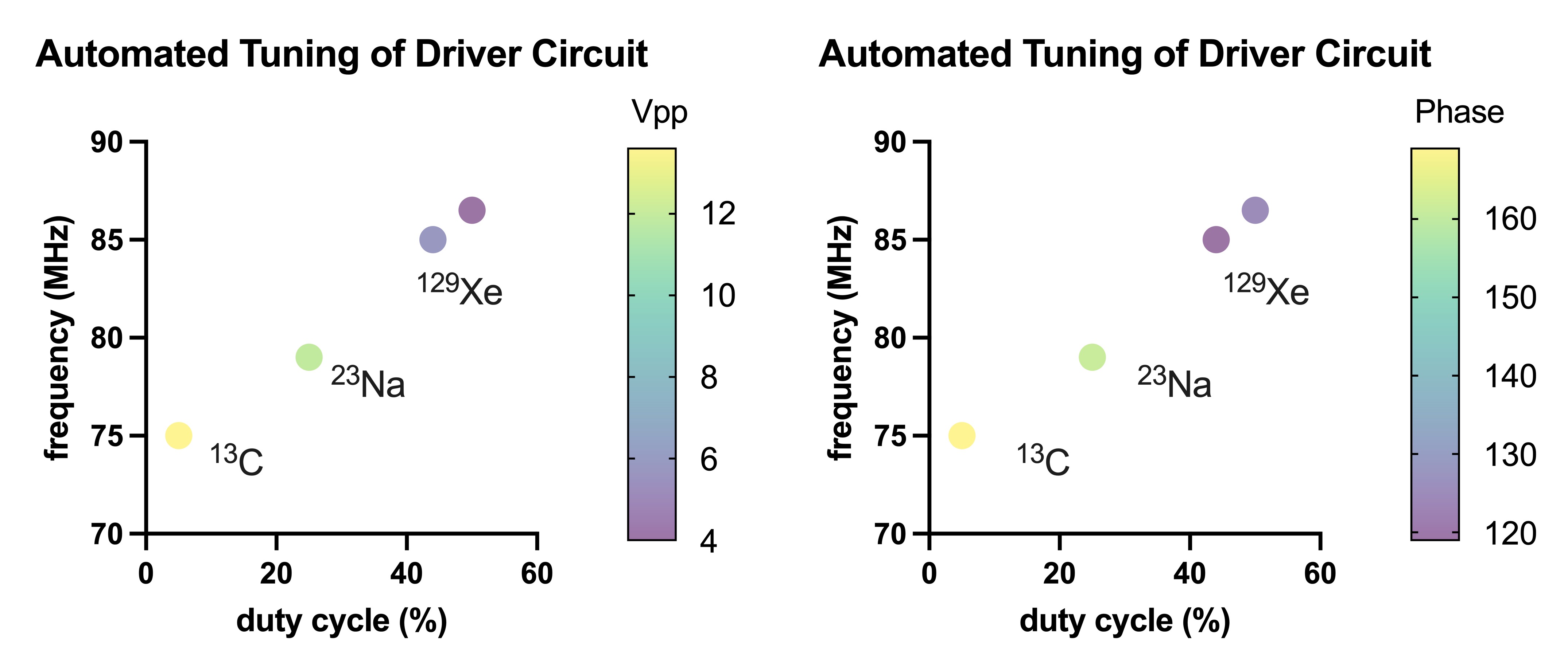
For this first implementation of the optically controlled tuning circuit, the range of selected frequencies includes 13C, 23Na and 129Xe (~75 MHz, ~79 MHz and ~83 MHz). Selected frequency (approximated to that of target X-nucleus), peak-to-peak voltage and phase difference of the preamplifier output signals as a function of the tuning signal duty cycle are shown in Fig. 3. After connection of the driver to the power stage, B1 generated at each of the selected frequencies was measured with a calibrated RF probe. In the presented amplifier-coil setup, B1+ amplitude at the coil per bias voltage of the CMCD stage ( VDD) was 3.5 μT/volt, 3.1 μT/volt and 1.5 μT/volt for 13C , 23Na and 1H respectively. Note the value for 1H was similar to that obtained with a single-tuned version of the amplifier8. Operation of all electronics in the scanner was successful and all targeted nuclei were detected. Proton images are shown in Fig. 4b, 23Na and 13C images are shown in Fig. 4c. High signal intensity is shown at the center of the image corresponding to the location of the inner compartment rich in X-nuclei.Discussion
This work demonstrates an adaptable transmit hardware that can excite several X-nuclei and 1H. The range of automatically selected X-nuclei could be extended by another implementation of the tunable device in the gate driver circuits of the preampfification and X-nuclei CMCD stages8. The X-nuclei receive coil was also tuned remotely by DC voltage. This technology combined with adaptable receive electronics can reduce the layers of hardware in the multinuclear MRI setup. Additionally, in a multichannel implementation, this technology benefits from the elimination of cables, matching networks and decoupling elements as it was shown for the 1H on-coil parallel transmission implementation9.Acknowledgements
This work was supported by the intramural research program of the National Institute of Neurological Disorders and Stroke, National Institutes ofHealth.References
- Henning A. Proton and multinuclear magnetic resonance spectroscopy in the human brain at ultra-high field strength: A review. Neuroimage. 2018;168:181-198.
- Poku LO, Phil M, Cheng Y, Wang K, Sun X. 23Na-MRI as a Noninvasive Biomarker for Cancer Diagnosis and Prognosis. Journal of Magnetic Resonance Imaging. 2021;53(4):995-1014.
- J. Dai, M. Gosselink, T. A. van der Velden, E. F. Meliadò, A. J. E. Raaijmakers, and D. W. J. Klomp, An RF coil design to enable quintuple nuclear whole-brain MRI. Magn Reson Med. 2023;89(5):2131–2141.
- Gudino N, Heilman JA, Riffe MJ, Heid O, Vester M, Griswold MA. On-coil multiple channel transmit system based on class-D amplification and pre-amplification with current amplitude feedback. Magn Reson Med. 2013;70(1):276-289.
- Gudino N, de Zwart JA, Duan Q, et al. Optically controlled on-coil amplifier with RF monitoring feedback. Magn Reson Med. 2018;79(5):2833-2841.
- Gudino N, Dodd S, Li S et al. Dual-Tuned Optically Controlled On-Coil Switch-Mode Amplifier. Proceedings of the 28th Annual meeting, ISMRM, Virtual 2020. ( Abstract 0751)
- Gudino N, Adaptable Dual-Tuned Optically Controlled On-Coil Amplifier for High-Field MRI Systems. US Patent Application Number 20230243905-A1
- Gudino N, Automated tuning of gate driver circuit using eGaN FETs. Proceedings of the 30th Annual meeting, ISMRM, London, UK 2022. (Abstract 1999)
- Gudino N, J. A. de Zwart, and J. H. Duyn, Eight-channel parallel transmit-receive system for 7 T MRI with optically controlled and monitored on-coil current-mode RF amplifiers. Magn Reson Med. 2020; 84(6):3494–3501.
Figures