0669
Brain CSF clearance measured by phase-contrast MRI and dynamic 18F-MK-6240 PET in Alzheimer's disease1Department of Radiology, Weill Cornell Medicine, New York, NY, United States, 2Amiens Picardy University Hospital, Amiens, France
Synopsis
Keywords: Neurofluids, Alzheimer's Disease
Motivation: Beta-amyloid (Aβ) in Alzheimer’s disease (AD) is caused by decreased glymphatic clearance function. As the main source of CSF in glymphatic system, the mechanism of CSF dynamic in ventricle system is of great importance.
Goal(s): Use dynamic 18F-MK-6240 PET and PC-MRI to study the relationship of CSF clearance in lateral ventricle (LV) and aqueduct.
Approach: CSF turnover rate in LV is derived from dynamic 18F-MK-6240 PET. Aqueduct parameters were calculated using PC-MRI data. Linear regression analysis was performed between PET and PC-MRI measurements.
Results: The CSF clearance measurements from both PET and MRI are consistent and show diagnostic group difference.
Impact: 18F-MK-6240 PET derived CSF turnover rate and phase-contrast MRI produced aqueduct CSF measurements are highly correlated. The data reveals diagnostic group difference after controlling for age and sex, indicating that the decreased CSF clearance is associated with Alzheimer’s disease.
INTRODUCTION
Alzheimer's disease (AD) represents a significant and growing public health challenge. As the search for effective treatments continues, understanding the pathophysiological mechanisms underlying AD is paramount in terms of the general glymphatic clearance system. Among these mechanisms, the clearance of metabolic waste from the brain, particularly through the cerebral spinal fluid (CSF), has emerged as a crucial area of study. Recent evidence suggests that the accumulation of neurotoxic proteins such as amyloid-beta, a hallmark of AD, may be due in part to impaired clearance mechanisms.1 The CSF system, including the lateral ventricles and cerebral aqueduct, plays a vital role in the brain's clearance process.2–4 To further understanding the CSF dynamic in both lateral ventricle and aqueduct, our study leverages the capabilities of dynamic positron emission tomography (PET) to measure CSF clearance in the lateral ventricles and PC-MRI to assess flow through the cerebral aqueduct. By integrating these modalities, we aim to provide a comprehensive picture of CSF clearance pathways and their alterations in AD. This approach holds promise not only for elucidating the role of CSF dynamics in AD pathology but also for identifying potential biomarkers that could facilitate early diagnosis and intervention. In this research, we try to evaluate the association between CSF clearance in the lateral ventricles and cerebral aqueduct, and see their impact on Alzheimer's disease. The findings from this study are expected to offer significant contributions to our understanding of AD, potentially informing the development of novel therapeutic strategies aimed at enhancing brain clearance mechanisms.METHODS
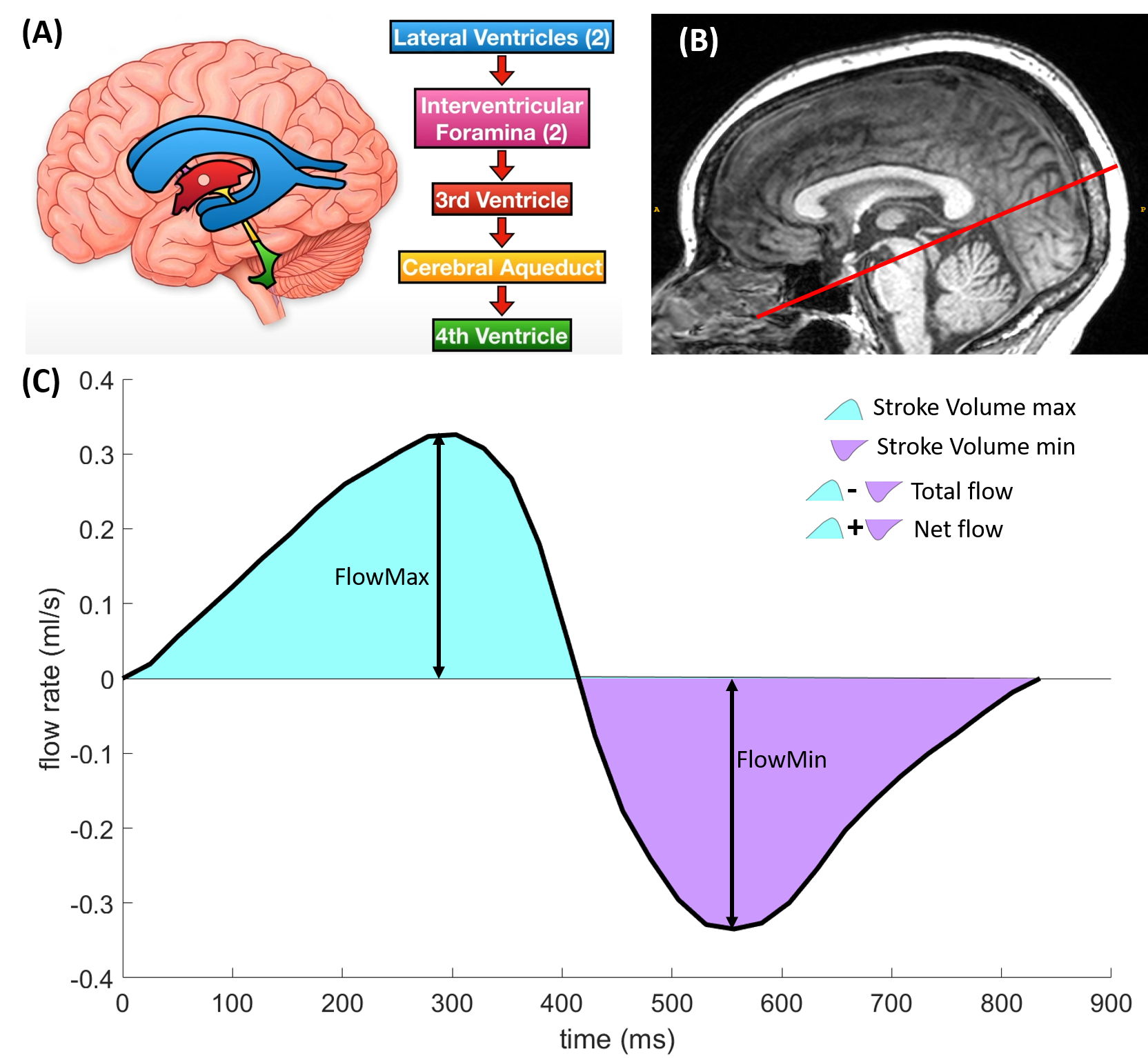
Ninety subjects (53 CN, age 70.07±8.49 years old, 34 Female; 37 MCI/AD, age 68.65±9.27, 19 female) underwent 3T MRI scan for T1w, T2w, PC-MRI and 18F-MK-6240 PET scan for tracer dynamic clearance. T1w was used for ROI parcellation and ROI value extraction.5 PC-MRI was used to calculate the CSF flow parameter in aqueduct using the Bio Flow Image Analysis software.6–8 The PC-MRI parameter for aqueduct including the total flow, stroke volume, maximum velocity, and crosssectional area as shown in Figure 1. The CSF turnover rate (vCSF) in the LV was calculated by the time activity curve (TAC) difference between 10 min to 30 min post tracer injection and normalized with the total tracer input in the brain using the dynamic 18F-MK-6240 PET following the formula in our previous works.2,9,10 The analysis focuses on the aging effect of PC-MRI parameters by diagnostic group and the association between PC-MRI parameters and vCSF by controlling for age, gender and intracranial volume.RESULTS
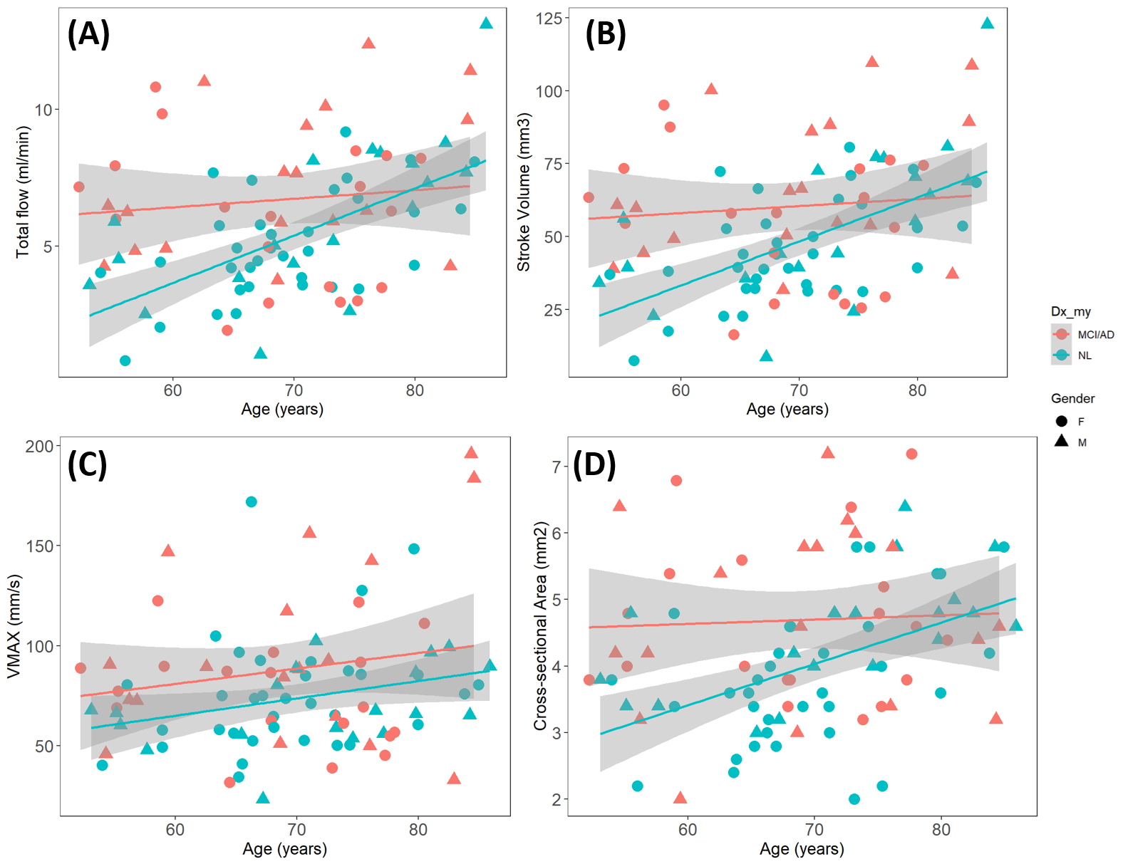
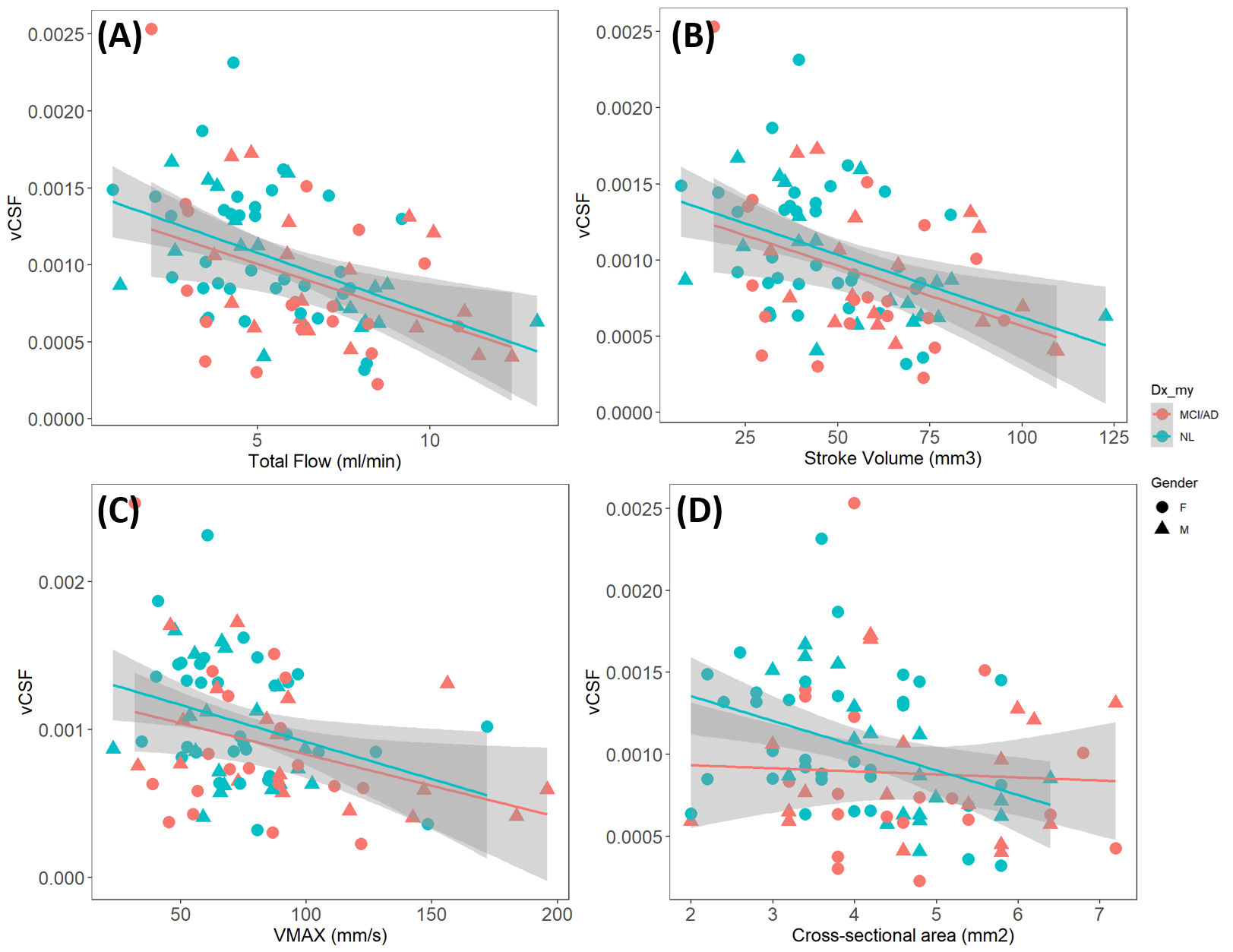
Figure 2 presented the aging effect of PC-MRI parameters in regression format controlling for age and gender. (A) show the relationship between total flow and age (age: t=3.718, p<0.05, Dx_CN: t=-2.68, p<0.05 with R2=0.202); (B) stroke volume vs age (age: t=3.451, p<0.05, Dx_CN: t=-2.602, p<0.05 with R2=0.191); (C) Vmax vs age (age: t=2.015, p<0.05, Dx_CN: t=-2.080, p<0.05 with R2=0.056); (D) cross-sectional area vs age (age: t=2.565, p<0.05, Dx_CN: t=-2.754, p<0.05 with R2=0.135).Figure 3 showed the relationship between vCSF and PC-MRI parameters. (A) vCSF vs total flow (age: t=-3.062, p<0.05, total flow: t=-3.488, p<0.05 with R2=0.250); (B) vCSF vs stroke volume (age: t=-3.192, p<0.05, stroke volume: t=-3.384, p<0.05 with R2=0.244); (C) vCSF vs Vmax (age: t=-3.804, p<0.05, Vmax: t=-2.919, p<0.05 with R2=0.220); (D) vCSF vs cross-sectional area (age: t=-3.814, p<0.05 with R2=0.165).
DISCUSSION
Our data showed that the PC-MRI parameters in aqueduct have strong aging effect and have diagnostic group difference. We also showed that PC-MRI parameters for aqueduct are associated with the CSF turnover rate in lateral ventricle. To the best of our knowledge, this is the first study to investigate the relationships of CSF dynamic in LV and aqueduct in the AD context.For all the four PC-MRI parameters including total flow, stroke volume, Vmax and crosssectional area show a positive correlation with age in CN group, but not significant increase in MCI/AD group. This could be due to the compensatory mechanism of the AD physiology.
All the four PC-MRI parameters are negatively correlated with vCSF, showing that decreased CSF clearance in LV is corresponding to the larger flow perturbation in aqueduct. The increase of aqueduct crosssectional area could due to the fluid stasis in the CSF system.
CONCLUSION
The dynamic of CSF clearance in lateral ventricle and aqueduct are significantly associated. Both of age and diagnosis are associated to the change of CSF clearance function.Acknowledgements
This work was supported by the National Institutes of Health (NIH) (R01 R56AG058913, R01 AG068398, AG057848, R01AG022374, RF1 AG057570).References
1. Mawuenyega KG, Sigurdson W, Ovod V, et al. Decreased Clearance of CNS β-Amyloid in Alzheimer’s Disease. Science. 2010;330(6012):1774-1774. doi:10.1126/science.1197623
2. Li Y, Rusinek H, Butler T, et al. Decreased CSF clearance and increased brain amyloid in Alzheimer’s disease. Fluids Barriers CNS. 2022;19(1):21. doi:10.1186/s12987-022-00318-y
3. Leon MJ de, Li Y, Okamura N, et al. Cerebrospinal Fluid Clearance in Alzheimer Disease Measured with Dynamic PET. J Nucl Med. 2017;58(9):1471-1476. doi:10.2967/jnumed.116.187211
4. Schubert JJ, Veronese M, Marchitelli L, et al. Dynamic 11C-PiB PET Shows Cerebrospinal Fluid Flow Alterations in Alzheimer Disease and Multiple Sclerosis. J Nucl Med Off Publ Soc Nucl Med. 2019;60(10):1452-1460. doi:10.2967/jnumed.118.223834
5. Fischl B. FreeSurfer. NeuroImage. 2012;62(2):774-781. doi:10.1016/j.neuroimage.2012.01.021
6. Balédent O, Gondry-Jouet C, Meyer ME, et al. Relationship between cerebrospinal fluid and blood dynamics in healthy volunteers and patients with communicating hydrocephalus. Invest Radiol. 2004;39(1):45-55. doi:10.1097/01.rli.0000100892.87214.49
7. Balédent O, Gondry-Jouet C, Stoquart-Elsankari S, Bouzerar R, Le Gars D, Meyer ME. Value of phase contrast magnetic resonance imaging for investigation of cerebral hydrodynamics. J Neuroradiol J Neuroradiol. 2006;33(5):292-303. doi:10.1016/s0150-9861(06)77287-x
8. Lokossou A, Metanbou S, Gondry-Jouet C, Balédent O. Extracranial versus intracranial hydro-hemodynamics during aging: a PC-MRI pilot cross-sectional study. Fluids Barriers CNS. 2020;17(1):1. doi:10.1186/s12987-019-0163-4
9. Zhou L, Nguyen TD, Wegiel J, de Leon MJ, Li Y. Multimodal Imaging Analysis for Brain Clearance: PET Imaging Based Clearance Slope, FASTT2 MRI-based Parenchyma CSF fraction, and DTI-based ALPS-index. Alzheimers Dement. 2022;18(S6):e068818. doi:10.1002/alz.068818
10. Zhou L, Butler TA, Wang XH, et al. Multimodal assessment of brain fluid clearance is associated with amyloid-beta deposition in humans. J Neuroradiol. Published online October 30, 2023. doi:10.1016/j.neurad.2023.10.009
Figures