0648
Measuring intravoxel incoherent motion (IVIM) using Spherical Tensor Encoding (STE) diffusion MRI1Academy of Medical Engineering and Translational Medicine, Tianjin University, Tianjin, China, 2Tianjin Key Laboratory of Brain Science and Neuroengineering, Tianjin, China, 3Haihe Laboratory of Brain-Computer Interaction and Human-Machine Intepration, Tianjin, China, 4Central Research Institute, United Imaging Healthcare Group, Shanghai, China, 5Central Research Institute, United Imaging Healthcare, Shanghai, China, 6State Key Laboratory of Oncology in South China, Guangdong Provincial Clinical Research Center for Cancer, Guangdong, China
Synopsis
Keywords: IVIM, Brain
Motivation: The assumption of Gaussian diffusion in the extravascular space in the IVIM model does not necessarily hold, especially for neuronal tissues.
Goal(s): To mitigate the impacts on IVIM estimation from complicated extravascular space diffusion such as in the crossing-fiber tissues.
Approach: We employed Spherical Tensor Encoding (STE) in place of the Linear Tensor Encoding (LTE) as in the conventional Stejskal-Tanner experiment to eliminate the orientational dependence of diffusion signal.
Results: The feasibility of the microscopic IVIM based on STE experiments was demonstrated in both healthy and diseased participants, with expected contrasts according to known anatomy/pathology.
Impact: A new framework of IVIM measurement was proposed based on the Linear Tensor Encoding diffusion experiment. The proposed approach can achieve diffusivity estimates in one excitation, which will otherwise require acquisition of multiple diffusion weighting directions.
The Intravoxel Incoherent Motion (IVIM) method1–3 separates diffusion MR signal into the perfusion components (i.e., flow of blood in vasculatures, also called pseudo diffusion) and the diffusion components (i.e., the gross total of extravascular space). The perfusion-related contributions can provide important information on micro-perfusion in the tissue, serving as a promising tool in studies of neurological and neurovascular diseases4. Typically, the water diffusion in the extravascular space is modeled as Gaussian diffusion. Under a Stejskal-Tanner experimental paradigm, a large number of b-values along three orthogonal diffusion weighting directions were sampled, and an ADC or MD was fitted for the extravascular space. However, in complex neuronal tissue environment, such as in the presence of crossing fibers, the assumption of Gaussian diffusion is not necessarily valid, which may yield erroneous estimation of IVIM signal and perfusion-related metrics. In this work, we aim to investigate the feasibility of IVIM measurement using spherical tensor encoding (STE)5 diffusion experiment in place of the conventional Linear Tensor Encoding (LTE), so that the orientational dependence of diffusion in neuronal tissues is mitigated.
Methods
Theory
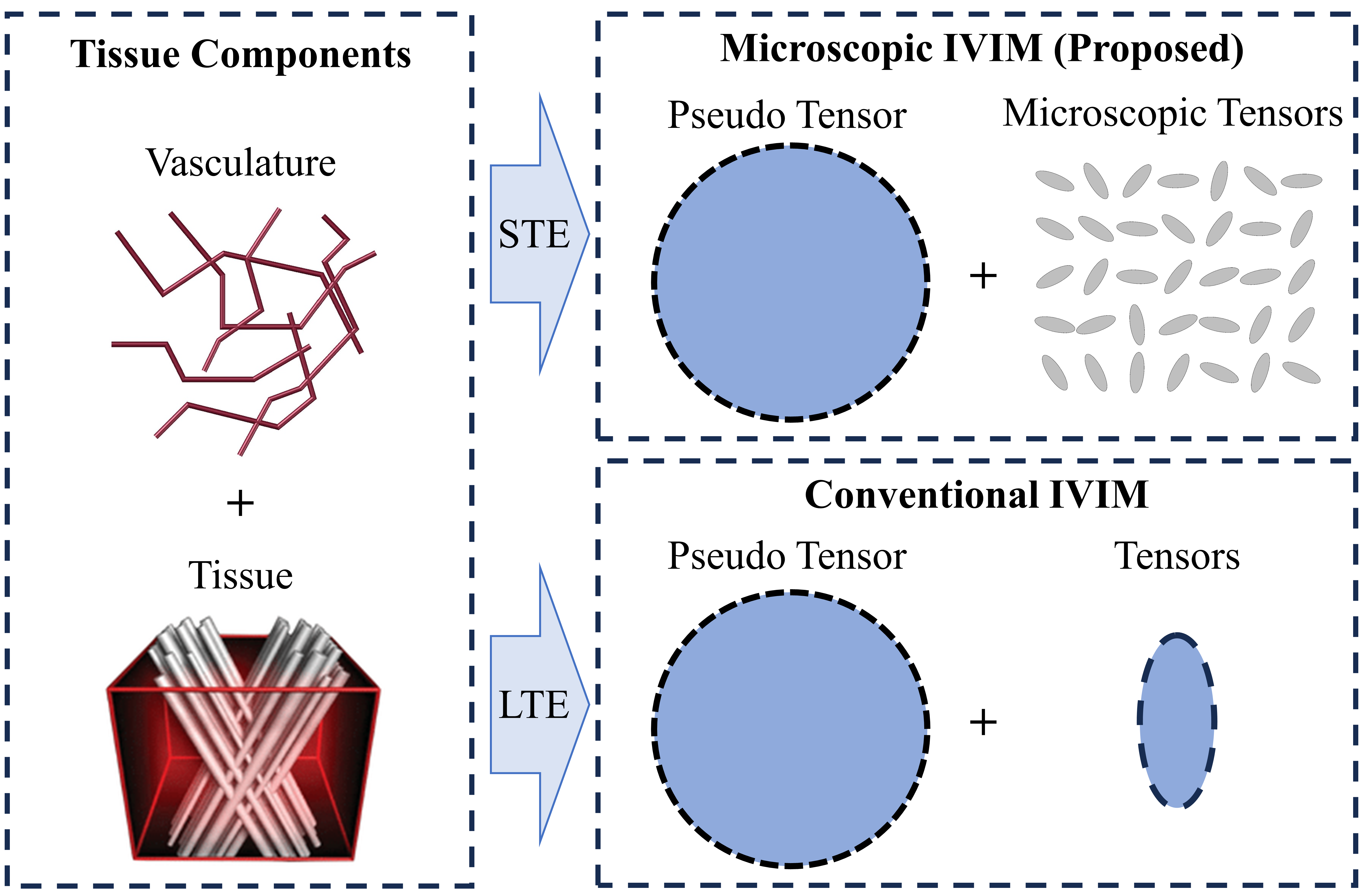
As shown in Figure 1, the STE version of IVIM model takes the similar form as in the LTE experiment::
$$\frac{S(b)}{S_0}=fe^{-bD^*}+(1-f)e^{-bD}$$
Where S(b) is the measured intravoxel STE diffusion signal, (1-f) is the signal contribution of diffusion, D is the mean diffusivity of microscopic tensor6, f is the perfusion-related signal contribution, and D* is the mean diffusivity of the microscopic pseudo-diffusion tensor.
In vivo Experiments
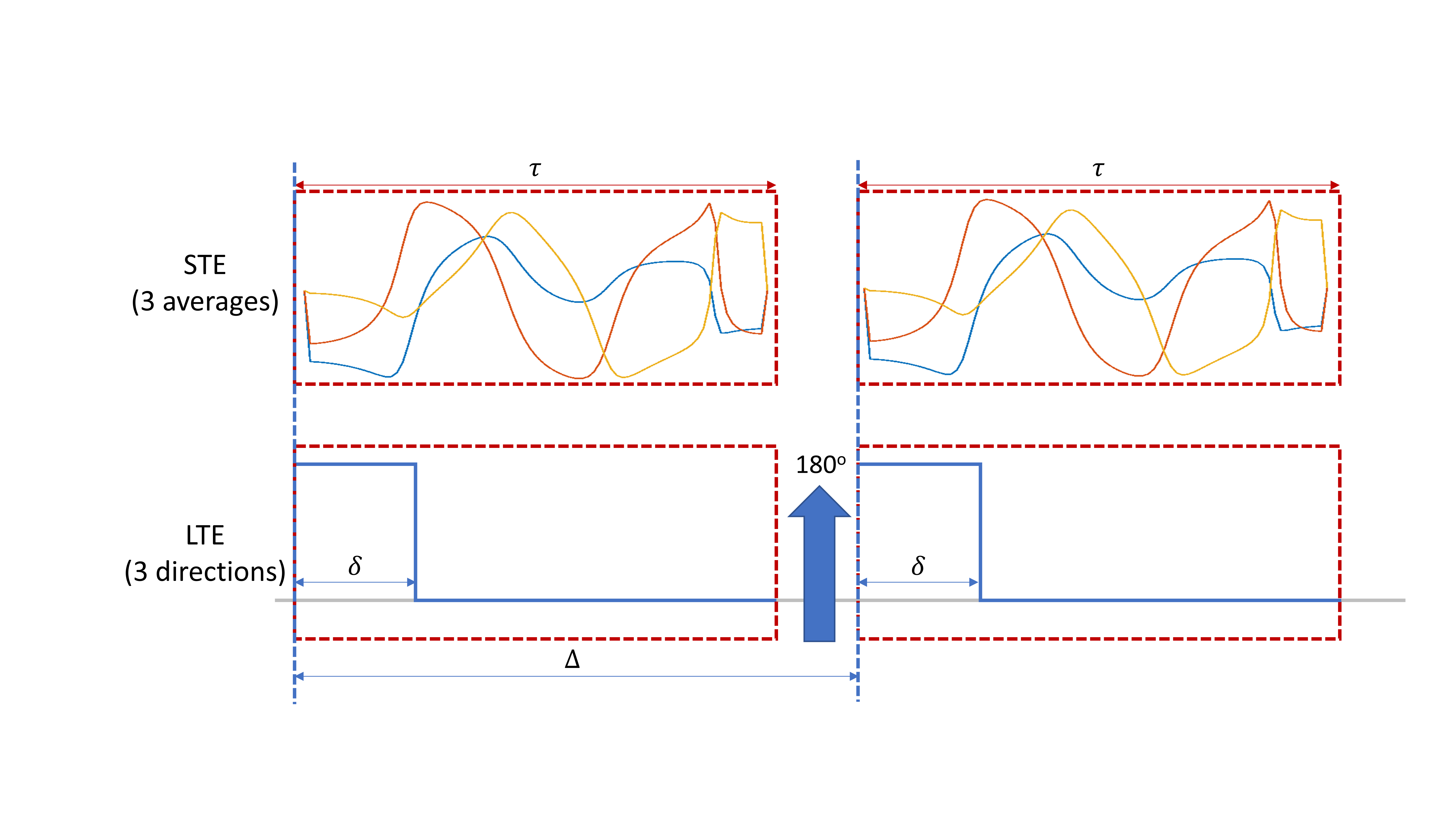
Data were acquired on a 3T United Imaging uMR890 scanner with a spin echo EPI sequence in two participants, one healthy male (34 yro) and one female patient with breast cancer brain metastases (BCBM) located in the cerebellum (55 yro). Two diffusion weighting waveforms were implemented as shown in Figure 2. 15 non-zero b-values (10, 20, 40, 80, 110, 140, 170, 200, 300, 400, 500, 600, 700, 800, 900 s/mm2) were acquired. The TR/TE is 6000/120.7ms, and voxel size is 2×2×4mm3. For linear tensor encoding, the δ is 11.25ms and Δ is 48.8ms. For STE, the onset time of the second diffusion encoding waveform was kept the same as that of the second pulse diffusion waveform of LTE, to ensure identical mixing time. The spherical encoding waveform is generated using the optimization toolbox of Sjölund7.
Data analysis
Data was pre-processed using TOPUP8,9, motion and eddy current correction were performed in FSL10,11. After the pre-processing, the data was fitted using the IVIM program in the multidimensional toolbox (https://github.com/markus-nilsson/md-dmri/tree/master).
Results
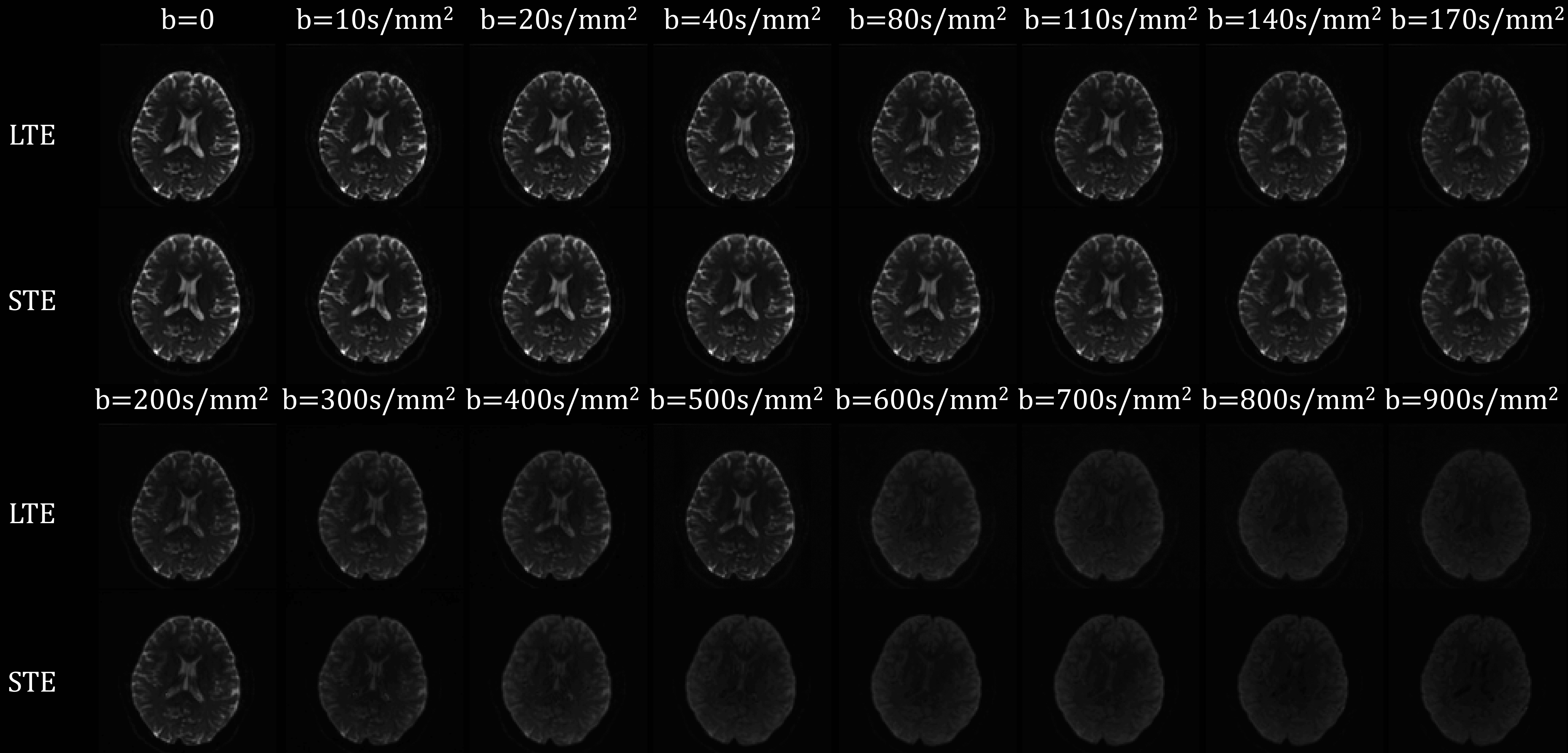
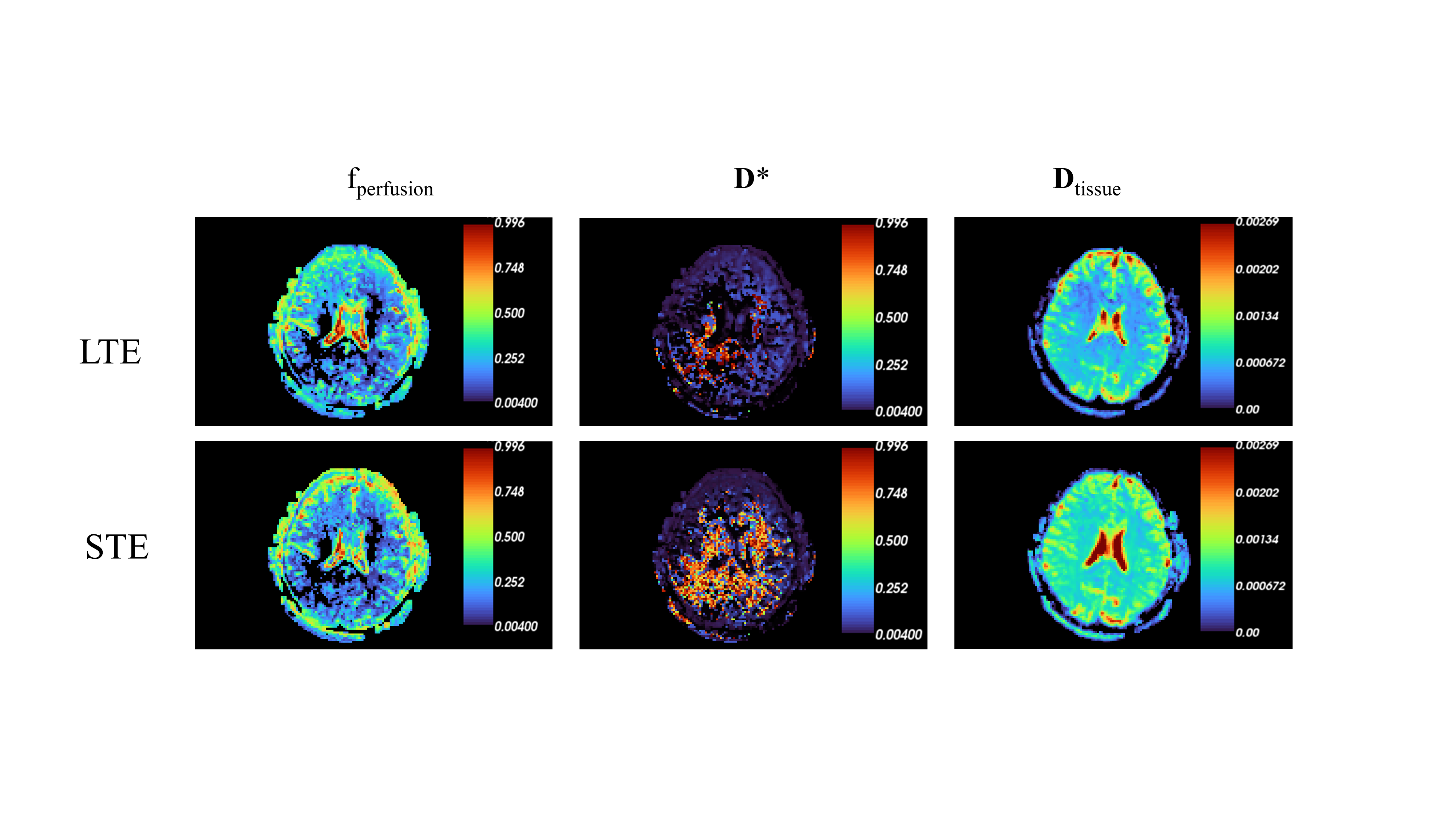
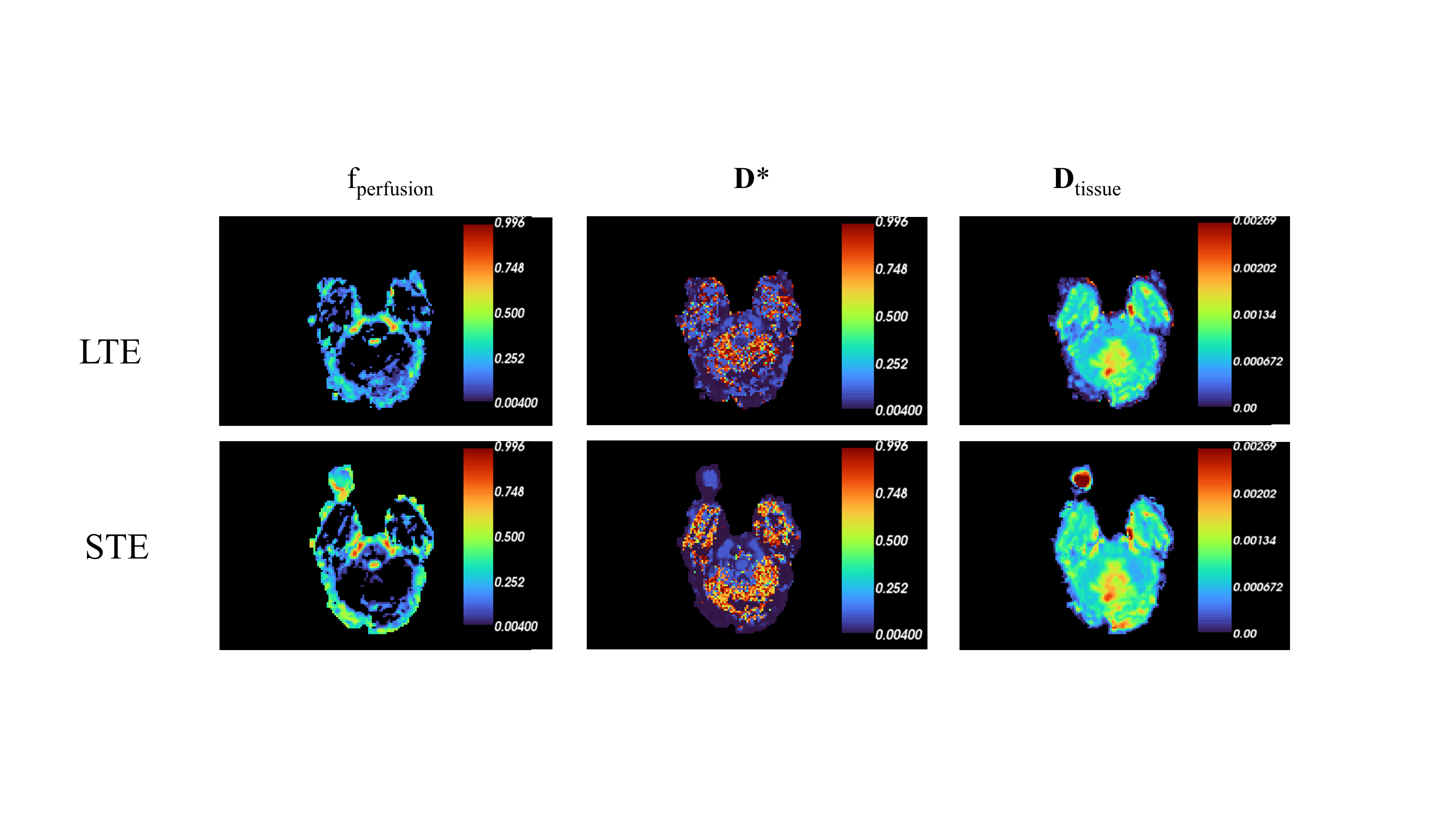
The mean DWI of the LTE diffusion images of three orthogonal DW directions and the 3 averages of STE diffusion images were shown in Figure 3, noticeable difference in signal intensity were observed as expected12. Figure 4 and 5 showed the IVIM fitting results using LTE and STE diffusion encoding respectively in one healthy subject, and one tumor patient.
Acknowledgements
This work was supported by the National Natural Scientific Foundation of China (82071994) and Key Project of Science and Technology of China (2023YFF1204300).References
1. Le Bihan, D. et al. MR imaging of intravoxel incoherent motions: application to diffusion and perfusion in neurologic disorders. Radiology 161, 401–407 (1986).
2. Le Bihan, D. et al. Separation of diffusion and perfusion in intravoxel incoherent motion MR imaging. Radiology 168, 497–505 (1988).
3. Le Bihan, D. Magnetic resonance imaging of perfusion. Magn. Reson. Med. 14, 283–292 (1990).
4. Paschoal, A. M., Leoni, R. F., Dos Santos, A. C. & Paiva, F. F. Intravoxel incoherent motion MRI in neurological and cerebrovascular diseases. NeuroImage Clin. 20, 705–714 (2018).
5. Topgaard, D. Isotropic diffusion weighting in PGSE NMR: Numerical optimization of the q-MAS PGSE sequence. Microporous Mesoporous Mater. 178, 60–63 (2013).
6. Szczepankiewicz, F. et al. The link between diffusion MRI and tumor heterogeneity: Mapping cell eccentricity and density by diffusional variance decomposition (DIVIDE). NeuroImage 142, 522–532 (2016).
7. Sjölund, J. et al. Constrained optimization of gradient waveforms for generalized diffusion encoding. J. Magn. Reson. 261, 157–168 (2015).
8. Andersson, J. L. R., Skare, S. & Ashburner, J. How to correct susceptibility distortions in spin-echo echo-planar images: application to diffusion tensor imaging. NeuroImage 20, 870–888 (2003).
9. Smith, S. M. et al. Advances in functional and structural MR image analysis and implementation as FSL. NeuroImage 23 Suppl 1, S208-219 (2004).
10. Glasser, M. F. et al. A multi-modal parcellation of human cerebral cortex. Nature 536, 171–178 (2016).
11. Graham, M. S., Drobnjak, I. & Zhang, H. Realistic simulation of artefacts in diffusion MRI for validating post-processing correction techniques. NeuroImage 125, 1079–1094 (2016).
12. Lasič, S., Szczepankiewicz, F., Eriksson, S., Nilsson, M. & Topgaard, D. Microanisotropy imaging: quantification of microscopic diffusion anisotropy and orientational order parameter by diffusion MRI with magic-angle spinning of the q-vector. Front. Phys. 2, (2014).
Figures