0644
Ultra-high gradient diffusion MRI on Connectome 2.0 reveals time-dependent diffusion and water exchange in human gray matter1Athinoula A. Martinos Center for Biomedical Imaging, Charlestown, MA, United States, 2Department of Radiology, Harvard Medical School, Boston, MA, United States, 3Donders Institute for Brain, Cognition and Behaviour, Nijmegen, Netherlands, 4Center of Functionally Integrative Neuroscience (CFIN) and MINDLab, Department of Clinical Medicine, Aarhus University, Aarhus, Denmark, 5Center for Biomedical Imaging, Department of Radiology, New York University School of Medicine, New York, NY, United States, 6Center for Advanced Imaging Innovation and Research (CAI2R), Department of Radiology, New York University School of Medicine, New York, NY, United States
Synopsis
Keywords: Microstructure, Microstructure
Motivation: In vivo mapping of exchange between intra-neurite and extracellular water in gray matter is challenging, as the required strong diffusion weighting significantly reduces the signal-to-noise ratio.
Goal(s): We aim to demonstrate the feasibility of in vivo neurite exchange imaging using the state-of-the-art Connectome 2.0 scanner equipped with high-performance gradient system (Gmax=500mT/m, (dG/dt)max=600T/m/s).
Approach: We acquired in vivo diffusion MRI measurements with multiple diffusion times (13-30ms) up to b-values of 17.5ms/μm2 on 5 human subjects. Anisotropic Kärger model (NEXI/SMEX) was used to estimate the exchange time from the diffusion data.
Results: The exchange time across the cortical ribbon is around 16ms.
Impact: The high performance gradient system on the Connectome 2.0 scanner enables in vivo mapping of water exchange time in gray matter, providing a tool to study neurite permeability in the healthy human brain and a variety of neuropsychiatric disorders.
Introduction
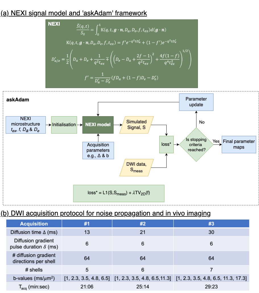
Probing gray matter(GM) microstructure using diffusion-weighted-imaging(DWI) offers unique opportunities to estimate cell dimensions in the brain1–5. Interestingly, the observed diffusion time-dependence at high b-value(10-100ms/μm2) signals in GM5,6 cannot be explained by a non-exchanging multi-compartment Gaussian model as in white matter, indicating a significant exchange effect between intra-neurite and extracellular water. This exchange effect can be modelled by extending the Kärger model to two exchanging anisotropic Gaussian compartments5–8. The Connectome 2.0 scanner(Gmax=500mT/m, (dG/dt)max=600T/m/s) allows the application of strong diffusion weightings at shorter time scales, increasing sensitivity to shorter exchange times and boosting the signal-to-noise ratio (SNR) with access to shorter TEs. Here, we imaged the exchange effect in vivo in human GM by employing diffusion weightings up to b=17.5ms/μm2 at short diffusion times(Δ=13-30ms) and fitted the anisotropic Kärger model to the spherical mean signals using a GPU-accelerated ‘askAdam’ processing framework(Fig.1a), enabling the estimation of water exchange time~10ms.Methods
Anisotropic Kärger model (NEXI/SMEX)We assumed two exchanging Gaussian compartments5-8: an anisotropic "stick"-like compartment (neurites) and local extra-cellular space (assumed isotropic for simplicity). The model has 4 parameters: neurite density(f), intra-neurite diffusivity(Da), extra-cellular diffusivity(De), and exchange time(tex). Here, we employed the narrow pulse solution of the orientationally-averaged anisotropic Kärger model(NEXI)6 for parameter estimation, as it provides similar numerical accuracy to the wide pulse solution(SMEX)5 and is more computationally efficient.
‘askAdam’ framework
‘askAdam’ is a model fitting tool9,10, leveraging the efficiency and suitability to handle large non-convex problems of stochastic gradient-descent based Adam optimizer commonly used in deep learning to perform network updates11. To assess its performance for NEXI, we compared 'askAdam' with conventional nonlinear least square(NLLS) fitting.
Noise propagation
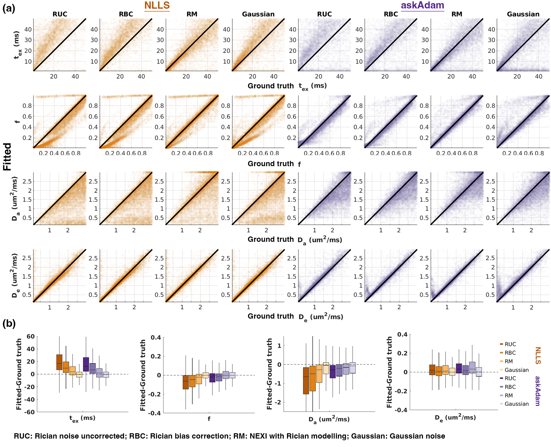
We evaluated the performance of NEXI model fitting using the voxel-wise NLLS and GPU-accelerated ‘askAdam’. We generated diffusion signals with the acquisition protocol in Fig.1b. We randomly chose NEXI parameters in 10,000 different combinations within the following range: tex=[1,50]ms, f=[0.01,0.99], Da=[0.1,3]μm2/ms, and De=[0.1,3]μm2/ms (Da≥De). We applied Rician or Gaussian noise to simulated signals at SNR=40 and fitted NEXI to the simulated signals. For Rician noise, we investigated two noise-floor-correction methods: correcting the noise floor before data fitting12 or integrating the noise floor within the signal model13.
In vivo MRI
Diffusion MRI data were acquired in 5 healthy volunteers(4F1M, 22-35years) on the 3T Connectome 2.0 scanner(MAGNETOM Connectom.X, Siemens Healthineers, Erlangen, Germany) using a custom-built 72-channel head coil14. Data acquisition included whole-brain T1-MPRAGE data of 0.9-mm isotropic resolutionand DWIs with the following protocol: SMS=2, 2mm isotropic, PF=6/8, GRAPPA=2, TR/TE=3600/54ms using the diffusion scheme in Fig.1b. We acquired interspersed b=0 images every 16 DWIs. DWI data were processed based on the DESIGNER pipeline15.
ROI analysis
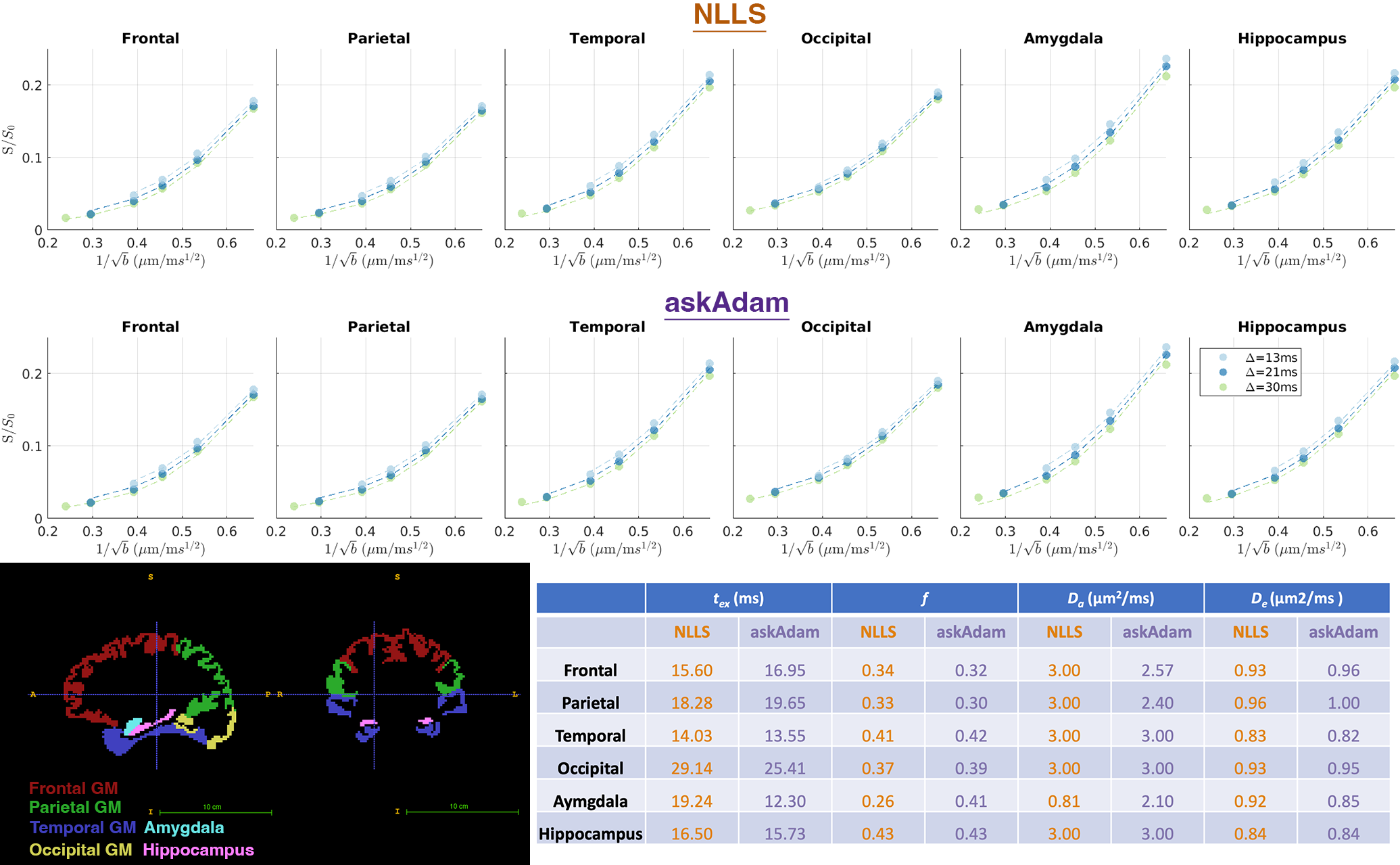
We used Freesurfer16 to segment GM ROIs in MPRAGE and reconstructed the cortical surface for each subject. The GM parcellation was transformed to the DWI space using nonlinear transformation17. We generated 6 ROIs from SynthSeg18, including 4 lobes(frontal, parietal, temporal, and occipital), amygdala, and hippocampus. The spherical mean DWI signals were averaged over each ROI and over all the subjects. We fitted NEXI using NLLS and ‘askAdam’. DWIs at b=1ms/μm2 were excluded from fitting given that restricted diffusion in the soma may still have non-negligible contributions to DWIs at low b-values.
Parameter mapping and cortical analysis
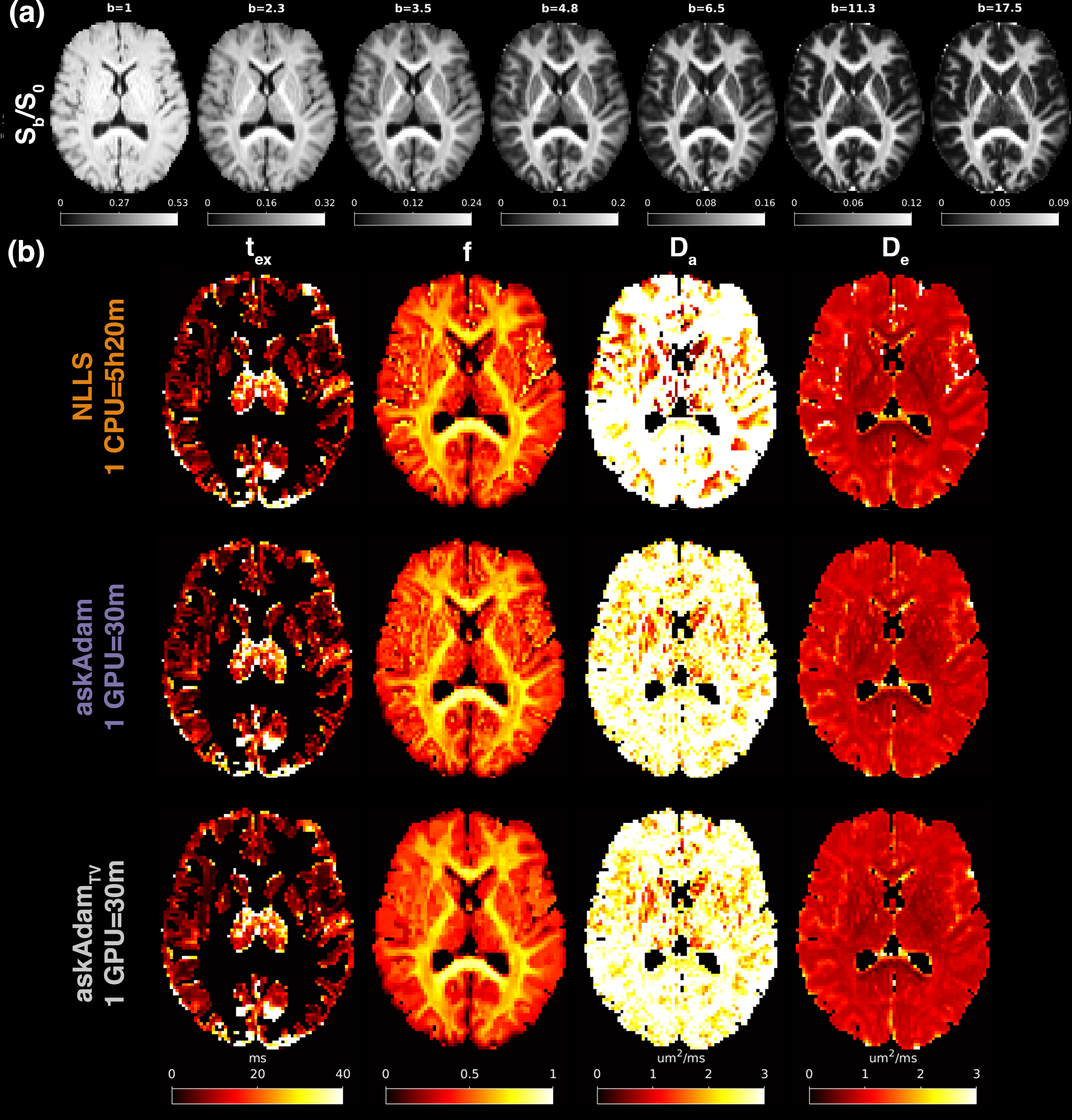
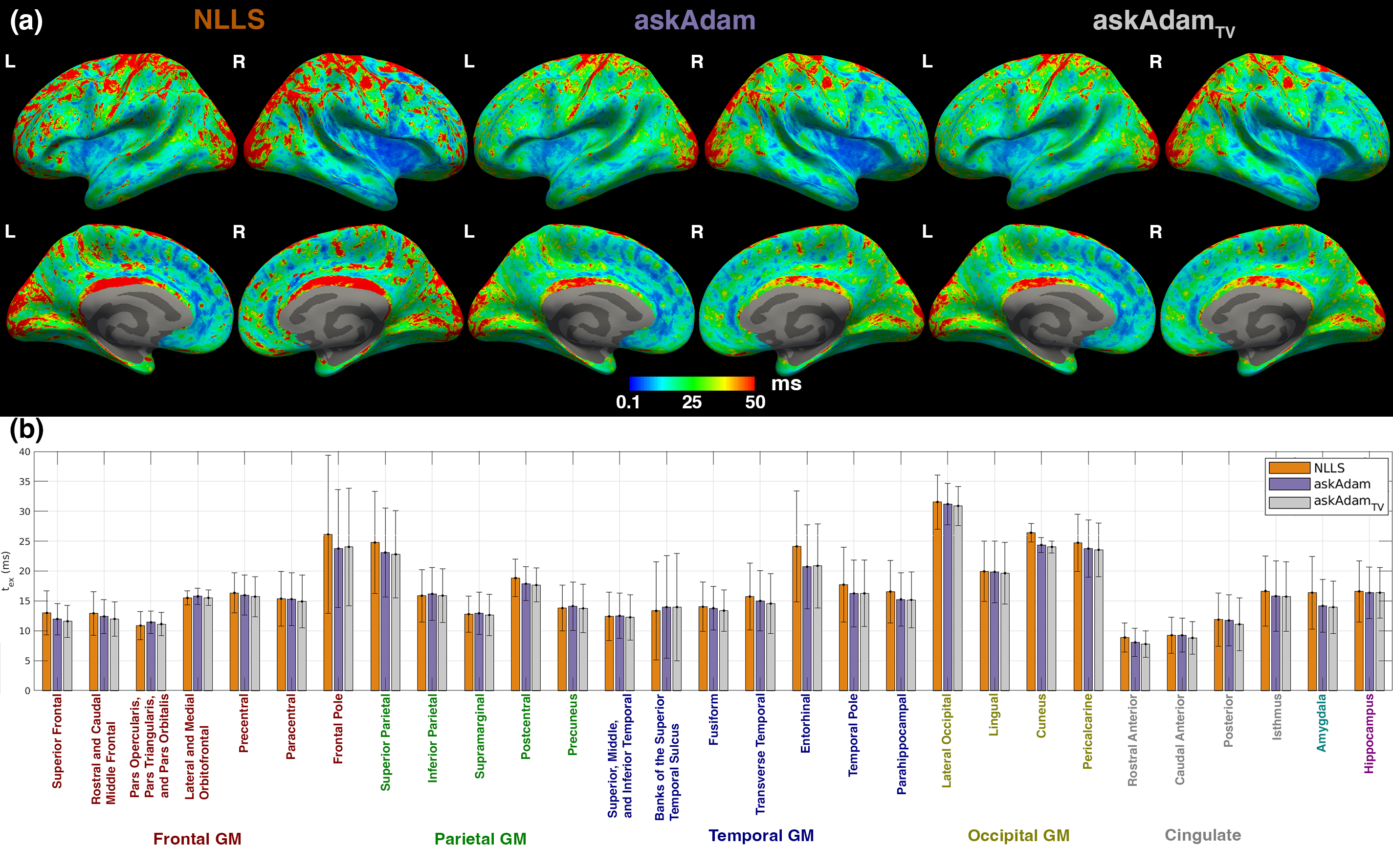
NEXI parametric maps were derived by applying the NLLS and ‘askAdam’ fitting. Additionally, we applied ‘askAdam’ with 2-dimensional spatial total-variation(TV) regularisation on the in-plane f map for NEXI fitting (askAdamTV, lambda=0.002). Parametric maps were transformed into the MPRAGE space and projected onto the cortical surface for visualization.
Results and Discussion
Incorporating noise correction methods reduces the bias in all tissue parameters(Fig.2); incorporating Rician model in NEXI or having Gaussian noise produced the best performance(Fig.2b). NLLS and ‘askAdam’ display similar noise performance.In vivo DWI signal decreases with diffusion time in all ROIs(Fig.3). The estimated exchange times ranged from 10.7ms(Amygdala) and 29.6ms(Occipital). NLLS and ‘askAdam’ had similar estimation performance, in accordance with the noise propagation results.
The b=0 images had an SNR=58.7 in the cortical ribbon(Fig.4a). ‘askAdamTV’ had the lowest inter-quartile range(IQR) on tex across the cortical ribbon amongst the three tested methods at the subject level(IQRNLLS:16.6ms; IQRaskAdam=14.4ms; IQRaskAdamTV=13.7ms, Fig.4b). The mean tex across the cortical ROIs was 17.0ms, 16.3ms and 16.0ms for NLLS, askAdam and askAdamTV, slightly longer than previously reported(~10ms, mouse spinal cord)19,20. Relatively longer tex was observed in the motor, somatosensory, visual and posterior cingulate cortices(Fig.5), in agreement with the pattern of myelin index in literatures21.
Conclusions
Ultra-high gradient diffusion MRI using Gmax=500mT/m on the Connectome 2.0 scanner enables probing water exchange effects in vivo in GM. NEXI fitting was accelerated by over 10x using ‘askAdam’.Acknowledgements
Acknowledgments This study is support by NIH under the award number: DP5OD031854, R01NS118187, P41EB015896, P41EB030006, U01EB026996, S10RR023401, S10RR019307, R21NS081230, R01NS088040, P41EB017183.References
1. Palombo M, Ianus A, Guerreri M, et al. SANDI: A compartment-based model for non-invasive apparent soma and neurite imaging by diffusion MRI. Neuroimage. 2020;215:116835.
2. Alexander DC, Hubbard PL, Hall MG, et al. Orientationally invariant indices of axon diameter and density from diffusion MRI. NeuroImage. 2010;52(4):1374-1389.
3. Huang SY, Tian Q, Fan Q, et al. High-gradient diffusion MRI reveals distinct estimates of axon diameter index within different white matter tracts in the in vivo human brain. Brain Struct Funct. 2020;225(4):1277-1291.
4. Veraart J, Nunes D, Rudrapatna U, et al. Nonivasive quantification of axon radii using diffusion MRI. Elife. 2020;9:e49855.
5. Olesen JL, Østergaard L, Shemesh N, Jespersen SN. Diffusion time dependence, power-law scaling, and exchange in gray matter. Neuroimage. 2022;251:118976.
6. Jelescu IO, Skowronski A de, Geffroy F, Palombo M, Novikov DS. Neurite Exchange Imaging (NEXI): A minimal model of diffusion in gray matter with inter-compartment water exchange. Neuroimage. 2022;256:119277.
7. Fieremans E, Novikov DS, Jensen JH, Helpern JA. Monte Carlo study of a two‐compartment exchange model of diffusion. NMR in Biomedicine. 2010 Aug;23(7):711-24.
8. Nilsson M, Alerstam E, Wirestam R, Sta F, Brockstedt S, Lätt J. Evaluating the accuracy and precision of a two-compartment Kärger model using Monte Carlo simulations. Journal of Magnetic Resonance. 2010 Sep 1;206(1):59-67.
9. Chan KS, Kim TH, Bilgic B, Marques JP. Semi-supervised learning for fast multi-compartment relaxometry myelin water imaging (MCR-MWI). In: Proceedings 30. Annual Meeting International Society for Magnetic Resonance in Medicine. London, United Kingdom.
10. Marques JP, Hollander DD, Chan KS. Improved R2* and QSM mapping for dummies - ask Adam. In: Proceedings 31. Annual Meeting International Society for Magnetic Resonance in Medicine. Toronto, Canada.
11. Kingma DP, Ba J. Adam: A Method for Stochastic Optimization. arXiv. 2014. doi:10.48550/arxiv.1412.6980
12. Koay CG, Basser PJ. Analytically exact correction scheme for signal extraction from noisy magnitude MR signals. J Magn Reson. 2006;179(2):317-322.
13. Uhl Q, Pavan T, Molendowska M, Jones DK, Palombo M, Jelescu I. Quantifying human gray matter microstructure using NEXI and 300 mT/m gradients. arXiv. 2023. doi:10.48550/arxiv.2307.09492
14. Mahmutovic M, Shrestha M, Ramos-Llordén G, et al. A 72-channel Head Coil with an Integrated 16-Channel Field Camera for the Connectome 2.0 Scanner. Submitted to ISMRM 2024.
15. Ades-Aron B, Veraart J, Kochunov P, et al. Evaluation of the accuracy and precision of the diffusion parameter EStImation with Gibbs and NoisE removal pipeline. NeuroImage. 2018;183:532-543.
16. Fischl B. FreeSurfer. NeuroImage. 2012;62(2):774-781.
17. Avants BB, Tustison NJ, Song G, Cook PA, Klein A, Gee JC. A reproducible evaluation of ANTs similarity metric performance in brain image registration. Neuroimage. 2011;54(3):2033-2044.
18. Billot B, Magdamo C, Cheng Y, Arnold SE, Das S, Iglesias JE. Robust machine learning segmentation for large-scale analysis of heterogeneous clinical brain MRI datasets. Proc National Acad Sci. 2023;120(9):e2216399120.
19. Williamson NH, Ravin R, Benjamini D, et al. Magnetic resonance measurements of cellular and sub-cellular membrane structures in live and fixed neural tissue. eLife. 2019;8:e51101. doi:10.7554/elife.51101
20. Williamson NH, Ravin R, Cai TX, Falgairolle M, O’Donovan MJ, Basser PJ. Water exchange rates measure active transport and homeostasis in neural tissue. PNAS Nexus. 2023;2(3):pgad056.
21. Ali TS, Lv J, Calamante F. Gradual changes in microarchitectural properties of cortex and juxtacortical white matter: Observed by anatomical and diffusion MRI. Magn Reson Med. 2022;88(6):2485-2503.
Figures