0643
Unbiasing the spherical variance of a diffusion-weighted MR signal: An application to intra-axonal T2 estimation1Laboratorio de Procesado de Imagen (LPI), ETSI Telecomunicación, Universidad de Valladolid, Valladolid, Spain, 2Department of Radiology, Center for Biomedical Imaging, Department of Radiology, New York University Grossman School of Medicine, New York, NY, United States
Synopsis
Keywords: Diffusion Modeling, Diffusion/other diffusion imaging techniques, intra-axonal T2, spherical variance, Rician bias
Motivation: The spherical variance (SV) from multiparametric diffusion MRI acquisitions enables the estimation of the axonal T2 relaxation time. The SV is prone to a noise-induced bias due to positively skewed Rician statistics, leading to overestimation in the axonal T2 parameter.
Goal(s): To derive a method to mitigate the Rician bias in the SV parameter map.
Approach: A closed-form formula to remove the Rician bias from the SV has been analytically derived and verified with in silico and in vivo data.
Results: The bias-corrected SV reduces the estimation error compared to the SV, translating to a less pronounced misestimation in the axonal T2 parameter.
Impact: The SV is a practical parameter to infer the properties of restricted compartments with diffusion MRI. This work shows a formula to remove the Rician bias from the SV. The correction can be used for other SV-based diffusion MRI measures.
Introduction
The intra-axonal T2 parameter can be estimated in numerous ways1-4, with the spherical mean (SM) based approach being one of the most straightforward ways4. Although the SM is easy to compute in an unbiased manner5,6 and its characteristics are well-studied7, it is sensitive to isotropic contributions from the diffusion MRI signal like those originating from the cell nuclei3,8. The spherical variance (SV) has been recently introduced as an alternative for SM to estimate the intra-axonal T2 relaxation time3,8. However, its computation introduces a larger positive noise-induced bias than the SM under Rician statistics, which has been de facto a standard noise assumption in diffusion MRI data10,11. This work derives a closed-form analytical formula to suppress this bias in the SV of a diffusion-weighted MRI signal, leading to a possible better estimate of the intra-axonal T2 relaxation.Methods and materials
Spherical variance (SV): Given a set of diffusion-weighted MR samples $$$\left\{S(\mathbf{g}_j,b) \right\}_{j=1}^{N_g}$$$ acquired under a b-value $$$b$$$, the SV is defined as:$$\mathcal{V}(b)=\eta\sum_{j=1}^{N_g}w_j\left( S(\mathbf{g}_j,b)-\sum_{k=1}^{N_g}w_kS(\mathbf{g}_k, b)\right)^2$$where $$$\eta=\left(1-\sum_{j=1}^{N_g}w^2_j\right)^{-1}$$$ and the weights $$$w_j$$$ compensate for the gradient sampling imperfections. The SV can be retrieved from the coefficients of spherical harmonics (SH) expansion of the signal up to order $$$L$$$ as9,13:$$\mathcal{V}(b)=\frac{1}{4\pi}\sum_{l=2,\mathrm{even}}^{L}\sum_{m=-l}^{l}c^2_{l,m}.$$
Unbiasing the SV: We assume the samples are Rician distributed, i.e, $$$S(\mathbf{g}_j,b)\sim \text{Rice}(A(\mathbf{g}_j,b), \sigma(b))$$$ with the amplitude parameter $$$A(\mathbf{g}_j,b)$$$ and the noise map $$$\sigma(b)$$$ given for the shell at b-value, $$$b$$$. The asymptotic expansion of the expectation operator of the random variable $$$S(\mathbf{g},b)$$$ is given by12 $$$\mathbb{E}\{S(\mathbf{g},b)\}\simeq A(\mathbf{g},b)+\frac{1}{2}\frac{\sigma^2(b)}{A(\mathbf{g},b)}+\frac{1}{8}\frac{\sigma^4(b)}{A^3(\mathbf{g},b)}$$$. Assuming the samples $$$S(\mathbf{g}_j,b)$$$ and $$$S(\mathbf{g}_k,b)$$$ are pairwise independent for $$$j\neq k$$$ and their second-order moments are finite, the closed-form formula for the unbiased SV can be analytically derived as:
$$\mathcal{V}_u(b)=\mathcal{V}(b)-\eta\sigma^2(b)\left(2-\overline{\overline{w}}-\overline{S}(b)\overline{S^{-1}}(b)\right)-\eta\frac{\sigma^4(b)}{4}\left(2\overline{\overline{S^{-2}}}(b)-\left(\overline{S^{-1}}(b)\right)^2\right)$$ with $$$\overline{\overline{w}}=\sum_{j=1}^{N_g}w_j^2$$$, and the weighted sample-moments $$$\overline{S^{p}}(b)$$$ and $$$\overline{\overline{S^{p}}}(b)$$$ given by:$$\overline{S^{p}}(b)=\sum_{j=1}^{N_g}w_jS^{p}(\mathbf{g}_j,b)\ \mathrm{and}\ \overline{\overline{S^{p}}}(b)=\sum_{j=1}^{N_g}w_j^2S^{p}(\mathbf{g}_j,b)\ \mathrm{with}\ p\in\mathcal{Z}.$$
Axonal T2 estimation: Given a strong diffusion-weighted MR signal acquired under a fixed b-value $$$b$$$ and two different echo times (TE), $$$TE_1$$$ and $$$TE_2$$$, we can estimate the axonal T2 parameter as follows3:$$T_{2a}=\frac{2(TE_2-TE_1)}{\ln\left(\mathcal{V}_u\left(b,TE_1\right)\right)-\ln\left(\mathcal{V}_u\left(b, TE_2\right)\right)}$$
where $$$\mathcal{V}_u\left(b,TE_i\right)$$$ is the unbiased SV of the signal at $$$b$$$ and $$$TE_i$$$.
In silico data: We generated synthetic data using a two-component representation that integrates the cellular $$$(1-f)$$$ and free-water $$$f=0.2$$$ components. The cellular component is modelled using two fibre bundles with an intra-axonal compartment modelled as a stick with perpendicular diffusivities $$$ \lambda^{\perp}=0$$$ (see the supplementary materials in14 for details). The ensemble signal is convolved with the fODF. The TEs equal $$$TE_1=50\ \mathrm{ms}$$$ and $$$TE_2=150\ \mathrm{ms}$$$.
In vivo data: We used the Human Connectome Project (HCP) MGH data17 acquired under $$$b=\{1,3,5,10\}\cdot 10^3\ \mathrm{s}/\mathrm{mm}^2$$$ and the MUDI dataset18,19 acquired under $$$b=3,000\ \mathrm{s}/\mathrm{mm}^2$$$ and $$$TE\in\{80,130\}\ \mathrm{ms}$$$.
Noise estimation: The spatially-variant noise maps for the in vivo data were estimated using the variance-stabilized homomorphic filter15,16 for each gradient direction separately, and then the gradient-specific maps were averaged to obtain a single map $$$\sigma(b)$$$ per $$$b$$$-value.
Results
Fig. 1 presents the synthetic experiment with SV estimated as a function of mean diffusion-weighted signal SNR and non-diffusion-weighted SNR under $$$b=\{1,3,5,10\}\cdot 10^3\ \mathrm{s}/\mathrm{mm}^2$$$ and $$$TE=80\ \mathrm{ms}$$$. We observe a significant error reduction of $$$\mathcal{V}_u(b)$$$ compared to $$$\mathcal{V}(b)$$$ for the whole range of SNRs.Fig. 2 depicts the histograms of SVs generated from $$$25^3$$$ experiment replicas. The bias-corrected histograms have been shifted negatively towards the reference SVs.
Fig. 3 illustrates SVs calculated from a single HCP subject with $$$\mathcal{V}(b)$$$ and its corrected version $$$\mathcal{V}_u(b)$$$. Again, we observe a positive noise-induced bias in $$$\mathcal{V}(b)$$$ that is compensated with the unbiased SV $$$\mathcal{V}_u(b)$$$.
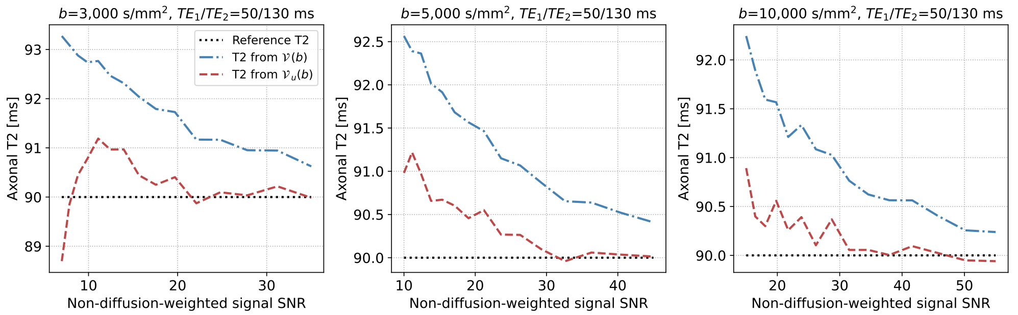
Fig. 4 compares the estimated intra-axonal T2 relaxation time from $$$\mathcal{V}(b)$$$ and $$$\mathcal{V}_u(b)$$$ in a synthetic scenario. We notice here a positive bias reduction in the estimated intra-axonal T2 from $$$\mathcal{V}_u(b)$$$.
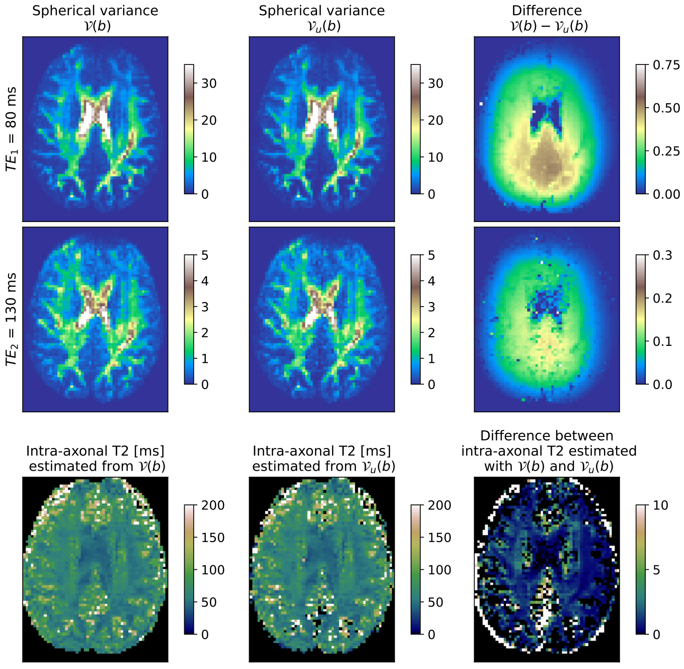
Fig. 5 shows the SV and intra-axonal T2 relaxation maps estimated from a single MUDI subject under $$$b=3,000\ \mathrm{s}/\mathrm{mm}^2$$$, with $$$\mathcal{V}(b)$$$ and $$$\mathcal{V}_u(b)$$$. We observe a positive bias compensation both in the SV and the intra-axonal T2 maps using $$$\mathcal{V}_u(b)$$$
Discussion and conclusions
We have analytically derived a closed-form solution to compensate for a Rician bias in the SV and verified it with in silico and in vivo data, leading to the following conclusions:- The proposal corrects the bias in SV and intra-axonal T2 for the whole range of reasonable SNRs.
- It requires a minimal computational load as it can be estimated from sample moments or the SH decomposition.
- It provides the potential for using the SV to estimate other (to be proposed) tissue relaxation or diffusion properties, which were previously not practicable due to a significant bias.
Acknowledgements
This work was supported by research grants PID2021-124407NB-I00, funded by MCIN/AEI/10.13039/501100011033/FEDER, UE, and TED2021-130758B-I00, funded by MCIN/AEI/10.13039/501100011033 and the European Union “NextGenerationEU/PRTR”. Tomasz Pieciak acknowledges the Polish National Agency for Academic Exchange for grant PPN/BEK/2019/1/00421 under the Bekker programme and the Ministry of Science and Higher Education (Poland) under the scholarship for outstanding young scientists (692/STYP/13/2018). Guillem París was funded by the Consejería de Educación de Castilla y León and the European Social Fund through the “Ayudas para financiar la contratación predoctoral de personal investigador - Orden EDU/1100/2017 12/12” program.References
- Veraart, J., Novikov, D. S., & Fieremans, E. (2018). TE dependent Diffusion Imaging (TEdDI) distinguishes between compartmental T2 relaxation times. NeuroImage, 182, 360-369.
- Barakovic, M., Tax, C. M., et al. (2021). Resolving bundle-specific intra-axonal T2 values within a voxel using diffusion-relaxation tract-based estimation. NeuroImage, 227, 117617.
- Pizzolato, M., Andersson, M., Canales-Rodríguez, E. J., Thiran, J. P., Dyrby, T. B. (2022). Axonal T2 estimation using the spherical variance of the strongly diffusion-weighted MRI signal. Magnetic resonance imaging, 86, 118-134.
- McKinnon, E. T., Jensen, J. H. (2019). Measuring intra-axonal T2 in white matter with direction-averaged diffusion MRI. Magnetic resonance in medicine, 81(5), 2985-2994.
- Afzali, M., Knutsson, H., Özarslan, E., & Jones, D. K. (2021). Computing the orientational-average of diffusion weighted MRI signals: A comparison of different techniques. Scientific Reports, 11(1), 1-16
- Pieciak T., Afzali M., Tristán-Vega A., Aja-Fernández S. (2022), Unbiasing orientationally-averaged diffusion MRI signal, Joint Annual Meeting ISMRM-ESMRMB & ISMRT, 3900, London, United Kingdom
- McKinnon, E. T., Jensen, J. H., Glenn, G. R., & Helpern, J. A. (2017). Dependence on b-value of the direction-averaged diffusion-weighted imaging signal in brain. Magnetic resonance imaging, 36, 121-127.
- Andersson, M., Kjer, H. M., et al. (2020). Axon morphology is modulated by the local environment and impacts the noninvasive investigation of its structure–function relationship. Proceedings of the National Academy of Sciences, 117(52), 33649-33659.
- Zucchelli, M., Deslauriers-Gauthier, S., & Deriche, R. (2020). A computational framework for generating rotation invariant features and its application in diffusion MRI. Medical image analysis, 60, 101597.
- Gudbjartsson, H., Patz, S. (1995). The Rician distribution of noisy MRI data. Magnetic resonance in medicine, 34(6), 910-914.
- Aja-Fernández, S., Vegas-Sánchez-Ferrero, G. (2016). Statistical analysis of noise in MRI. Switzerland: Springer International Publishing.
- Pieciak, T., Rabanillo-Viloria, I., Aja-Fernández, S. (2018, April). Bias correction for non-stationary noise filtering in MRI. In 2018 IEEE 15th International Symposium on Biomedical Imaging (ISBI 2018) (pp. 307-310). IEEE.
- Descoteaux, M., Angelino, E., Fitzgibbons, S., Deriche, R. (2007). Regularized, fast, and robust analytical Q-ball imaging. Magnetic resonance in medicine, 58(3), 497-510
- Pieciak, T., París, G., Beck, D., Maximov, I. I., Tristán-Vega, A., de Luis-García, R., Westlye, L.T., Aja-Fernández, S. (2023). Spherical means-based free-water volume fraction from diffusion MRI increases non-linearly with age in the white matter of the healthy human brain. NeuroImage, 279, 120324.
- Pieciak, T., Aja-Fernandez, S., Vegas-Sanchez-Ferrero, G. (2017). Non-stationary Rician noise estimation in parallel MRI using a single image: a variance-stabilizing approach. IEEE transactions on pattern analysis and machine intelligence, 39(10), 2015-2029.
- Aja-Fernandez, S., Pieciak, T., Vegas-Sánchez-Ferrero, G. (2015). Spatially variant noise estimation in MRI: A homomorphic approach. Medical image analysis, 20(1), 184-197.
- Fan, Q., Witzel, T., Nummenmaa, A., et al. (2016). MGH–USC Human Connectome Project datasets with ultra-high b-value diffusion MRI. Neuroimage, 124, 1108-1114.
- Pizzolato, M., Palombo, M., et al. (2020). Acquiring and predicting multidimensional diffusion (MUDI) data: an open challenge. In Computational Diffusion MRI: MICCAI Workshop, Shenzhen, China, October 2019 (pp. 195-208). Springer International Publishing.
- Pieciak, T., Afzali, M., Bogusz, F., Ciupek, D., Jones, D. K., & Pizzolato, M. (2021). Is the inversion time important? A study of the reciprocal influence of inversion time and b-value on diffusion and longitudinal relaxation in MRI. In International Society for Magnetic Resonance Imaging (ISMRM 2021) (p. 209).
Figures
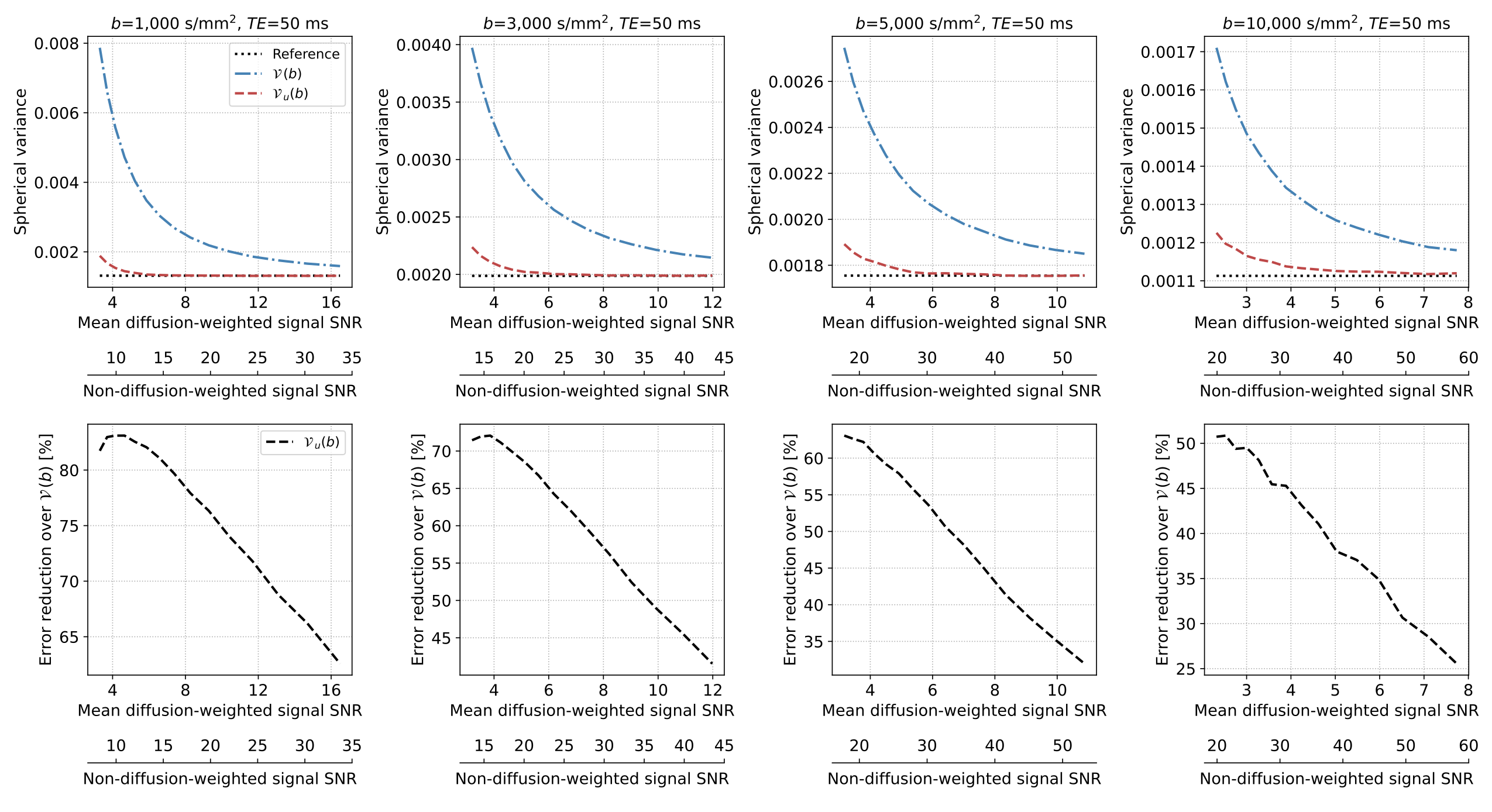
Fig. 1 The in silico results present the spherical variance under different $$$b$$$-values and a fixed $$$TE=50 \ \mathrm{ms}$$$ as a function of mean diffusion-weighted signal SNR and non-diffusion-weighted SNR (i.e., baseline SNR at $$$b=0$$$). The bottom row presents the error reduction of $$$\mathcal{V}_u(b)$$$ over $$$\mathcal{V}(b)$$$. The plots present median values calculated over $$$25^3$$$ experiment replicas for each SNR level.
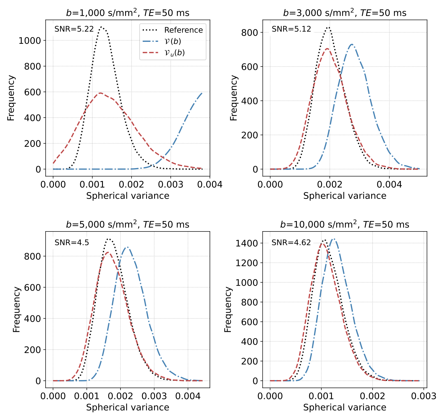
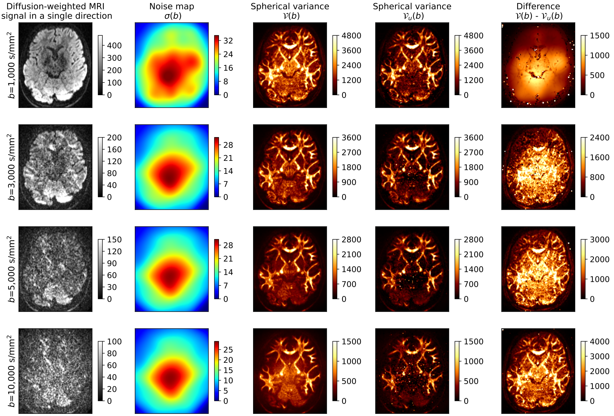
Fig. 3 Spherical variance maps obtained at four different $$$b$$$-values from the Human Connectome Project MGH data (subject 1032, slice 40). The first column presents the noisy slice at a random gradient direction, the second depicts the local SNR map $$$\sigma(b)$$$, the third and fourth show $$$\mathcal{V}(b)$$$ and $$$\mathcal{V}_u(b)$$$, respectively, and the last one presents the difference between $$$\mathcal{V}(b)$$$ and $$$\mathcal{V}_u(b)$$$.