0636
Articular Cartilage 3D T2* Mapping at 7 Tesla in Patients with Medial Meniscus Posterior Root Tears1CMRR/Radiology, University of Minnesota, Minneapolis, MN, United States, 2Biomedical Engineering, Lemer Research Institute, Cleveland Clinic, Cleveland, OH, United States, 3Colby College, Waterville, ME, United States, 4Twin Cities Orthopedics, Edina, MN, United States, 5Radiology, Ohio State University, Columbus, OH, United States
Synopsis
Keywords: Cartilage, Cartilage, Osteoarthritis
Motivation: Meniscal root tears are increasingly recognised as the ‘root cause’ of osteoarthritis with articular cartilage loss as the hallmark of the disease.
Goal(s): Utilising SNR gains at 7T UHF MRI to detect early changes in cartilage composition.
Approach: Compare high resolution quantitative cartilage 3D T2* relaxation times at 7T in patients with arthroscopically confirmed medial meniscal posterior root tears (MMPRT’s) to matched controls.
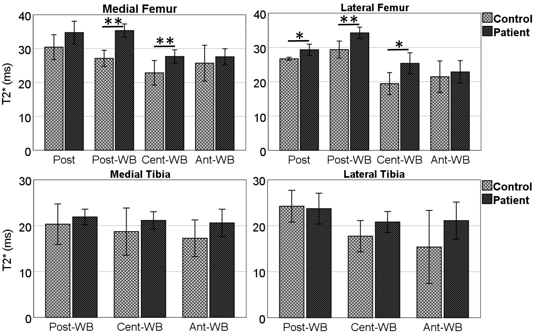
Results: Prolonged T2* relaxation times were found in the weight bearing medial and lateral cartilage of the femur in patients with meniscal tears, when compared with controls. No changes were seen in the tibial cartilage.
Impact: Identifying early cartilage degeneration using high resolution T2* at 7 Tesla could inform timely treatments, such as prompt meniscal repair, potentially preventing the onset of osteoarthritis.
Introduction
Meniscal root tears are increasingly recognized as the ‘root cause’ of osteoarthritis (OA) and considered a previously unrecognized ‘silent epidemic’ 1. Medial meniscus posterior root tears (MMPRTs) in particular, are one of the strongest risk factors for the development and progression of knee OA2. Meniscal tears cause meniscal extrusion3. This is thought to lead to accelerated cartilage breakdown2. Previous studies of T2 and T2* relaxation time mapping demonstrated a strong correlation with collagen architecture and histologic zones of articular cartilage4, 5. The aim of this study was to utilize the signal-to-noise ratio (SNR) and resolution gains at 7T for the evaluation of compositional changes in the cartilage of patients with arthroscopically verified MMPRTs using 3D T2* mapping. We hypothesize that the T2* values will increase in the cartilage of the patients, suggestive of degenerative changes in the tissue due to the MMPRTs.Methods
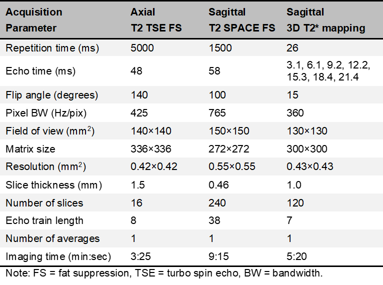
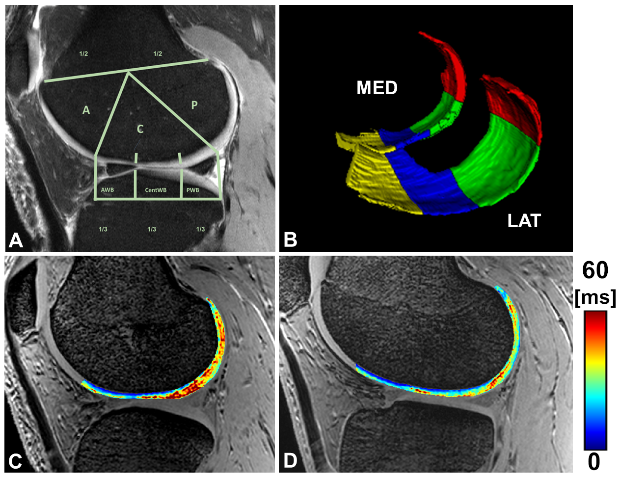
This is an IRB approved and HIPPA compliant study. Ten patients with unilateral MMPRTs (mean age: 52 years; age range: 36-62 years; mean body mass index (BMI): 28 kg/m2, 8 females) and five asymptomatic controls (mean age: 48 years; age range: 34-60 years; mean BMI: 28 kg/m2, 3 females) were included in the study. MRI was conducted on a 7T system using a birdcage transmit and 28-channel receive phased-array knee coil. The MRI protocol included morphological sequences and a quantitative 3D multi-echo gradient recalled echo (GRE) sequence with seven echo times (TE) between 3.1 and 21.4 ms (Fig 1). The T2* maps of the articular cartilage were calculated by fitting a mono-exponential signal decay of the multi-echo T2* data with a two-parametric least-square fitting routine in Matlab. The fitting accuracy was evaluated by calculating the root mean square error (RMSE) normalized to the estimated signal intensity at the echo time of 0 ms. The 3D segmentation of the medial and the lateral femoral and tibial cartilage was manually performed on 120 high resolution T2*-weighted images using ITK-SNAP. Each compartment of the femoral cartilage was subdivided into lateral and medial regions and then into four subregions: anterior weight bearing (anterior WB), central WB, and posterior WB and posterior regions6 (Fig. 2). A linear mixed effect model was used to compare the T2* values of the cartilage regions between the controls, and the corresponding regions in the MMPRT patients. Extrusion of the medial and lateral menisci in patients and controls was measured by three musculoskeletal radiologists, blinded to T2* results, as described previously7. Arthroscopic evaluation at the time of meniscal repair confirmed the presence of MMPRT in all patients and cartilage was intraoperatively assessed.Results
Quantitative T2* evaluation: Longer T2* relaxation times were found in the distal femoral articular cartilage in the central WB (P < 0.05) and posterior WB (P < 0.01) regions in both the medial and lateral femoral compartments. In addition, the posterior region of the lateral femoral condyle revealed significantly higher T2* values (P < 0.05) when compared to the same regions in the controls (Figure 3). The highest differences were found in the posterior WB region of the medial femoral condyle. No statistical differences were found in the medial and lateral tibial plateau. The mean normalised RMSE was below 5% for all the analysed regions, which demonstrates a good reliability of the fitting results. Meniscus extrusion: Positive extrusion criteria were met in 10 patients medial meniscus, criteria were not met in in lateral menisci and control group. Arthroscopic evaluation: Arthroscopy evaluation revealed grades I to III cartilage lesions on the femoral, tibial and patellar cartilage. MMPRT was confirmed in all patients.Discussion
In the medial and lateral knee joint compartment, elevated T2* values were found in the weight bearing areas of articular cartilage in patients with MMPRT’s. Previous studies of T2 and T2* relaxation time mapping demonstrated a strong correlation with alteration of collagen architecture and association with osteoarthritis8, 9 10. It has been shown that biomechanical consequences of a posterior root tear in the medial meniscus are similar to total meniscectomy, which significantly increases articular cartilage contact pressure in the medial as well as the lateral compartment11. Our findings of prolonged T2* relaxation times in both, the medial and lateral compartments in patients emphasise the significant impact of the medial meniscal posterior root tear on the entire knee joint health.Conclusion
The results support the hypothesis that MMPHRT’s are the 'root cause' of OA as evidenced by prolonged cartilage T2* relaxation times in patients. Thus, identifying early cartilage degeneration could inform timely treatments, potentially preventing the onset of OA.Acknowledgements
National Institute of Biomedical Imaging and Bioengineering (P41 EB027061: Technology to Realize the Full Potential of UHF MRI), S10 OD025256 (7 Tesla Terra Instrument) and R01EB034575.References
1. Cinque ME, Chahla J, Moatshe G, Faucett SC, Krych AJ, LaPrade RF. Meniscal root tears: a silent epidemic. Br J Sports Med. 2018;52(13):872-6. Epub 2018/03/27. doi: 10.1136/bjsports-2017-098942. PubMed PMID: 29574455.2. Krych AJ, Hevesi M, Leland DP, Stuart MJ. Meniscal Root Injuries. J Am Acad Orthop Surg. 2020;28(12):491-9. doi: 10.5435/JAAOS-D-19-00102. PubMed PMID: 31693530.3. Fox AJ, Bedi A, Rodeo SA. The basic science of human knee menisci: structure, composition, and function. Sports Health. 2012;4(4):340-51. doi: 10.1177/1941738111429419. PubMed PMID: 23016106; PMCID: PMC3435920.4. Einarsson E, Svensson J, Folkesson E, Kestila I, Tjornstrand J, Peterson P, Finnila MAJ, Hughes HV, Turkiewicz A, Saarakkala S, Englund M. Relating MR relaxation times of ex vivo meniscus to tissue degeneration through comparison with histopathology. Osteoarthr Cartil Open. 2020;2(2). Epub 2021/05/12. doi: 10.1016/j.ocarto.2020.100061. PubMed PMID: 33972933; PMCID: PMC7610736.5. Nieminen MT, Rieppo J, Toyras J, Hakumaki JM, Silvennoinen J, Hyttinen MM, Helminen HJ, Jurvelin JS. T2 relaxation reveals spatial collagen architecture in articular cartilage: a comparative quantitative MRI and polarized light microscopic study. Magn Reson Med. 2001;46(3):487-93. doi: 10.1002/mrm.1218. PubMed PMID: 11550240.6. Hunter DJ, Guermazi A, Lo GH, Grainger AJ, Conaghan PG, Boudreau RM, Roemer FW. Evolution of semi-quantitative whole joint assessment of knee OA: MOAKS (MRI Osteoarthritis Knee Score). Osteoarthritis Cartilage. 2011;19(8):990-1002. Epub 20110523. doi: 10.1016/j.joca.2011.05.004. PubMed PMID: 21645627; PMCID: PMC4058435.7. Costa CR, Morrison WB, Carrino JA. Medial meniscus extrusion on knee MRI: is extent associated with severity of degeneration or type of tear? AJR Am J Roentgenol. 2004;183(1):17-23. doi: 10.2214/ajr.183.1.1830017. PubMed PMID: 15208101.8. Zarins ZA, Bolbos RI, Pialat JB, Link TM, Li X, Souza RB, Majumdar S. Cartilage and meniscus assessment using T1rho and T2 measurements in healthy subjects and patients with osteoarthritis. Osteoarthritis Cartilage. 2010;18(11):1408-16. Epub 20100807. doi: 10.1016/j.joca.2010.07.012. PubMed PMID: 20696262; PMCID: PMC2975868.9. Link TM, Joseph GB, Li X. MRI-based T(1rho) and T(2) cartilage compositional imaging in osteoarthritis: what have we learned and what is needed to apply it clinically and in a trial setting? Skeletal Radiol. 2023;52(11):2137-47. Epub 20230331. doi: 10.1007/s00256-023-04310-x. PubMed PMID: 37000230.10. Newbould RD, Miller SR, Toms LD, Swann P, Tielbeek JA, Gold GE, Strachan RK, Taylor PC, Matthews PM, Brown AP. T2* measurement of the knee articular cartilage in osteoarthritis at 3T. J Magn Reson Imaging. 2012;35(6):1422-9. Epub 20120207. doi: 10.1002/jmri.23598. PubMed PMID: 22314961; PMCID: PMC4351813.11. Allaire R, Muriuki M, Gilbertson L, Harner CD. Biomechanical consequences of a tear of the posterior root of the medial meniscus. Similar to total meniscectomy. J Bone Joint Surg Am. 2008;90(9):1922-31. doi: 10.2106/JBJS.G.00748. PubMed PMID: 18762653.Figures