0634
Quantitative analysis of induced tendon degeneration using magic angle insensitive ultra-short echo time magnetization transfer1Department of Radiology, Balgrist University Hospital, Zurich, Switzerland, 2Swiss Center for Musculoskeletal Imaging, Balgrist Campus, Zurich, Switzerland, 3Institute of Veterinary Pathology, University of Zurich, Zurich, Switzerland, 4Swiss Center for Musculoskeletal Imaging (SCMI), Balgrist Campus, Zurich, Switzerland, 5Advanced Clinical Imaging Technology (ACIT), Siemens Healthineers International AG, Zurich, Switzerland
Synopsis
Keywords: Tendon/Ligament, Tendon/Ligament
Motivation: Ultra-short echo time (UTE) imaging combined with magnetization transfer (MT) allows for magic angle effects (MAE)-insensitive quantification of short T2 tissues, but clinically feasible applications are rare.
Goal(s): The objectives of this study were to qualitatively and quantitatively assess changes in bovine flexor tendons before and after collagen degradation.
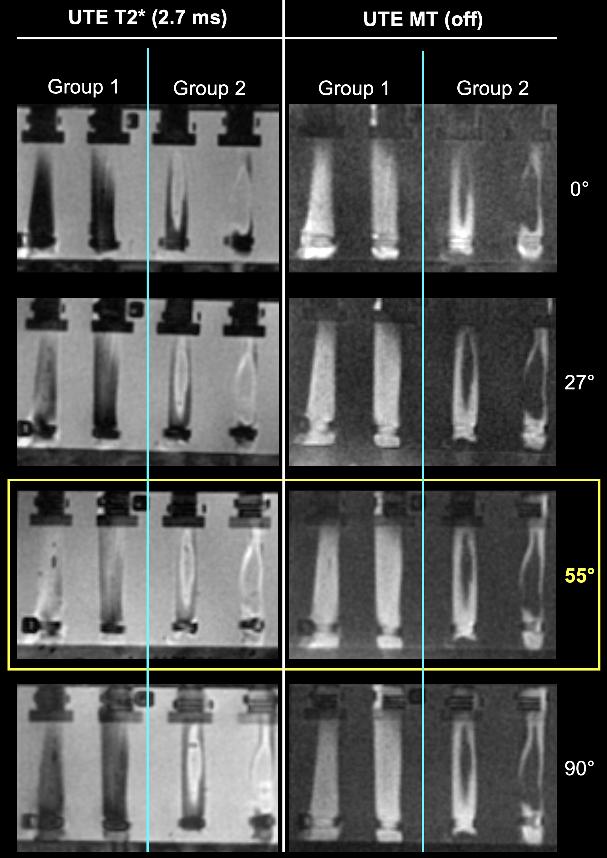
Approach: 3D UTE MT imaging was compared to UTE T2* mapping at 0°, 27°, 55° and 90° relative to the B0 field within a clinically feasible acquisition time.
Results: Compared to UTE T2* mapping, UTE MT imaging was significantly less affected by the MAE and demonstrated a better performance in differentiating partial tendon tears.
Impact: UTE MT imaging is a reliable and reproducible method for quantifying tendon degeneration that is robust to the magic angel effect, acquired within a clinically feasible scan time, and could therefore be a useful tool in the diagnosis of tendinopathies.
Introduction
Magnetic resonance imaging is the modality of choice for the assessment of tendon and soft tissue pathologies1. However, the assessment of tendon pathologies can often be challenging, particularly in the shoulder or ankle, due to the magic angle effect (MAE), which creates a false positive signal enhancement of the tendon, mimicking tears or degenerative changes 2-4.Ultra-short echo time (UTE) imaging combined with a magnetization transfer (MT) preparation allows for MAE-insensitive quantitative assessment of tissues with very short T2 5, 6. However, there are still only a few applications that are feasible for use in routine clinical diagnostics. The aim of this study was to qualitatively and quantitatively assess changes in bovine flexor tendons before and after collagen degradation and at different angles in relation to the static B0field using 3D UTE MT imaging within a clinically feasible acquisition time.
Methods and Materials
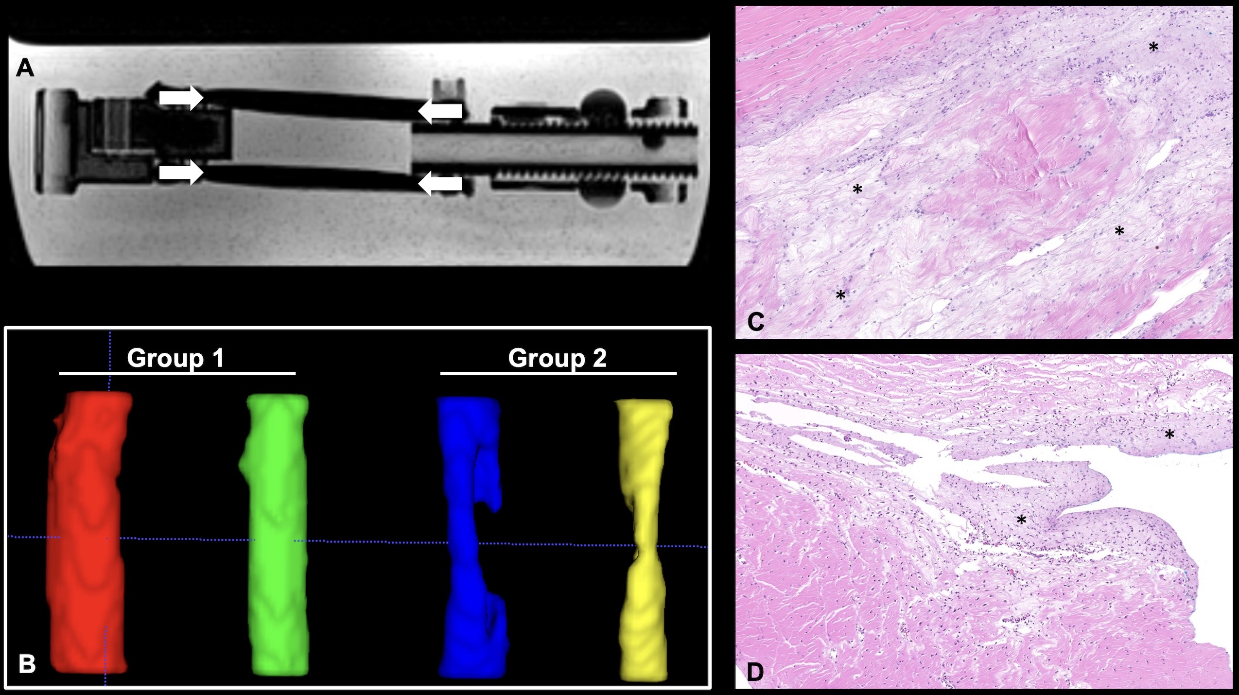
Eight bovine flexor tendons were examined at 3T MRI (MAGNETOM Prisma, Siemens Healthineers, Erlangen, Germany) before and after enzyme-induced degradation. The tendons were divided into two groups: Group 1 (controls) treated with phosphate-buffered saline (PBS) and Group 2 treated with collagenase I to induce collagen degradation. MR imaging was repeated at 0°, 27°, 55° and 90° with respect to the B0 field. Standard T1- and T2-weighted spin-echo sequences were used to assess qualitative tendon integrity. In addition, a 3D UTE research application sequence with and without MT magnetization preparation was acquired (acquisition time 4:04 min) and for comparison, a 3D UTE multi-echo research application sequence was acquired twice for T2* mapping with 4 in-phase and 4 out-of-phase echoes (acquisition time 6:38 min, each). UTE MT ratios (MTR) were computed in Matlab (The MathWorks Inc., MATLAB version 9.5 - R2018b) and to calculate quantitative tissue properties, all tendons were semi-automatically segmented and changes in quantitative UTE T2* and UTE MTR were compared at different angles and between groups. Receiver operating characteristic curves were used to assess the performance of UTE MTR and UTE T2* values in differentiating between healthy and degenerated tendons using histopathology as reference standard. In addition to descriptive statistics, the coefficient of variation was calculated in the segmented tendon ROIs to compare UTE MT and UTE T2* imaging.Results
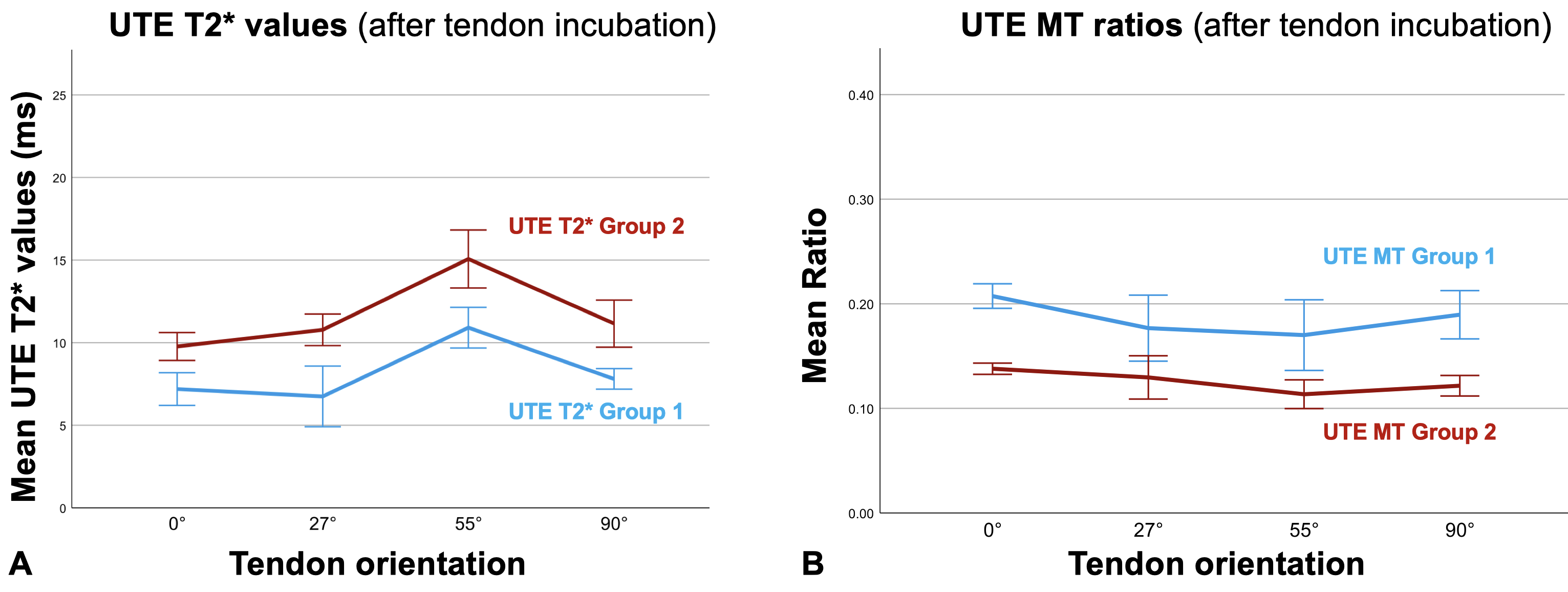
UTE MTR showed a significantly lower coefficient of variation compared to UTE T2* values, indicating a more robust imaging method (UTE MTR 9.64-11.25%, UTE T2* 18.81-24.06%, P < .001) (Figure 1). Both methods showed good performance in detecting degenerated tendons using histopathology as reference standard, with UTE MT imaging having a slightly better area under the curve than UTE T2* mapping (0.918 vs. 0.865) (Figure 2). UTE T2* mapping showed highly amplified relaxation times for all tendons at an angle of 55° to the B0 field due to the magic angle effect, while the ultra-short echo time magnetization transfer ratios remained robust (Figure 3).Conclusion and Discussion
In this study, UTE MT imaging has shown to be a reliable and reproducible method to quantify tendon degradation using histopathology as the reference standard. The reliability of UTE MT imaging was assessed in a reproducible phantom model using bovine flexor tendons and enzymatic modification to provide a controlled setup. In addition, scans were repeated at different angles of tendon orientation relative to the static magnetic field B0 to assess the MAE. Compared to UTE T2* mapping, UTE MT imaging was significantly less affected by the MAE and showed better performance in differentiating partial tendon tears. Both methods showed good performance in detecting degenerative changes in the enzymatically modified tendons. Ultimately, UTE MT imaging proved to be a reliable method of assessing tendon degradation that was robust to the MAE and could be acquired within a clinically feasible time.Acknowledgements
No acknowledgement found.References
1. Zubrod, C.J. and M.F. Barrett, 2007, Magnetic Resonance Imaging of Tendon and Ligament Injuries. Clinical Techniques in Equine Practice. 6(3): p. 217-229.
2. Richardson, M.L., B. Amini, and T.L. Richards, 2018, Some new angles on the magic angle: what MSK radiologists know and don't know about this phenomenon. Skeletal Radiol. 47(12): p. 1673-1681.
3. Timins, M.E., S.J. Erickson, L.D. Estkowski, et al., 1995, Increased signal in the normal supraspinatus tendon on MR imaging: diagnostic pitfall caused by the magic-angle effect. AJR Am J Roentgenol. 165(1): p. 109-14.
4. Singh, D.R., M.S. Chin, and W.C. Peh, 2014, Artifacts in musculoskeletal MR imaging. Semin Musculoskelet Radiol. 18(1): p. 12-22.
5. Zhu, Y., X. Cheng, Y. Ma, et al., 2018, Rotator cuff tendon assessment using magic-angle insensitive 3D ultrashort echo time cones magnetization transfer (UTE-Cones-MT) imaging and modeling with histological correlation. J Magn Reson Imaging. 48(1): p. 160-168.
6. Yang, J., H. Shao, Y. Ma, et al., 2020, Quantitative ultrashort echo time magnetization transfer (UTE-MT) for diagnosis of early cartilage degeneration: comparison with UTE-T2* and T2 mapping. Quant Imaging Med Surg. 10(1): p. 171-183.
Figures