0633
Association of Patella Bone Shape and MR-Diagnosed Patellar Tendinopathy with Patellar Cartilage T2/T1ρ in Elite Basketball Players1Radiology, Stanford University, Stanford, CA, United States, 2University of Sydney, Sydney, Australia, 3Bioengineering, Stanford University, Stanford, CA, United States, 4Human Biology, Stanford University, Stanford, CA, United States, 5Electrical Engineering, Stanford University, Stanford, CA, United States, 6Computer Science, Stanford University, Stanford, CA, United States, 7Department of Radiology and Biomedical Imaging, University of California San Francisco, San Francisco, CA, United States, 8Hospital for Special Surgery, New York, NY, United States
Synopsis
Keywords: Tendon/Ligament, Tendon/Ligament, Bone shape, T2 mapping, T1ρ mapping
Motivation: Patellar tendinopathy (PT) is a common injury in basketball that can lead to early retirement. Basketball influences bone shape and cartilage composition, yet the association between these factors and PT is unknown.
Goal(s): We examined the association between PT, bone shape, and patellar cartilage composition in collegiate basketball players.
Approach: We developed a measure of bone shape indicative of PT and investigated whether PT-associated bone shape is associated with patellar cartilage T2/T1ρ.
Results: We effectively separated grades of PT using bone shape and identified bone shape features associated with PT. We found patellar cartilage composition is independent of PT and bone shape.
Impact: We developed a measure to identify varying grades of PT based on bone shape in collegiate basketball players. Future work will determine the association of our PT-bone shape score with MR-identifiable measures to identify athlete specific PT risk factors.
Introduction
Patellar tendinopathy (PT) is a common injury in sports that involve repetitive jumping1. Up to 32% of elite basketball players experience current symptoms of PT2, and over 50% of athletes with PT retire early from sport3. MRI detects PT as a thickened tendon with increased signal intensity4.Patella bone shape is highly variable, and recent studies indicate that there are differences in shape between basketball players and athletes in low-knee impact sports5. qMRI techniques such as T2 and T1ρ can quantify early degenerative changes in articular cartilage and detect stress induced by basketball6. Given that high patellar tendon loads produce high patellofemoral compression, it is possible that PT, bone shape, and patellar cartilage composition are inter-related. In this study, we developed a measure of PT-bone shape and subsequently applied this measure to determine whether PT-bone shape features are associated with patellar cartilage T2/T1ρ.
Methods
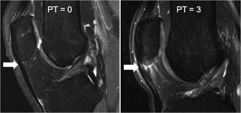
In this multicenter study, National Collegiate Athletic Association (NCAA) basketball players underwent an MRI proceeding their season using a GE 3T SIGNA Premier scanner (GE Healthcare, Milwaukee, WI) and an 18-channel flexible phased-array, transmit/receive coil (Quality Electrodynamics, Mayfield Village, OH). The complete imaging protocol can be found in (Fig.1A).A musculoskeletal radiologist graded 2D FSE knee images using Modified Noyes criteria for PT on a scale of 0-3, where higher scores indicate increased severity of PT (Fig.2).
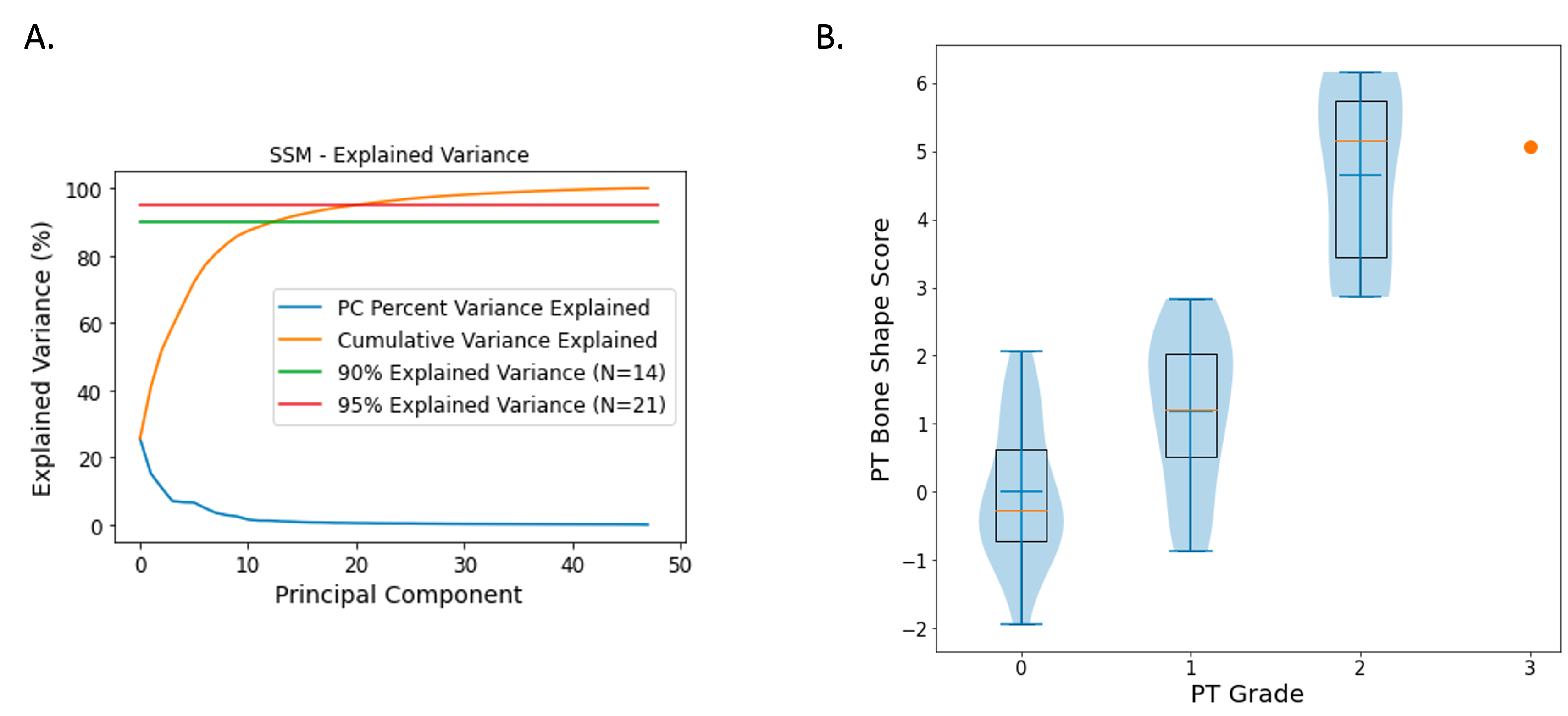
To build a statistical shape model (SSM), patella bones in 48 knees were manually segmented from the 3D FSE images. Binary segmentation masks were Gaussian smoothed, and surfaces were extracted and resampled to have 5,000 points. A template bone was non-rigidly registered to all others using established methods, and principal components analyses (PCA) were used to extract shape features (Fig.4A)7. The first 21 features (95% explained variance) were used to generate a scalar PT-bone shape score for each subject using methods proposed previously for knee osteoarthritis8. Each bone was projected onto a vector from the average bone shape of knees with PT grade 0 to the average bone shape of knees with PT grade 2/3; projected points were normalized using mean and standard deviation of knees with PT grade 0. The first 14 shape features (90% explained variance) were retained for correlation analyses.
Patellar cartilage was manually segmented using a subset of 30 of the 48 knees from the MAPSS images. T2/T1ρ relaxation times were computed from MAPSS images using a mono-exponential fit of signal data acquired at various echo/spinlock (TE/TSL) times (Fig.1A)9. An open-source software was used to automatically sub-divide cartilage into medial/lateral regions10.
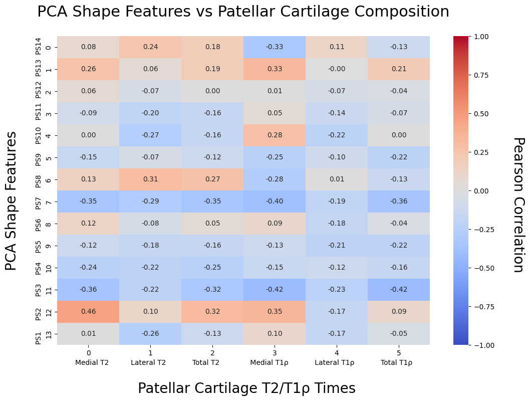
Pearson correlation coefficients (PCCs) were used to determine association between average patellar cartilage subregion T2/T1ρ and PT-bone shape scores, PT grades, and individual SSM bone shape features (Fig.3). Individual SSM shape feature correlations were Bonferroni corrected (p<0.008).
Results
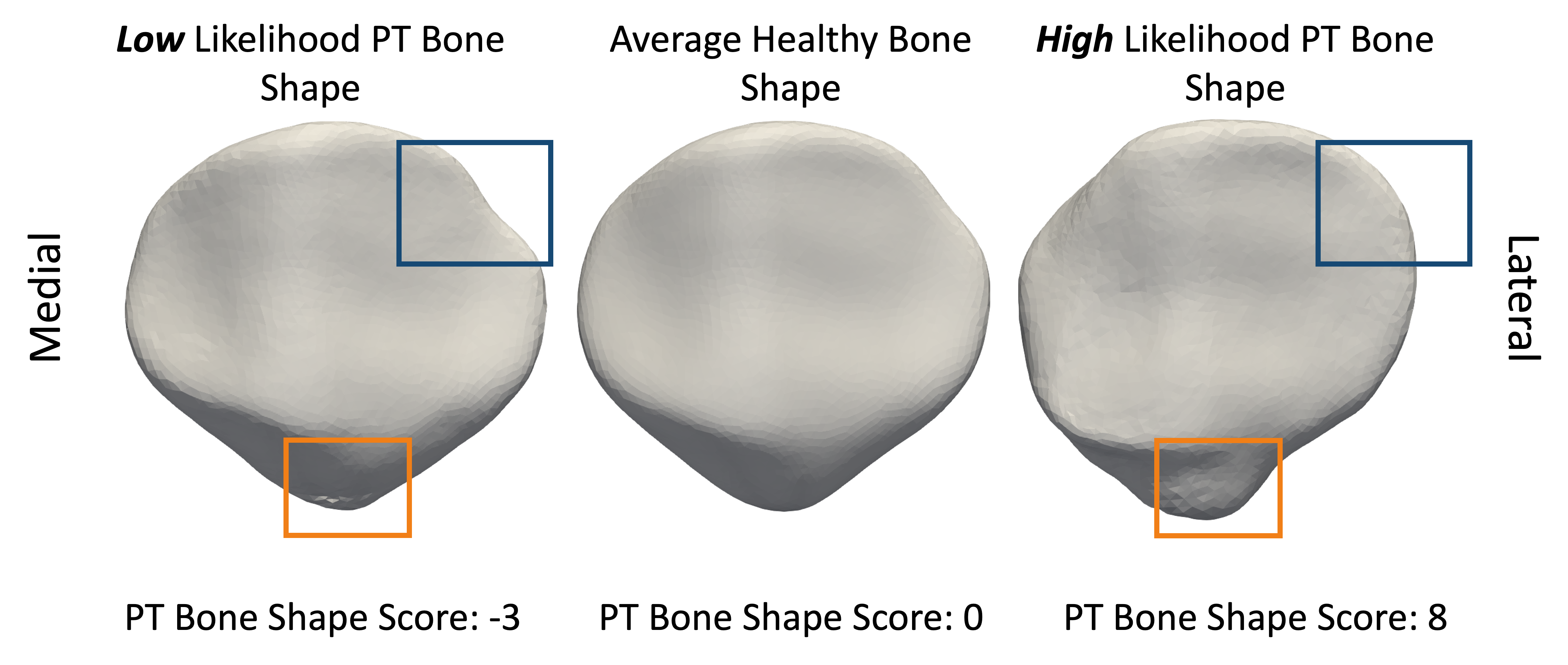
Our PT-bone shape score progressively increases with increasing PT grade (Fig.4B) and highlights features of bone shape that are related to PT (Fig.5). No significant associations were found between patellar cartilage T2/T1ρ and PT-bone shape score, PT grade, or individual SSM shape features (Fig.3).Discussion
Our PT-bone shape score effectively separates grades of PT in NCAA basketball players. Increasing PT-bone shape score reveals specific shape features in athletes with PT, including changes at the inferior pole of the patella, potentially indicating mechanical remodeling has occurred (Fig.5). MRI has identified changes in athletes with PT at the junction of the inferior pole and the patellar tendon, a region commonly assessed in clinical PT diagnosis, which suggests further investigation is warranted11,12.Despite observed bony changes, we found no association between PT-bone shape score, PT grades, or SSM shape features and patellar cartilage T2/T1ρ. This suggests that patellar cartilage composition may be independent of PT and patella shape in collegiate basketball players. Prior studies have demonstrated cartilage changes in patients with advanced PT, however our athletes were young with primarily mild-moderate PT13.
Further investigation of PT-bone shape features may identify athletes at a higher risk of developing PT. Future work will apply our PT-bone shape score to evaluate association with MR-identifiable factors such as patellar tendon size/composition and muscle symmetry. Better characterization of factors involved in PT pathogenesis may enable clinicians to customize rehabilitation plans to individual athlete anatomy.
Conclusion
We classified varying grades of PT based on bone shape and identified bone shape features associated with PT in elite basketball players. We found no association between PT/bone shape and patellar cartilage composition. Future work will apply this PT-bone shape measure to investigate other features of PT pathogenesis and identify athlete specific risk factors.Acknowledgements
This work was supported by GE Healthcare, National Basketball Association, National Institutes of Health (R01EB002524, K24AR062068), Wu Tsai Human Performance Alliance, Canadian Institutes of Health Research.References
1. Theodorou A, Komnos G, Hantes M. Patellar tendinopathy: an overview of prevalence, risk factors, screening, diagnosis, treatment and prevention. Arch Orthop Trauma Surg. 2023 Nov;143(11):6695-6705.
2. Lian OB, Engebretsen L, Bahr R. Prevalence of jumper's knee among elite athletes from different sports: a cross-sectional study. Am J Sports Med. 2005 Apr;33(4):561-7.
3. Kettunen JA, Kvist M, Alanen E, Kujala UM. Long-term prognosis for jumper's knee in male athletes. A prospective follow-up study. Am J Sports Med. 2002 Sep-Oct;30(5):689-92.
4. Campbell RS, Grainger AJ. Current concepts in imaging of tendinopathy. Clin Radiol. 2001 Apr;56(4):253-67.
5. Gao KT, Pedoia V, Young KA, Kogan F, Koff MF, Gold GE, Potter HG, Majumdar S. Multiparametric MRI characterization of knee articular cartilage and subchondral bone shape in collegiate basketball players. J Orthop Res. 2021 Jul;39(7):1512-1522.
6. Rubin EB, Mazzoli V, Black MS, Young K, Desai AD, Koff MF, Sreedhar A, Kogan F, Safran MR, Vincentini DJ, Knox KA, Yamada T, McCabe A, Majumdar S, Potter HG, Gold GE. Effects of the Competitive Season and Off-Season on Knee Articular Cartilage in Collegiate Basketball Players Using Quantitative MRI: A Multicenter Study. J Magn Reson Imaging. 2021 Sep;54(3):840-851.
7. Gatti AA, Keir PJ, Noseworthy MD, Maly MR. Investigating acute changes in osteoarthritic cartilage by integrating biomechanics and statistical shape models of bone: data from the osteoarthritis initiative. MAGMA. 2022 Oct;35(5):861-873.
8. Bowes MA, Kacena K, Alabas OA, Brett AD, Dube B, Bodick N, Conaghan PG. Machine-learning, MRI bone shape and important clinical outcomes in osteoarthritis: data from the Osteoarthritis Initiative. Ann Rheum Dis. 2021 Apr;80(4):502-508.
9. Li X, Han ET, Busse RF, Majumdar S. In vivo T(1rho) mapping in cartilage using 3D magnetization-prepared angle-modulated partitioned k-space spoiled gradient echo snapshots (3D MAPSS). Magn Reson Med. 2008 Feb;59(2):298-307.
10. Desai AD, Caliva F, Iriondo C, Mortazi A, Jambawalikar S, Bagci U, Perslev M, Igel C, Dam EB, Gaj S, Yang M, Li X, Deniz CM, Juras V, Regatte R, Gold GE, Hargreaves BA, Pedoia V, Chaudhari AS; IWOAI Segmentation Challenge Writing Group. The International Workshop on Osteoarthritis Imaging Knee MRI Segmentation Challenge: A Multi-Institute Evaluation and Analysis Framework on a Standardized Dataset. Radiol Artif Intell. 2021 Feb 10;3(3):e200078. arXiv:2004.14003.
11. Ramos LA, Carvalho RT, Garms E, Navarro MS, Abdalla RJ, Cohen M. Prevalence of pain on palpation of the inferior pole of the patella among patients with complaints of knee pain. Clinics (Sao Paulo). 2009;64(3):199-202.
12. van Wilgen P, van der Noord R, Zwerver J. Feasibility and reliability of pain pressure threshold measurements in patellar tendinopathy. J Sci Med Sport. 2011 Nov;14(6):477-81.
13. Little RD, Smith SE, Cicuttini FM, Tanamas SK, Wluka AE, Hussain SM, Urquhart DM, Jones G, Wang Y. Association between increased signal intensity at the proximal patellar tendon and patellofemoral geometry in community-based asymptomatic middle-aged adults: a cross-sectional study. BMC Musculoskelet Disord. 2020 Aug 22;21(1):571.
Figures