0631
Temporal Structured Low-Rank Reconstruction for First-Pass Myocardial Perfusion Imaging1Biomedical Imaging Research Institute, Cedars-Sinai Medical Center, Los Angeles, CA, United States, 2Department of Radiological Sciences, David Geffen School of Medicine at UCLA, Los Angeles, CA, United States, 3Department of Bioengineering, UCLA, Los Angeles, CA, United States
Synopsis
Keywords: Sparse & Low-Rank Models, Perfusion, First-pass myocardial perfusion; structured low-rank
Motivation: First-pass myocardial perfusion imaging is a powerful tool for assessing coronary artery disease, but needs high levels of undersampling to achieve sufficient spatial coverage, spatiotemporal resolution, and motion robustness.
Goal(s): To develop efficient temporal image reconstruction models which can leverage linear time-invariant models of dynamic contrast enhancement without identifying an arterial input function or assuming tissue transfer function shapes.
Approach: We propose a novel temporal structured low-rank modeling technique to implicitly leverage linear time-invariant models of dynamic contrast enhancement.
Results: Temporal structured low-rank modeling outperforms conventional low-rank methods, especially as a local constraint.
Impact: Temporal structured low-rank modeling has the potential to improve spatial coverage, spatial resolution, and/or motion robustness for first-pass myocardial perfusion MRI.
Introduction
First-pass myocardial perfusion (FPP) imaging is powerful for assessing coronary artery disease. Accelerated perfusion reconstruction has previously used low-rank (LR) constraints to exploit spatiotemporal correlations, but the need for increased motion robustness, spatial coverage, and spatiotemporal resolution motivates even more efficient temporal models to handle higher undersampling. Dynamic contrast enhancement (DCE) employs linear time-invariant (LTI) system modeling for quantification1–3, but integrating these models into reconstruction requires arterial input function (AIF) identification and may overly restrict tissue transfer function (TTF) shapes.Structured low-rank modeling of k-space neighborhoods has proven useful for reconstructing images produced by linear shift-invariant spatial transformations, such as coil-sensitivity-modulated images in parallel imaging4–6. Analogously, here we propose temporal structured low-rank (tSLR) modeling for FPP imaging. This method implicitly leverages LTI DCE modeling without identifying/assuming an AIF or a particular tissue function family. We compared the proposed method to conventional LR constraints in simulations and in vivo experiments.
Methods
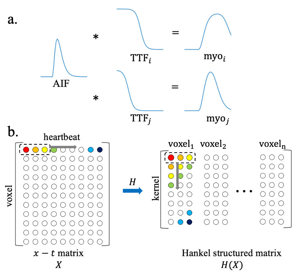
TheoryFPP imaging assumes a convolutional model wherein an AIF, typically contrast concentration curve in left ventricle(LV), is convolved with a TTF(e.g., a Fermi model2) to produce myocardial contrast concentration(Fig.1a):
$$AIF(t)\ast TTF_i(t)=Myo_i(t) [1]$$
$$AIF(t)\ast TTF_j(t)=Myo_j(t) [2]$$
where $$$i$$$ and $$$j$$$ denote different voxels. Semi-quantitative perfusion ignores non-linearity between $$$\Delta R1$$$ ($$$\propto $$$contrast concentration) and T1-weighted MR signals, analyzing T1-weighted signals instead. Eqs 1&2 reveal an annihilation relation:
$$Myo_i (t)*TTF_j (t)-Myo_j (t)*TTF_i (t)=0 [3]$$
The TTFs/convolution kernels are compact, so Eq3 can be reformulated as matrix multiplication, using the block-Hankel structured matrix $$$H(Myo)$$$ constructed from the 1D myocardial curve:
$$H(Myo_i )⋅\overrightarrow{TTF_j} -H(Myo_j )⋅\overrightarrow{TTF_i} =0 [4]$$
$$\begin{bmatrix} H(Myo_i )& H(Myo_j)\end{bmatrix}×\begin{bmatrix} \overrightarrow{TTF_j}\\ \overrightarrow{-TTF_i}\end{bmatrix}=0 [5]$$
Eq 5 suggests the composite matrix $$$\begin{bmatrix} H(Myo_i )& H(Myo_j)\end{bmatrix}$$$ is rank-deficient; the TTFs manifest as its null space vector. Extension to more voxels defines a low-rank, structured matrix from the whole image(Fig.1b). Note that unlike other structured low-rank reconstructions where Hankel matrices are constructed in k-space, here they are constructed from the 1D time dimension.
Image reconstruction
A general SENSE-based tSLR reconstruction can be formulated as:
$$\underset{x}{\operatorname{argmin}} ‖E(X)-Y‖_F^2+\lambda‖H(X)‖_* [6]$$
where image series $$$X\in \mathbb{C} ^{n \times t}$$$ has $$$n$$$ voxels and $$$t$$$ heartbeats. $$$E$$$ is the forward model and $$$Y$$$ is the acquired k-space. $$$\left\| \right\|_*$$$ denotes nuclear norm. The operator $$$ H\in \mathbb{C}^{n \times t} \to \mathbb{C}^{l \times kn}$$$ constructs and concatenates multiple Hankel matrices along the column direction, where $$$k$$$ is the kernel length and $$$l=t-k+1$$$ is the number of overlapping kernels along the time dimension.
Since $$$t$$$ is typically small(<60) in FPP, $$$H(X)$$$ will have an extremely small row size compared to its column size. Thus, we also consider locally-tSLR, which constructs structured matrices on each local block of the image:
$$\underset{x}{\operatorname{argmin}} ‖E(X)-Y‖_F^2+\lambda\sum_{R_b\in \Omega}^{}‖H(R_bX)‖_* [7]$$
where $$$R_b\in \Omega$$$ extracts the $$$b_{th}$$$ block from $$$X$$$. For kernel length $$$k=1$$$, Eqs 6&7 reduce to conventional global- and locally-LR reconstructions.
Experiments
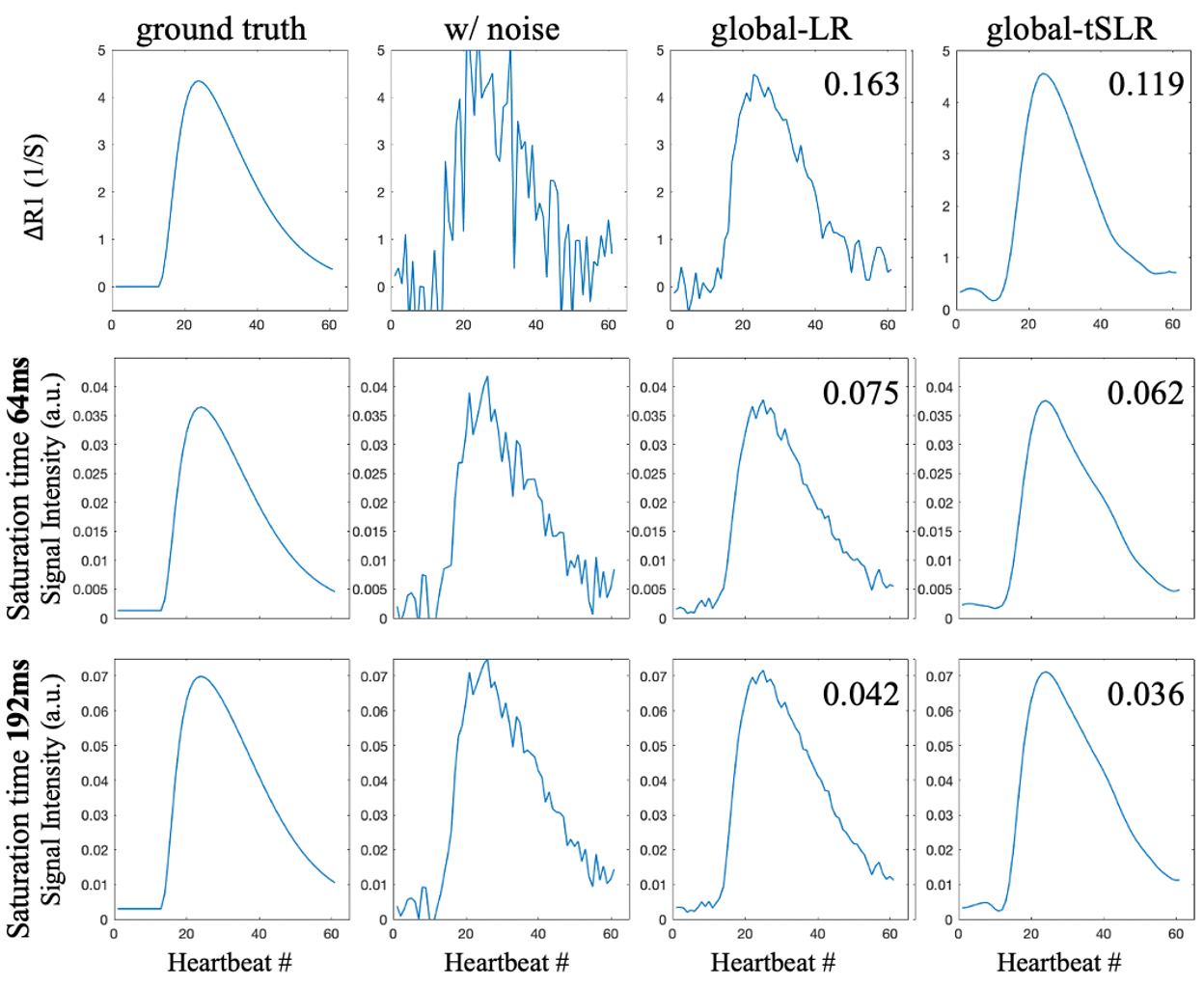
We simulated 50 noisy myocardial ΔR1 and T1-weighted signal curves at two saturation times by passing the same AIF through different Fermi functions. The denoising performance of global-LR and tSLR were compared(each at its lowest NRMSE respectively).
In vivo perfusion data were collected at 3T(Siemens Biograph mMR) with breath-holding and cardiac gating, with $$$1.4 \times 1.4 \times 8 mm^3 $$$spatial resolution, t=37 heart beats, and tPAT=3. Additional retrospective acceleration yielded R=6 uniform $$$k_y$$$ under-sampling(cycling along $$$k_y$$$ at different heart beats). Global/locally- LR/tSLR reconstructions were performed, selecting each $$$\lambda$$$ to achieve matching data consistency between methods, which also corresponded to the lowest NRMSE for each method. Kernel length $$$k=11$$$. Local block size $$$=10 \times 10$$$. The SENSE reconstruction at R=3 was used as the reference for NRMSE calculations, so NRMSE of an ideal denoiser should be well above zero.
Results
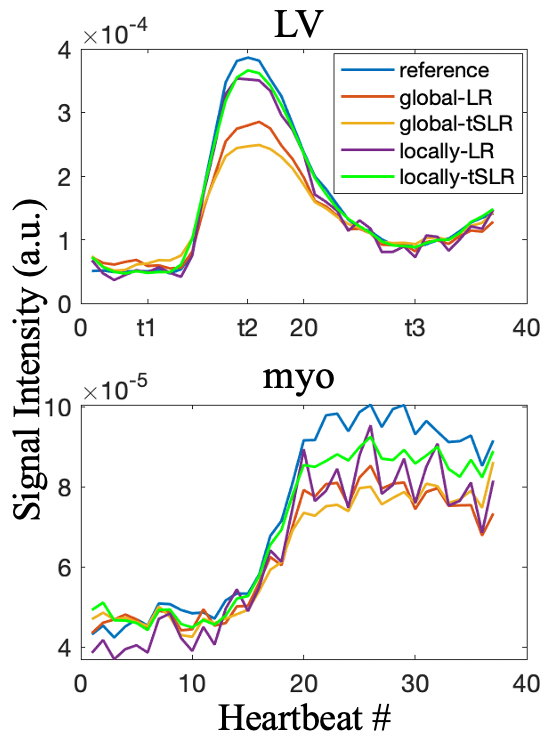
Fig. 2 shows that tSLR is a more effective denoiser than LR for both ΔR1 and T1-weighted signal intensity at different saturation times.Fig. 3 compares average signal intensity curves of two ROIs in the LV and myocardium. Locally-tSLR produced the smoothest curves and the highest similarity to reference curves. Global tSLR doesn’t work well in this scenario, possibly due to extremely unbalanced row and column size of the Hankel matrix(27x 541200).
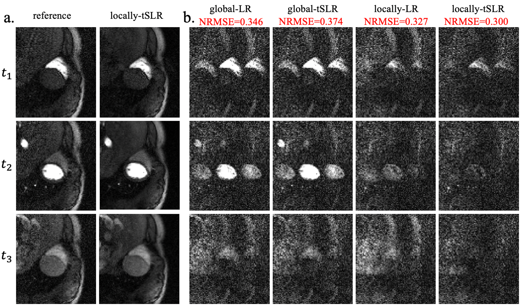
Fig. 4 shows images and difference maps in a cardiac ROI; locally-tSLR has the lowest NRMSE and the least structure in difference maps.
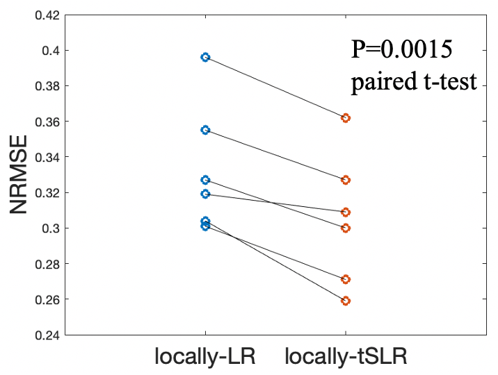
Fig.5 shows locally-tSLR achieves a significantly lower NRMSE than locally-LR in all 6 subjects (p=0.0015, paired t-test).
Conclusion
Temporal structured low-rank reconstruction generalizes and outperforms low-rank reconstruction for FPP imaging, and performs especially well as a local constraint. Its reconstruction fidelity may have potential to be traded for higher SNR, more spatial coverage, or higher spatiotemporal resolution.Acknowledgements
This work was partially supported by NIH R01 HL124649 and NIH R01 EB032801.References
- Axel L. Tissue mean transit time from dynamic computed tomography by a simple deconvolution technique. Invest Radiol. 1983;18(1):94-99. doi:10.1097/00004424-198301000-00018.
- Jerosch-Herold M, Wilke N, Stillman AE, Wilson RF. Magnetic resonance quantification of the myocardial perfusion reserve with a Fermi function model for constrained deconvolution. Med Phys. 1998;25(1):73-84. doi:10.1118/1.5981633.
- Jerosch-Herold M, Swingen C, Seethamraju RT. Myocardial blood flow quantification with MRI by model-independent deconvolution. Med Phys. 2002;29(5):886-897. doi:10.1118/1.14731354.
- Lee J, Jin KH, Ye JC. Reference-free single-pass EPI Nyquist ghost correction using annihilating filter-based low rank Hankel matrix (ALOHA): Reference-Free Single-Pass EPI Nyquist Ghost Correction using ALOHA. Magn Reson Med. 2016;76(6):1775-1789. doi:10.1002/mrm.260775.
- Haldar JP. Low-rank modeling of local k-space neighborhoods (LORAKS) for constrained MRI. IEEE Trans Med Imaging. 2014;33(3):668-681. doi:10.1109/TMI.2013.22939746.
- Chen X, Wu W, Chiew M. Improving robustness of 3D multi-shot EPI by structured low-rank reconstruction of segmented CAIPI sampling for fMRI at 7T. NeuroImage. 2023;267:119827. doi:10.1016/j.neuroimage.2022.1198277.
Figures