0629
Time-Resolved Cardiac function: Myocardium Strain and First-Pass Perfusion Using MR-MOTUS1Computational Imaging Group for MR Therapy and Diagnostics, University Medical Center Utrecht, Utrecht, Netherlands, 2UMC Utrecht Cancer Center, Department of Radiotherapy, University Medical Center Utrecht, University Medical Center Utrecht, Utrecht, Netherlands, 3Department of Radiology, University Medical Centre Utrecht, University Medical Center Utrecht, Utrecht, Netherlands, 4School of Biomedical Engineering and Imaging Sciences, King's College London, London, United Kingdom, 5Centre for Marine Sciences (CCMAR), Faro, Portugal
Synopsis
Keywords: Myocardium, Image Reconstruction, First-Pass Myocardial Perfusion; Motion estimation; Motion correction; Low-Rank & Sparse; Time-resolved imaging; Cine; Dynamic; Perfusion; Cardiac
Motivation: We aim at improving accuracy and reliability in cardiac imaging for coronary artery disease diagnosis and management. We address motion artifacts in first-pass myocardial perfusion MR imaging.
Goal(s): To develop and validate motion correction techniques for motion field accuracy and strain quantification in non-ECG triggered myocardial first pass perfusion examinations.
Approach: We use a modified MR-MOTUS framework for motion separation and reconstruction in patient data.
Results: Our approach demonstrates higher accuracy in respiratory/cardiac motion field estimation with additional strain analysis.
Impact: Our findings have the potential to improve patient care by enabling free-breathing and non-ECG triggered examinations of myocardial first-pass perfusion simultaneously with strain analysis. We also open avenues for further research in cardiac imaging and motion correction techniques.
Introduction
Cardiac strain analysis and first-pass myocardial perfusion MR imaging are pivotal indicators in diagnosing and managing coronary artery disease1. To deal with physiological motion during imaging, patients are equipped with an ECG for triggered acquisitions and instructed to perform difficult breath-holds. The complex blood flow and motion dynamics during first-pass myocardial perfusion examinations make it challenging for conventional image registration-based motion correction techniques.2Previously, we introduced a modified MR-MOTUS framework3,4, which enabled the separation of respiratory/cardiac motion and contrast dynamics from in-silica time-resolved data.4 In this study, we apply this framework to multiple patient datasets of 2D free breathing (FB) first-pass myocardial perfusion without ECG triggering; we reconstruct cines of the contrast inflow and quantify myocardial strain with accurate motion fields simultaneously.
Theory
$$\min_{\mathbf{\Phi}\mathbf{\Psi}^T=[\mathbf{D}_1,...,\mathbf{D}_M]}\sum_t^M\parallel \mathbf{F}(\mathbf{D}_t\mid\mathbf{q}_t)-\mathbf{s}_t\parallel_2^2 +\lambda_{\mathrm{TV}}\mathbf{TV}(\mathbf{D})+\lambda_{J}\parallel\mathbf{J}(\mathbf{D})-1\parallel_2^2\tag{1}$$$$\min_{\mathbf{Q}=[\mathbf{L}+\mathbf{S}]}\sum_t^M\parallel \mathbf{F}(\mathbf{q}_t\mid\mathbf{D}_t)-\mathbf{s}_t\parallel_2^2 +\lambda_{\mathrm{L}}\parallel\mathbf{L}\parallel_*+\lambda_{\mathrm{S}}\parallel\mathcal{F}\mathbf{S}\parallel_1\tag{2}$$
The two problems are solved in an alternating scheme. $$$\mathbf{F}$$$ denotes the MR-MOTUS signal model5, which enables the estimation of low-rank motion fields $$$\mathbf{\Phi}\mathbf{\Psi}^T=\mathbf{D}$$$ from highly undersampled data $$$\mathbf{s}$$$ (1). Subsequently we use $$$\mathbf{D}$$$ to reconstruct a motion-corrected image $$$\mathbf{Q}$$$ decomposed6 by a Low Rank $$$\mathbf{L}$$$ and Sparse component $$$\mathbf{S}$$$ (2). $$$\mathbf{M}$$$ denotes the total number of frames, $$$\mathbf{t}$$$ denotes the frame index, $$$\mathcal{F}$$$ denotes the temporal Fourier transform, $$$\mathbf{J}$$$ the Jacobian determinant operator, and $$$\lambda$$$ is the regularization parameters. An overview of the pipeline is presented in figure 1.
Methods
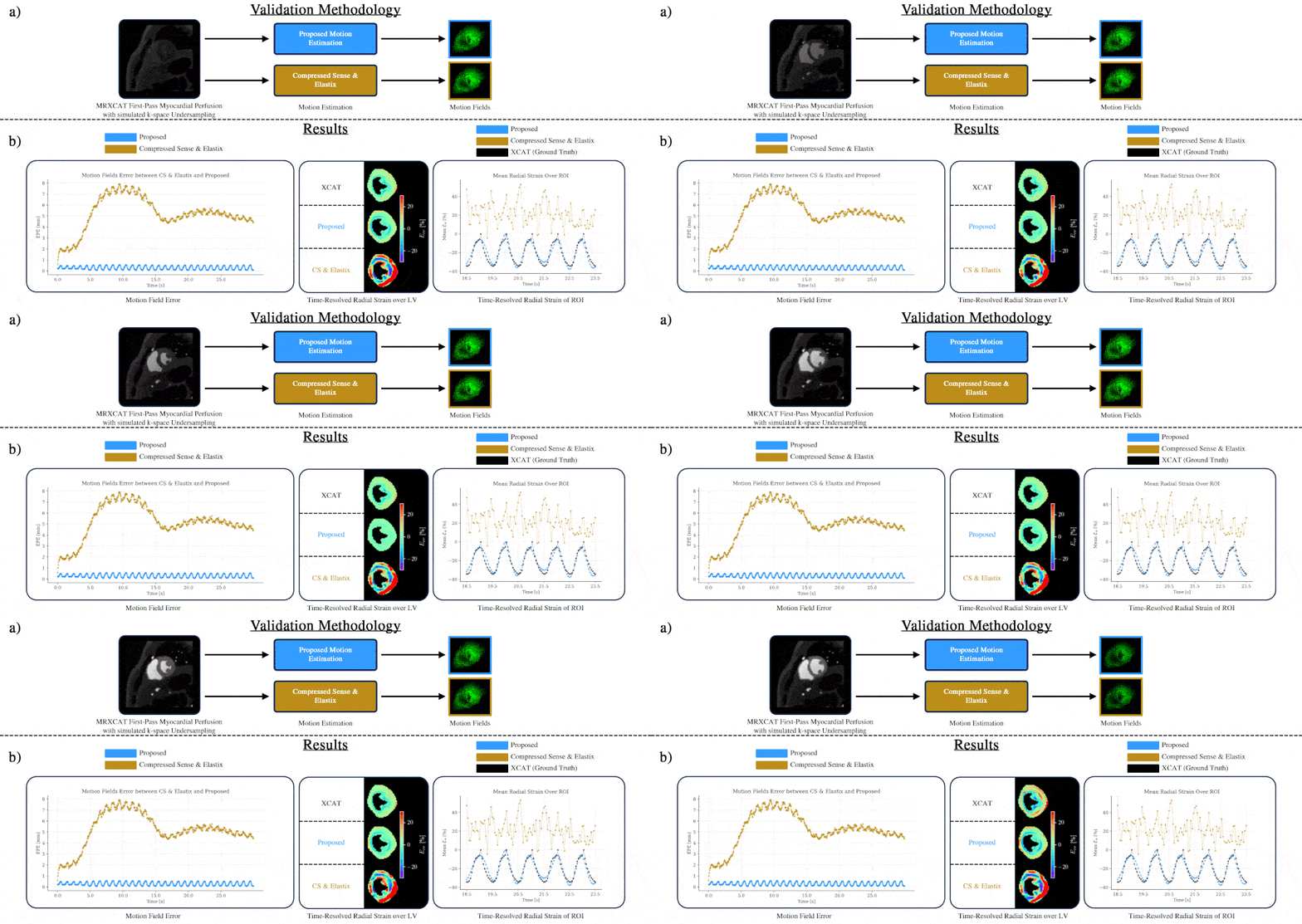
Test 1: Digital cardiac perfusion phantomWe validated our method using a numerical phantom7 for simulating 2D myocardial perfusion, using a time-resolved 10-fold undersampled Cartesian8 acquisition.
Reconstructions
- Proposed MR-MOTUS technique: We perform the proposed alternating reconstructions on the same data and explicitly enforce rank one for both the $$$\mathbf{D}$$$ and $$$\mathbf{L}$$$. Radial strain9 was computed based on the reconstructed MR-MOTUS motion fields.
- State of the art: We performed a compressed sensing (CS) reconstruction for comparison, using BART10 with spatial wavelet and temporal total variation regularizations. Elastix11 was used to compute motion fields using the first frame as the fixed.
- Image quality: The image quality between the motion-resolved reconstructions (MR-MOTUS and CS) and the ground-truth MRXCAT frames was determined using the structural similarity metric (SSIM)12.
- Motion fields and strain: The end point error (EPE) w.r.t. ground truth XCAT motion fields were determined for the proposed MR-MOTUS fields, and the motion fields were estimated with Elastix after CS reconstruction. The DICE score between the ground-truth XCAT segmentations and the warped segmentations from the fixed image was determined for both motion estimation methods. The radial strain9 was determined for each motion field over the myocardial walls.
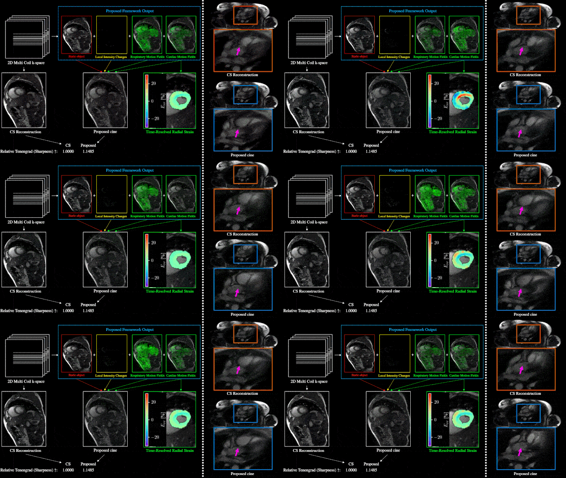
We used two publicly-available prospectively-undersampled 2D FB bSSFP cardiac MRI cine datasets13, covering short-axis (SAX) and long-axis (LAX) views. We reconstructed both datasets using our proposed method. We assessed myocardial sharpness using the Tenengrad metric14,15 and calculated the myocardial radial strain in the SAX view.
Test 3: In vivo BH ECG triggered first-pass myocardial perfusion
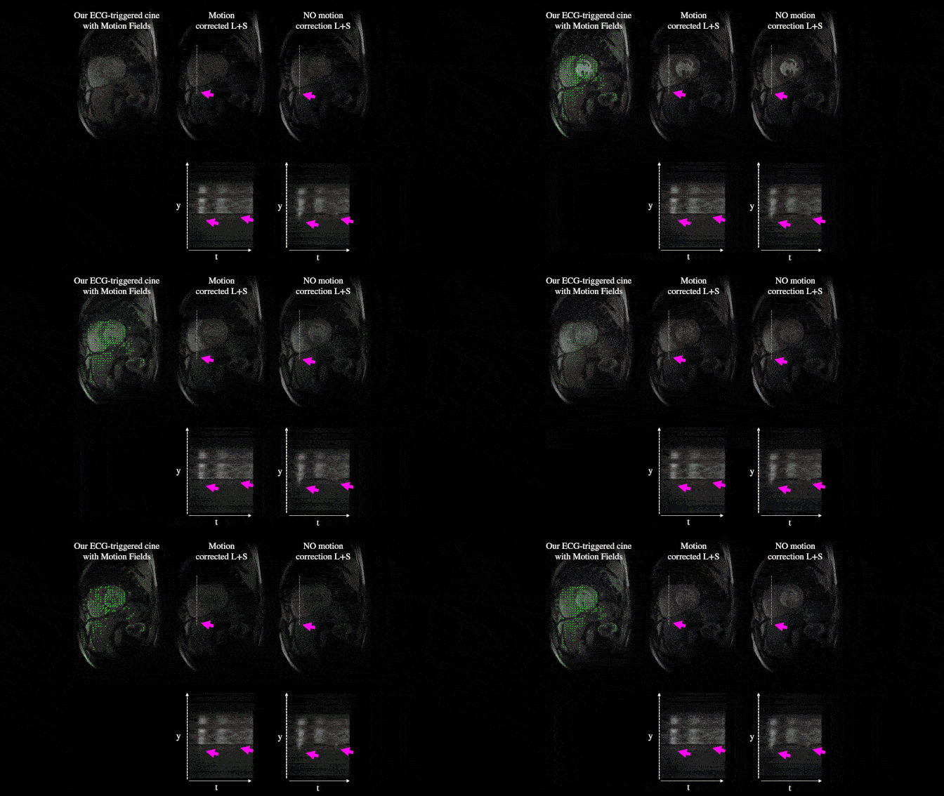
One patient dataset with ECG-triggered first-pass myocardial perfusion MRI featured imperfect breath-holds causing motion artifacts. Radial sampling was used with 10 spokes per frame. We compared standard L+S reconstruction with our time-resolved alternating reconstruction to assess the impact of motion correction on the results.
Test 4: In vivo time-resolved first-pass myocardial perfusion
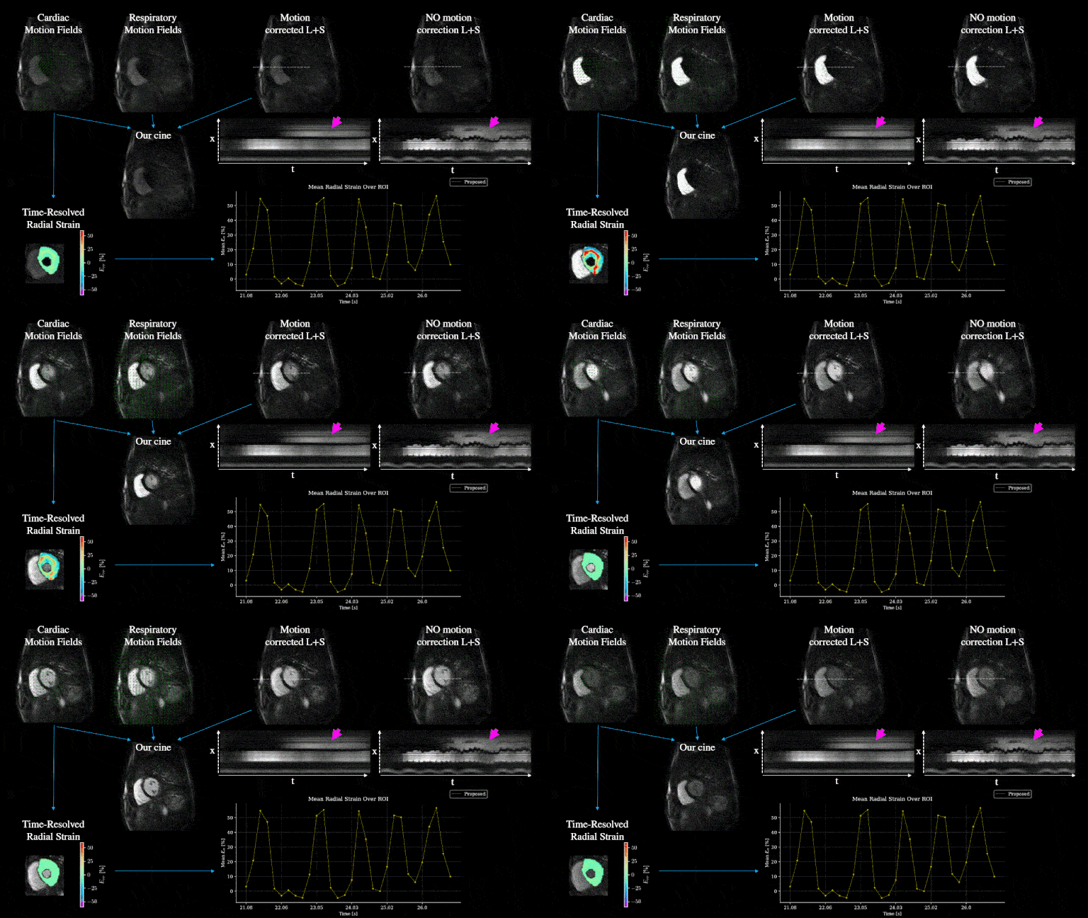
One patient dataset of a time-resolved (free-breathing and no triggering) first-pass myocardial perfusion MRI was used for the same analysis as in test 3 but including the radial strain.
Results
In Test 1, our proposed method achieved a higher DICE score (0.9302) for warped segmentations compared to the CS + Elastix approach (0.8913). The proposed method and CS reconstruction had similar SSIM values (0.9292 and 0.9298, respectively), indicating comparable cine image quality. However, MR-MOTUS additionally provides accurate motion field extraction. Strain and EPE are found in figure 1.In the other tests, MR-MOTUS provides sharper images than CS, as indicated by the Tenengrad metric (Figure 3), and better image quality than L+S reconstructions due to the motion correction (Figure 4,5).
Discussion & Conclusion
Our proposed method enables motion-corrected, time-resolved first-pass perfusion imaging of the myocardium without needing BH or ECG. Explicit motion modeling enables the separation of motion and enhancement allowing spatial alignment of all contrast-enhanced dynamics. In addition, our framework can separate breathing and cardiac dynamics, and quantify strain providing important diagnostic information.This was showcased with two patient examples, consisting of non-compliant breath hold as well as FB acquisitions without ECG triggering first-pass myocardial perfusion examinations. In conclusion, we showcased our new proposed MR-MOTUS framework with time-resolved in vivo measurements of motion-resolved myocardial first-pass perfusion imaging and cardiac strain quantification.
Acknowledgements
This research is funded by the Netherlands Organisation for Scientific Research (NWO), domain Applied and Engineering Sciences, Grant number 19003.References
1. Ismail, T. F. et al. Cardiac MR: From Theory to Practice. Front. Cardiovasc. Med. 9, 826283 (2022).
2. Scannell, C. M., Villa, A. D. M., Lee, J., Breeuwer, M. & Chiribiri, A. Robust Non-Rigid Motion Compensation of Free-Breathing Myocardial Perfusion MRI Data. IEEE Trans. Med. Imaging 38, 1812–1820 (2019).
3. Huttinga, N. R. F., van den Berg, C. A. T., Luijten, P. R. & Sbrizzi, A. MR-MOTUS: model-based non-rigid motion estimation for MR-guided radiotherapy using a reference image and minimal k -space data. Phys. Med. Biol. 65, 015004 (2020).
4. Olausson, T. E., Beijst, C., Sbrizzi, A., van den Berg, C. A. T. & Huttinga, N. R. F. Time-Resolved Cardiac Imaging and Motion Analysis Using a Multi-Scale Dynamics Decomposition. in Proc. Inti. Soc. Mag. Reson. Med. 0287 (2023).
5. Huttinga, N. R. F., Bruijnen, T., Berg, C. A. T. & Sbrizzi, A. Nonrigid 3D motion estimation at high temporal resolution from prospectively undersampled k‐space data using low‐rank MR‐MOTUS. Magn. Reson. Med. 85, 2309–2326 (2021).
6. Otazo, R., Candès, E. & Sodickson, D. K. Low-rank plus sparse matrix decomposition for accelerated dynamic MRI with separation of background and dynamic components: L+S Reconstruction. Magn. Reson. Med. 73, 1125–1136 (2015).
7. Wissmann, L., Santelli, C., Segars, W. P. & Kozerke, S. MRXCAT: Realistic numerical phantoms for cardiovascular magnetic resonance. J Cardiovasc Magn Reson 16, 63 (2014).
8. Joshi, M., Pruitt, A., Chen, C., Liu, Y. & Ahmad, R. Technical Report (v1.0)--Pseudo-random Cartesian Sampling for Dynamic MRI. Preprint at http://arxiv.org/abs/2206.03630 (2022).
9. Ghadimi, S., Abdi, M. & Epstein, F. H. Improved computation of Lagrangian tissue displacement and strain for cine DENSE MRI using a regularized spatiotemporal least squares method. Front. Cardiovasc. Med. 10, 1095159 (2023).
10. Blumenthal, M. et al. mrirecon/bart: version 0.8.00. (2022) doi:10.5281/ZENODO.592960.
11. Klein, S., Staring, M., Murphy, K., Viergever, M. A. & Pluim, J. elastix: A Toolbox for Intensity-Based Medical Image Registration. IEEE Trans. Med. Imaging 29, 196–205 (2010).
12. Xue, W., Zhang, L., Mou, X. & Bovik, A. C. Gradient Magnitude Similarity Deviation: A Highly Efficient Perceptual Image Quality Index. IEEE Trans. on Image Process. 23, 684–695 (2014).
13. Chen, C. et al. OCMR (v1.0)--Open-Access Multi-Coil k-Space Dataset for Cardiovascular Magnetic Resonance Imaging. Preprint at http://arxiv.org/abs/2008.03410 (2020).
14. Spieker, V. et al. Deep Learning for Retrospective Motion Correction in MRI: A Comprehensive Review. IEEE Trans. Med. Imaging 1–1 (2023) doi:10.1109/TMI.2023.3323215.
15. Krotkov, E. Focusing. Int J Comput Vision 1, 223–237 (1988).
Figures
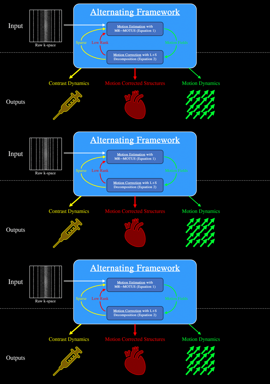
Figure 1 gif: Proposed MR-MOTUS workflow. The top-left shows time-resolved raw k-space as the input into the proposed framework (center). The outputs of Motion Estimation (Motion Fields) and Motion Correction (Low Rank and Sparse components) are fed into each other. The outputs (bottom) of the alternating framework at convergence are contrast dynamics (Sparse component), motion- corrected structures (Low Rank component), and motion dynamics (Motion Fields).