0628
Towards ultrafast submillimeter T2* and QSM quantification at 3T using spherical Echo Planar Time Resolved Imaging (sEPTI)1Radiology Department, Stanford University, Stanford, CA, United States, 2Electrical Engineering, Stanford University, Stanford, CA, United States
Synopsis
Keywords: Image Reconstruction, Image Reconstruction
Motivation: To further improve the image quality, SNR, and reduce scan time by reducing averages for submillimeter T2* and QSM at 3T
Goal(s): To achieve whole-brain 0.75-mm T2* and QSM quantification using sEPTI within a single average scan
Approach: We developed: (1) an iterative data-driven B0 update pipeline for accurate and high SNR B0 map; (2) data-driven eddy-current correction approach to reduce artifacts; (3) a physics-informed unrolled network to boost the SNR of the reconstructed image to achieve 2X acceleration by reducing the need of averages.
Results: sEPTI achieved whole-brain 0.75-mm T2* and QSM quantification within 84 seconds with the potential for wide applications
Impact: The work presented synergetic improvements in B0 update, eddy-current correction, and unrolled-network based SNR-boosted reconstruction for sEPTI, which achieves whole-brain 0.75-mm distortion-free and blurring-free T2* and QSM quantification at 3T in 84 seconds with the potentials for wide applications.
Introduction
Echo Planar Time Resolved Imaging (EPTI) is a distortion-free rapid quantitative technique with exciting neuroimaging applications1-5. Last year, we developed a 3D spherical-EPTI (sEPTI)6 achieving 1.4X additional acceleration compared to already-fast EPTI with improved image quality for the >100-echo T2*-weighted images and T2* maps. However, SNR has always been a problem for T2* and QSM due to the rapidly decreased signal intensity at long TE. Averages are usually necessary, resulting in doubled or tripled scan time. In this work, we further improved the image quality and reduced averages (scan time) innovatively from 3 aspects: (1) an improved data-driven B0 update pipeline for high-quality B0 map; (2) a widely-applicable eddy-current correction pipeline that significantly reduces the artifacts; (3) a physics-informed unrolled network for subspace reconstruction7 to improve the SNR by 2X times. The optimized sEPTI achieved whole-brain T2* and QSM quantifications at 0.75 mm resolution on 3T within a single-average 84-second scan.Methods
Prototype acquisition and reconstruction: All data were acquired with gradient echo sequence with sEPTI sampling. The reconstruction was performed using low-rank subspace framework through two steps. First step is to obtain a data-updated high resolution B0 map2 from a low-resolution B0,low from calibration data using:$$\widehat{\mathbf{U}_{\boldsymbol{B}}}=\arg\min_{\mathbf{U}_{\boldsymbol{B}}}\left\|\Omega\mathrm{FSB_l}\mathbf{U}_{\boldsymbol{B}}\boldsymbol{\Phi}_{\boldsymbol{B}}-\mathbf{d}\right\|_2^2+R\left(\mathbf{U}_{\boldsymbol{B}}\right),(1)$$
where $$$\mathbf{d}$$$ is the acquired data, $$$\mathrm{B_l}$$$ is the B0,low-induced phase, $$$\boldsymbol{\Phi}_{\boldsymbol{B}}$$$ is temporal subspace containing both T2* decay and phase evolution with off-resonance [-50,50]Hz, and $$$\mathbf{U}_{\boldsymbol{B}}$$$ is the spatial coefficients to be solved. The reconstructed images $$$\mathbf{U}_{\boldsymbol{B}}\boldsymbol{\Phi}_{\boldsymbol{B}}$$$ provides a residual phase of $$${\Delta}{B_0}$$$ to achieve $$$B_0=B_{0,low}+{\Delta}{B_0}$$$. In the second step, the reconstruction is performed as6:
$$\widehat{\mathbf{U}}=\arg\min_{\mathbf{U}}\|\Omega\mathrm{FSB}\mathbf{U}\boldsymbol{\Phi}-\mathbf{d}\|_2^2+R(\mathbf{U}),(2)$$
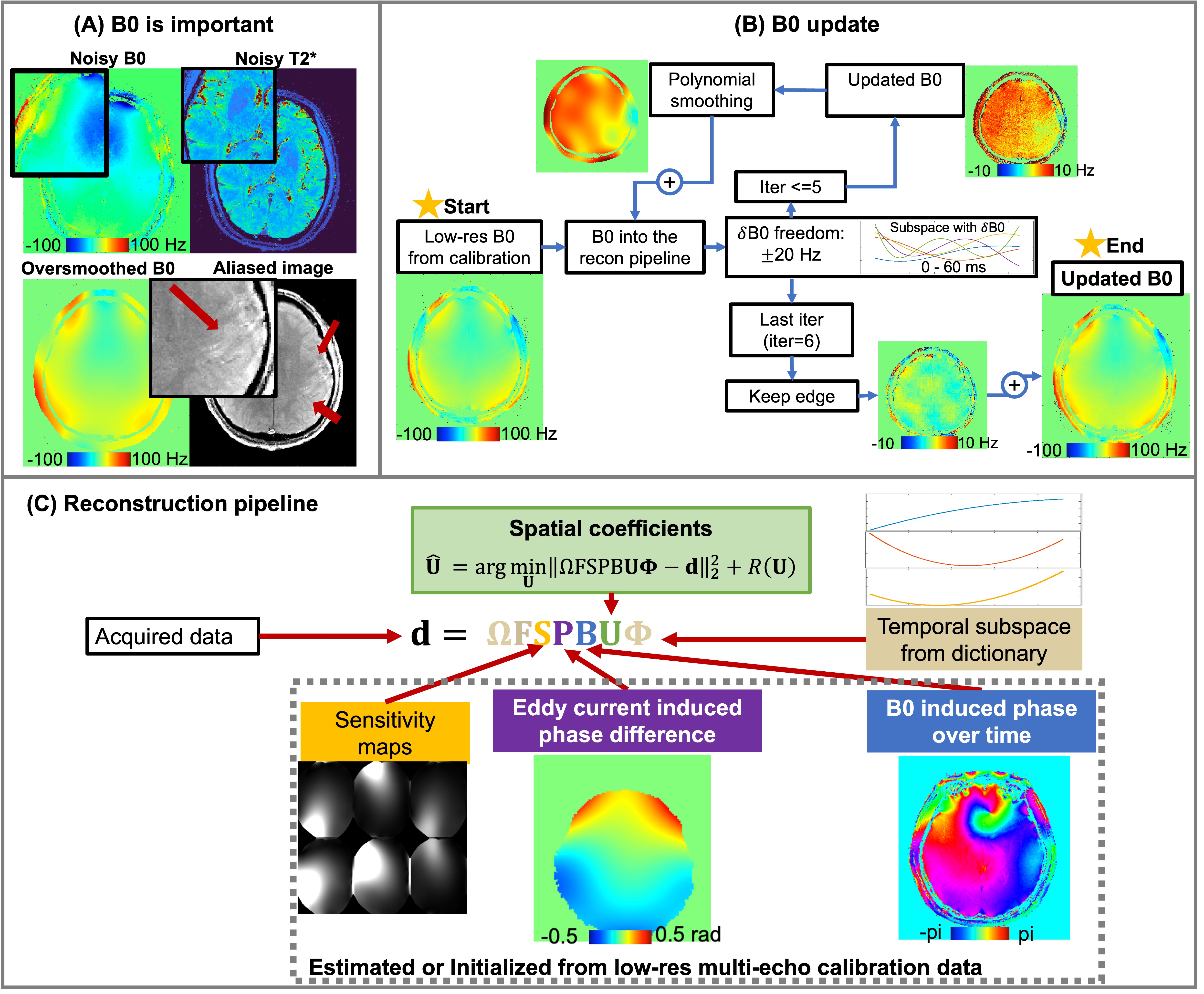
Improved B0 updating: Reduced SNR at 0.75 mm results in noisy $$${\Delta}{B_0}$$$ from Eq.(1) and even noisier images. Polynomial fitting can smooth $$${\Delta}{B_0}$$$, but tends to oversmooth the edge and leads to aliasing (Fig.1A). To resolve this issue, we developed an iterative B0 updating-smoothing framework. In each iteration, a $$$\pm$$$20 Hz $$${\Delta}{B_0}$$$ is updated and smoothed through polynomial fitting; the last iteration without polynomial fitting is to preserve the sharp edge (Fig.1B).
Eddy current correction: Eddy current induced gradient non-linearity is a fundamental issue with all EPI-based sampling8 including EPTI. To compensate this effect, a phase difference $$$P$$$ between odd and even echoes was estimated from the low-resolution calibration data, which is four-echo GRE sequences providing sensitivity maps, initial B0,low, and eddy current phase. The reconstruction model was then updated as (Fig.1C):
$$\widehat{\mathbf{U}}=\arg\min_{\mathbf{U}}\|\Omega\mathrm{FSPB}\mathbf{U}\boldsymbol{\Phi}-\mathbf{d}\|_2^2+R(\mathbf{U}),(3)$$
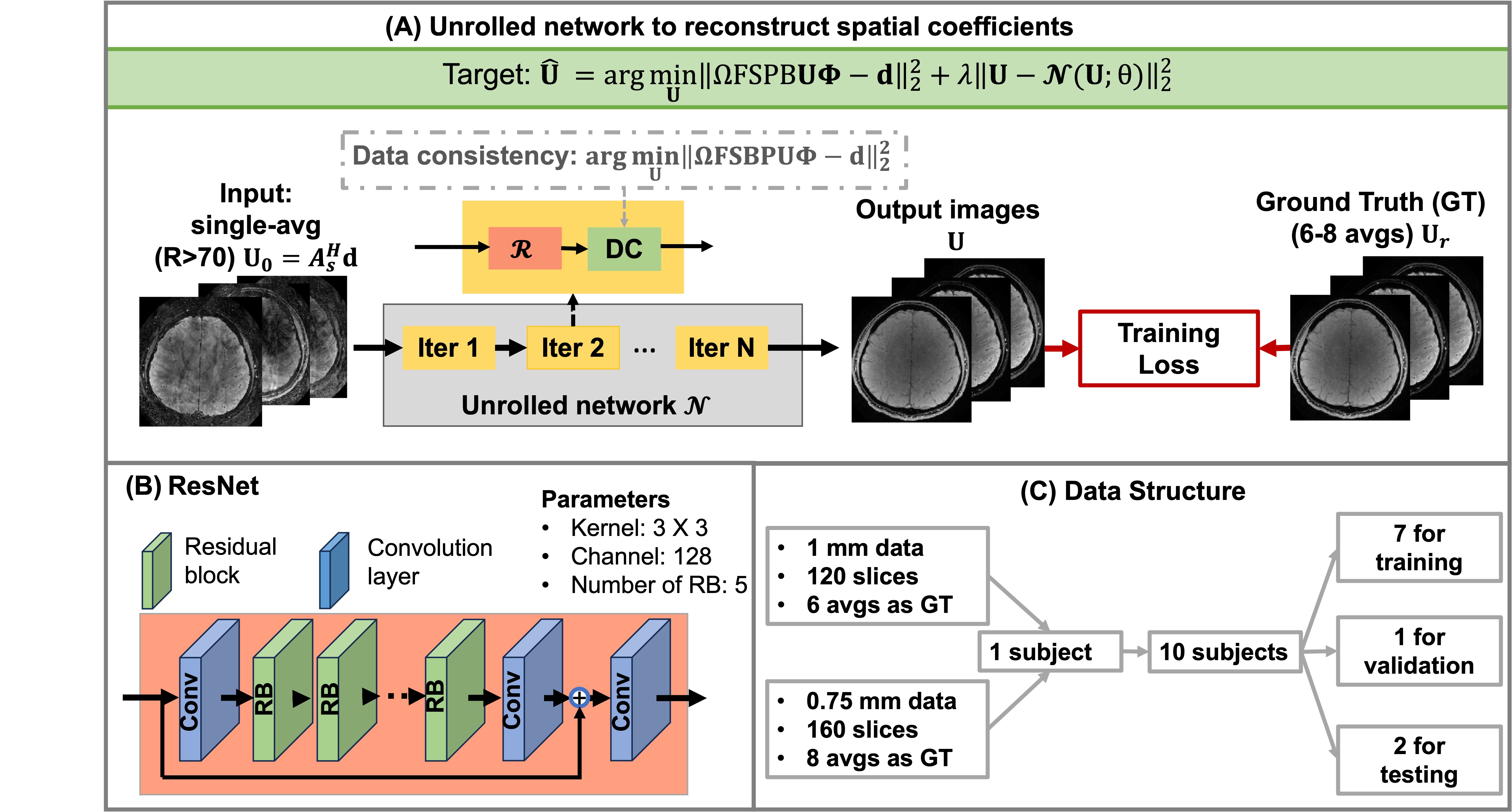
Unrolled network for subspace reconstruction (Figure 2): Data with 0.75-mm resolution suffers from 40% SNR loss compared to 1 mm. To further improve the SNR and reduce the need of averages, an unrolled network was developed for the subspace reconstruction9:
$$\widehat{\mathbf{U}}=\arg\min_{\mathbf{U}}\|\Omega\text{FSBPU}\mathbf{\Phi}-\mathbf{d}\left\|_2^2+\lambda\right\|\mathbf{U}-N(\mathbf{U};\theta)\|_2^2,(4)$$
where $$$N(\mathbf{U};\theta)$$$ is the convolutional neural network (CNN)-based denoiser with trainable parameters $$$\theta$$$. The network was trained through a supervised manner with the acquired single-average sEPTI as input and the spatial coefficients from multi-averaged sEPTI as ground truth (GT). To ensure the GT is of good quality, each average was reconstructed using Eq.(2) separately, co-registered to the same motion state, corrected with background phase differences10, and then averaged to boost SNR. The data were acquired at 1 mm and 0.75 mm, and the networks were trained on data with a single resolution or both.
In vivo experiments: Ten volunteers were studied on a 3T system (Premier, GE Healthcare) with a 48-channel head-coil. Sagittal images were acquired with: FOV=240x240x216mm3, TEs=5-60ms, TR=65ms, flip angle=20°. The 1.0-mm data was acquired 45 seconds per average and 6 averages were acquired; the 0.75-mm data was acquired 84 seconds per average and 8 averages were acquired. Data from 7 subjects were used for training, 1 for validating, and 2 for testing. To test generalizability, 1-mm data were acquired on a pediatric patient with epilepsy where the T2* contrast is different from training population.
Results
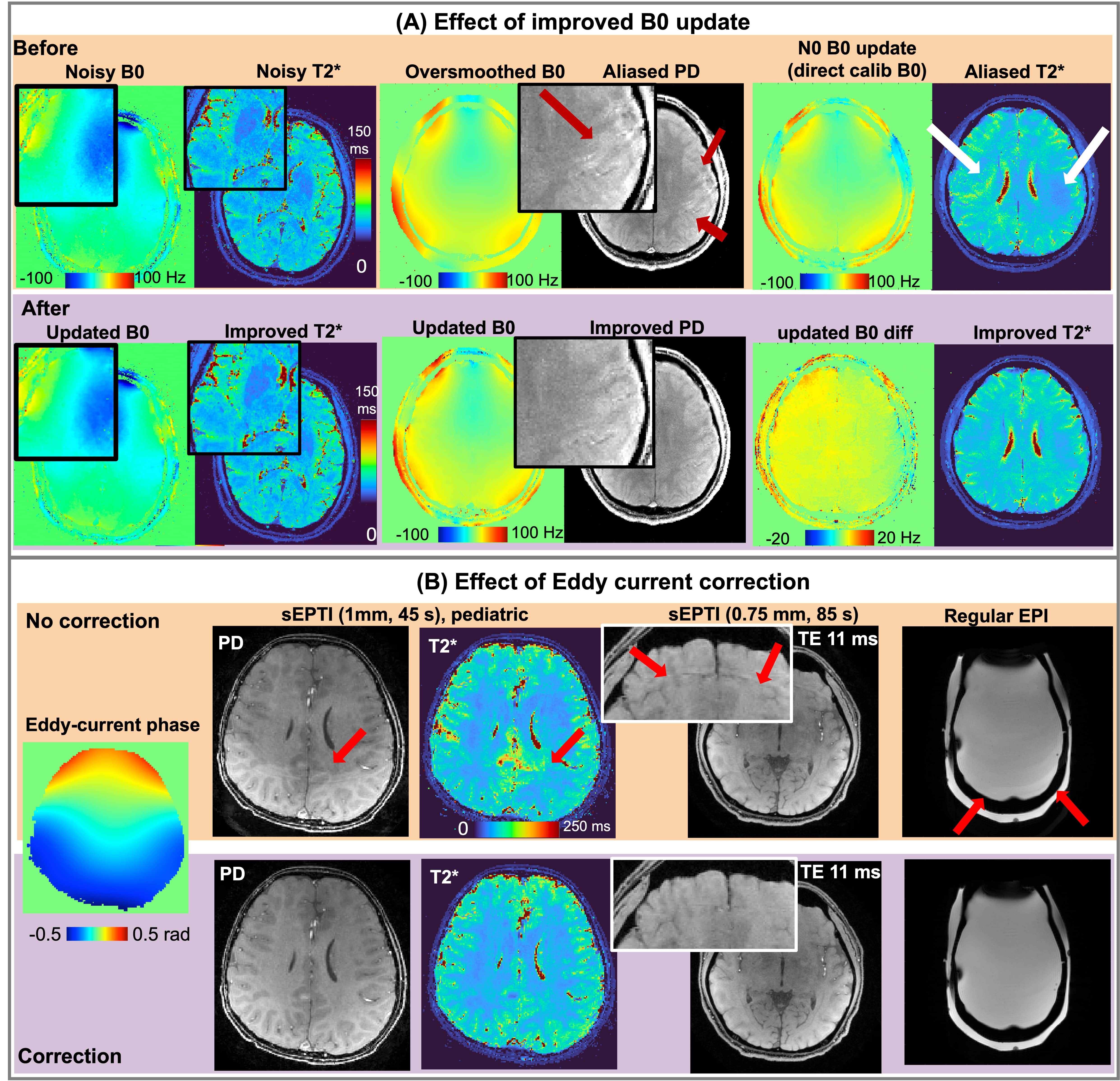
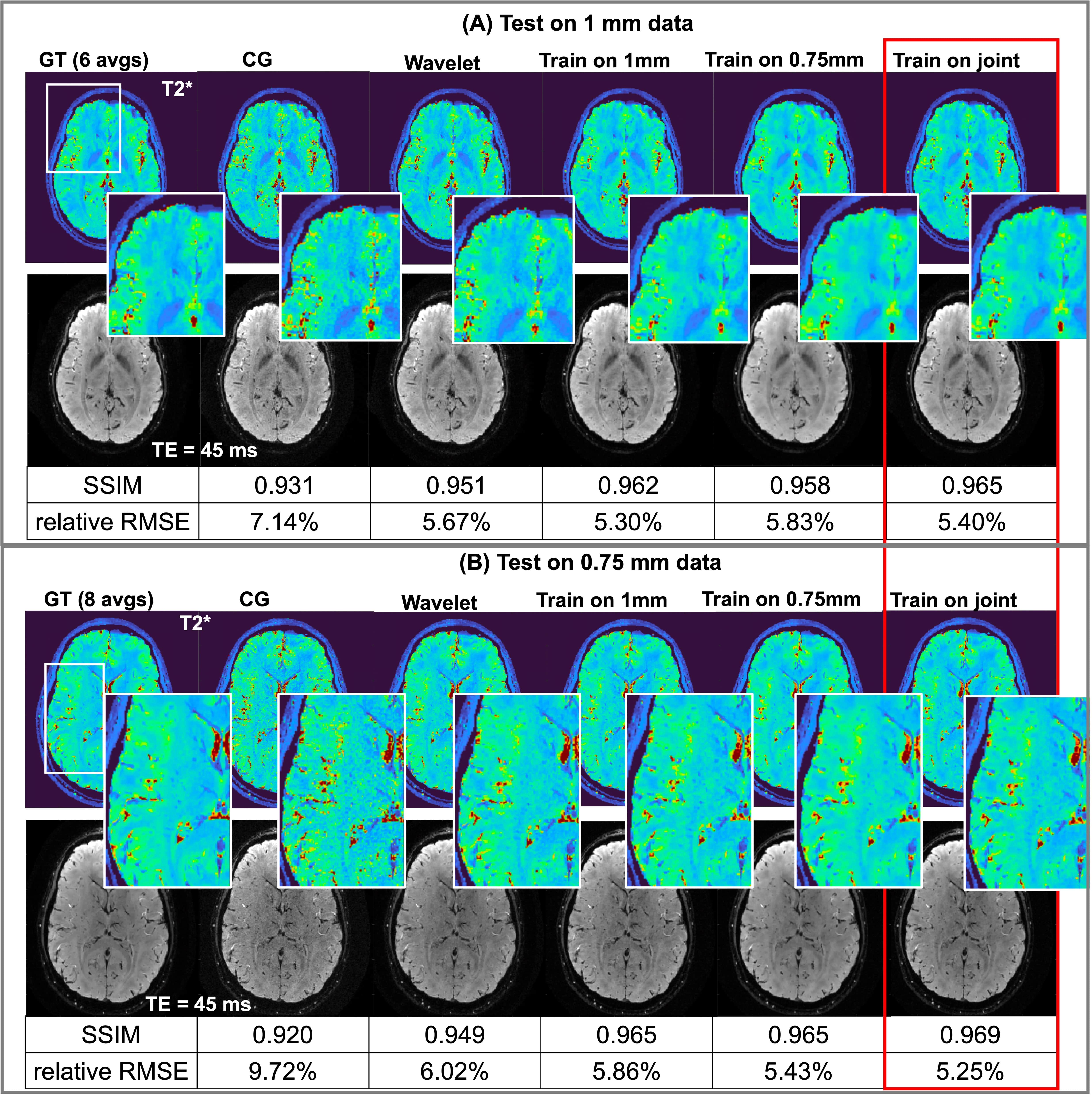
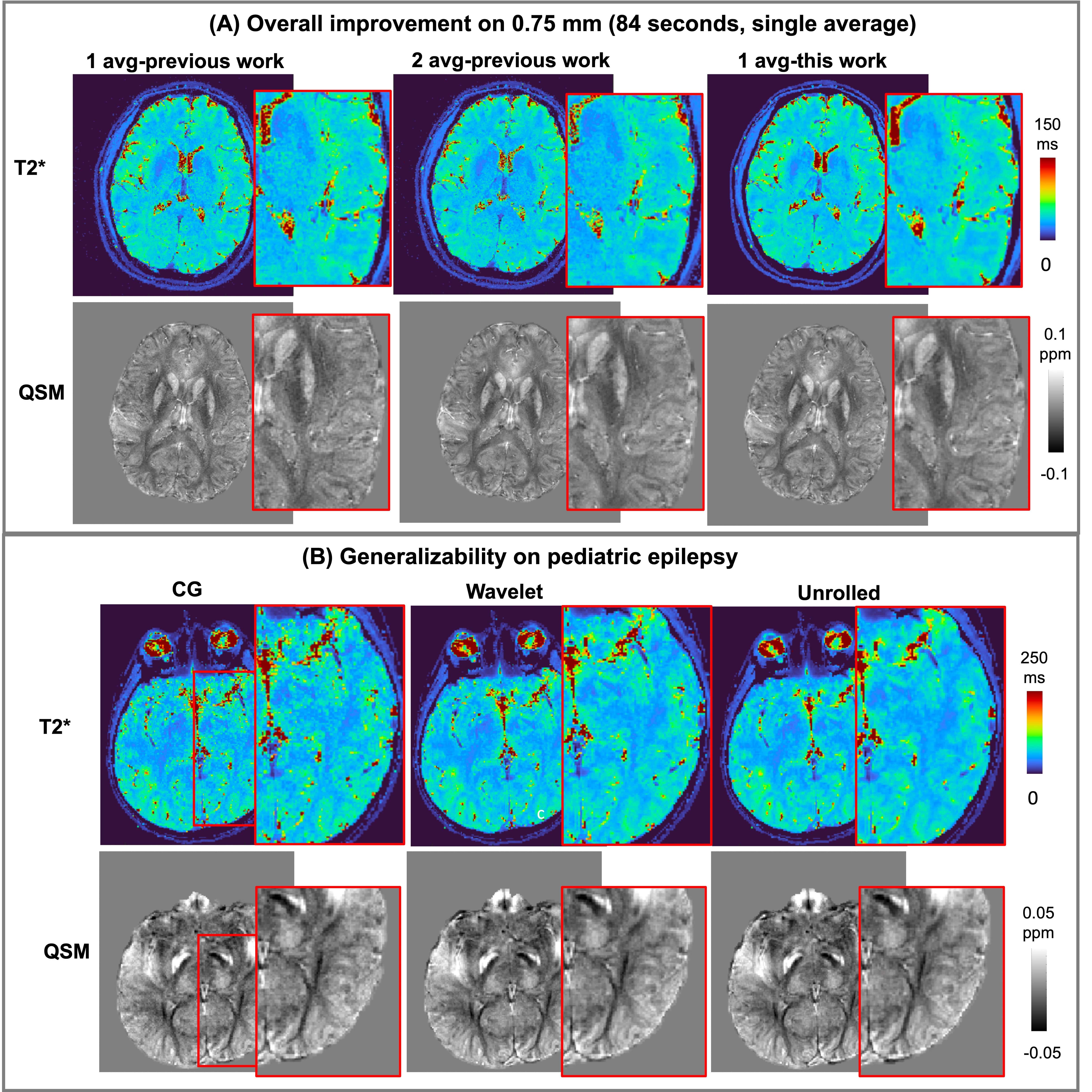
Figure 3 demonstrated visible SNR improvement and artifacts reduction from improved B0 update and eddy current correction. The reconstruction of unrolled network further improves the SNR by 2 times (Fig.4), and the model trained on both resolution provides the best outcome. The test showed that the improvements in this work achieved better image quality compared to 2-average data (Fig.5A); the pipeline can be extended for wide applications with different contrasts (Fig.5B).Conclusion
With the improvement in the B0 update, eddy current correction, and integration of deep learning, sEPTI achieved additional 2-times acceleration for 0.75-mm T2* and QSM quantification on 3T within 84 seconds. This opens the potential for submillimeter T2* and QSM on clinical field strength.Acknowledgements
This work is partially supported by R01MH116173, R01EB019437, U01EB025162, P41EB030006, R01EB033206, U24NS129893References
1. Wang, Fuyixue, Zijing Dong, Timothy G. Reese, Berkin Bilgic, Mary Katherine Manhard, Jingyuan Chen, Jonathan R. Polimeni, Lawrence L. Wald, and Kawin Setsompop. "Echo planar time‐resolved imaging (EPTI)." Magnetic resonance in medicine 81, no. 6 (2019): 3599-3615.
2. Dong, Zijing, Fuyixue Wang, Timothy G. Reese, Berkin Bilgic, and Kawin Setsompop. "Echo planar time‐resolved imaging with subspace reconstruction and optimized spatiotemporal encoding." Magnetic resonance in medicine 84, no. 5 (2020): 2442-2455.
3. Wang, F., Z. Dong, T. G. Reese, L. L. Wald, and K. Setsompop. "3D‐EPTI for ultra‐fast multi‐contrast and quantitative imaging." Proc Intl Soc Mag Reson Med. Montreal(2019): 944.
4. Dong, Zijing, Fuyixue Wang, Lawrence Wald, and Kawin Setsompop. "SNR-efficient distortion-free diffusion relaxometry imaging using ACcelerated Echo-train shifted EPTI (ACE-EPTI)." bioRxiv (2021).
5. Dong, Zijing, Fuyixue Wang, Kwok-Shing Chan, Timothy G. Reese, Berkin Bilgic, José P. Marques, and Kawin Setsompop. "Variable flip angle echo planar time-resolved imaging (vFA-EPTI) for fast high-resolution gradient echo myelin water imaging." NeuroImage 232 (2021): 117897.
6. Wang N et al. “Spherical Echo-Planar Time-resolved Imaging (sEPTI) for 3D highly-accelerated, distortion-free, time-resolved whole-brain T2* mapping”, ISMRM2023 P0119
7. Hosseini, Seyed Amir Hossein, et al. "Dense recurrent neural networks for accelerated MRI: History-cognizant unrolling of optimization algorithms." IEEE Journal of Selected Topics in Signal Processing 14.6 (2020): 1280-1291.
8. Hoge, W. Scott, and Jonathan R. Polimeni. "Dual‐polarity GRAPPA for simultaneous reconstruction and ghost correction of echo planar imaging data." Magnetic resonance in medicine 76.1 (2016): 32-44.
9. Yaman, Burhaneddin, et al. "Self‐supervised learning of physics‐guided reconstruction neural networks without fully sampled reference data." Magnetic resonance in medicine 84.6 (2020): 3172-3191.
10. Van, Anh T., Diego Hernando, and Bradley P. Sutton. "Motion-induced phase error estimation and correction in 3D diffusion tensor imaging." IEEE transactions on medical imaging 30.11 (2011): 1933-1940.
Figures