0627
Modeling Phase Errors for Robust and Efficient Multidimensional MR Fingerprinting for Simultaneous Relaxation and Diffusion Mapping1Department of Biomedical Engineering, Case Western Reserve University, Cleveland, OH, United States, 2Imaging Institute, Cleveland Clinic, Cleveland, OH, United States, 3Medical Radiation Physics, Clinical Sciences Lund, Lund University, Lund, Sweden, 4Department of Radiology, Case Western Reserve University, Cleveland, OH, United States, 5Cardiff University Brain Research Imaging Centre (CUBRIC), School of Psychology, Cardiff University, Cardiff, United Kingdom
Synopsis
Keywords: MR Fingerprinting, MR Fingerprinting
Motivation: Diffusion MRI can be corrupted by phase errors due to physiological motion, bulk motion, eddy currents, and other system imperfections, which makes its efficient embedding into MR Fingerprinting challenging.
Goal(s): To develop a new approach to correct artifacts in multidimensional MR Fingerprinting (mdMRF) for simultaneous relaxation and diffusion quantification, that obviates cardiac gating, motion compensation, navigators, or data removal.
Approach: Modeling potential phase errors using phase offset and phase dispersion during dictionary generation, then quantifying and correcting measured phase errors in dictionary matching.
Results: The proposed approach significantly mitigates artifacts in mdMRF diffusion parameter mapping.
Impact: Phase error-induced artifacts due to physiological motion, bulk motion, and eddy currents is a key limitation in diffusion MRI. We develop an approach to improve robustness and efficiency of artifact correction in multidimensional MR Fingerprinting for relaxation and diffusion mapping.
Introduction
Multidimensional MR Fingerprinting (mdMRF)1–3 enables simultaneous relaxation and diffusion quantification. The sequence structure consists of multiple segments, each starting with a preparation module (T1, T2, or diffusion preparation), followed by continuous data acquisition and a randomized wait time (Figure 1A). mdMRF utilizes a diffusion-preparation (DP) scheme4,5 for diffusion encoding (Figure 1B) followed by SSFP for data acquisition (DP-SSFP).Unfortunately, DP-SSFP is susceptible to phase errors stemming from physiological motion (e.g., cardiac pulsation, CSF flow),6 bulk motion, and eddy currents, leading to image corruption (including attenuation and phase variation) and artifacts in diffusion parameters.4 Existing strategies to correct such artifacts fall into two broad categories. The first seeks to prevent phase error generation, through the use of ECG or peripheral pulse gating,2 motion-compensated gradients,7 and eddy current-compensated gradients.8 The second strategy attempts to cancel out the effects caused by phase errors by employing phase-cycling and sum-of-squares (PC-SoS),9 and magnitude stabilizer (MS).10,11 However, both approaches have drawbacks where scan efficiency and/or SNR is reduced.
We propose a novel approach to address such artifacts, by modeling potential phase errors using two additional parameters for each diffusion-encoded segment in mdMRF dictionary generation, and quantifying and correcting measured phase errors in dictionary matching. Additionally, two phase error maps for each diffusion-encoded segment can be quantified simultaneously, along with T1, T2, and FA maps.
Method
Experiments:Each mdMRF scan consisted of 118 segments: one T1-prepared segment, nine T2-prepared segments, and 108 diffusion-prepared segments, with 96 TRs (TR=6 ms) within each segment. Scan time was approximately 2 minutes. Diffusion-prepared segments were structured into 6 distinct sets, comprising of 18 segments each, with a different diffusion encoding direction and a b-value of 700 s/mm2. FOV was 300×300 mm2, in-plane resolution was 1.5×1.5 mm2 and slice thickness was 5 mm.
Reconstruction:
Self-calibrated low-rank subspace reconstruction3 was employed to reconstruct aliasing-free and time-resolved images, including both non-corrupted images and those corrupted by phase errors.
Dictionary Generation:
Two additional parameters – phase offset (α) and phase dispersion (β) within a single voxel, were introduced to characterize accumulated phase errors during diffusion preparation (before the 90° tip-up pulse) for each diffusion-encoded segment (Figure 1C). Figure 1D shows simulated mdMRF signal evolutions with and without phase errors. The phase errors caused signal attenuation and phase variation, with varying effects from phase offset and phase dispersion. The addition of waiting time between segments prevented phase error effects from propagating to subsequent segments.
Dictionary Matching:
We developed an iterative dictionary matching method for T1-, T2-, and diffusion-prepared segments along a specific axis, with three steps per iteration: 1) match α and β while fixing T1, T2, M0 and allowing ADC to search in a small range; 2) generate phase error-free images for each diffusion-encoded segment (by setting α and β to 0); and 3) perform global dictionary matching on generated phase error-free images to update T1, T2, M0, and ADC. The algorithm was applied to quantify ADC maps for 6 diffusion encoding directions, which were used to calculate fractional anisotropy (FA) and directional encoded color maps (color-FA maps).
Results
Figure 2 shows reconstructed diffusion-weighted images that are affected by phase error effects including both magnitude and phase, the quantified phase offset maps, and phase dispersion maps across all diffusion encoding directions.Figure 3 compares ADC mapping using different methods. Without error removal or modeling, ADC maps suffer from severe shading artifacts. By only removing the corrupted segments as previously described,3 artifacts can be significantly mitigated. However, some artifacts still persist. The best performance was observed for our proposed method, which mitigated artifacts more completely.
Figures 4 and 5 show the T1, T2, FA and color-FA maps from two healthy subject scans. Without error removal or modeling, the FA and color-FA maps are markedly corrupted. By removing the corrupted segments,3 the maps can be improved, but still suffer from some artifacts. The best performance was observed for our proposed method.
Discussion and Conclusion
We proposed and evaluated a novel method to correct phase error-induced artifacts in mdMRF diffusion parameter mapping without the need for cardiac gating, motion compensation, navigators, or data removal, along with simultaneous T1 and T2 mapping. Additionally, phase error maps can be quantified, turning the “error” into “information.” The computation time of iterative dictionary matching can be reduced to the same order of magnitude as in conventional matching of only T1, T2, and ADC using parallel computing. Some artifacts still persist in diffusion maps using the proposed approach in this study and need further improvement.Acknowledgements
This work was supported by Siemens Healthineers, NIH grants R01 CA269604, R01 CA282516, R01 NS109439, UKRI MR/W031566.
References
1. Ma D, Gulani V, Seiberlich N, et al. Magnetic resonance fingerprinting. Nature. 2013;495(7440):187-192. doi:10.1038/nature11971
2. Afzali M, Mueller L, Sakaie K, et al. MR Fingerprinting with b‐Tensor Encoding for Simultaneous Quantification of Relaxation and Diffusion in a Single Scan. Magnetic Resonance in Med. 2022;88(5):2043-2057. doi:10.1002/mrm.29352
3. Qiu Z, Hu S, Zhao W, et al. Simultaneous Quantification of Relaxation and Diffusion using MR Fingerprinting with Self-Calibrated Subspace Reconstruction. Proc Intl Soc Mag Reson Med. Published online 2023:0426.
4. Van AT, Cervantes B, Kooijman H, Karampinos DC. Analysis of phase error effects in multishot diffusion-prepared turbo spin echo imaging. Quant Imaging Med Surg. 2017;7(2):238-250. doi:10.21037/qims.2017.04.01
5. Lu L, Erokwu B, Lee G, et al. Diffusion‐prepared fast imaging with steady‐state free precession (DP‐FISP): A rapid diffusion MRI technique at 7 T. Magnetic Resonance in Med. 2012;68(3):868-873. doi:10.1002/mrm.23287
6. Jones D, Pierpaoli C. Contribution of cardiac pulsation to variability of tractography results. Proceedings of the 13th Annual Meeting of the ISMRM. Published online 2005:222.
7. Szczepankiewicz F, Sjölund J, Dall’Armellina E, et al. Motion‐compensated gradient waveforms for tensor‐valued diffusion encoding by constrained numerical optimization. Magnetic Resonance in Med. 2021;85(4):2117-2126. doi:10.1002/mrm.28551
8. Alexander AL, Tsuruda JS, Parker DL. Elimination of eddy current artifacts in diffusion‐weighted echo‐planar images: The use of bipolar gradients. Magnetic Resonance in Med. 1997;38(6):1016-1021. doi:10.1002/mrm.1910380623
9. Thomas DL, Pell GS, Lythgoe MF, Gadian DG, Ordidge RJ. A quantitative method for fast diffusion imaging using magnetization‐prepared turboFLASH. Magnetic Resonance in Med. 1998;39(6):950-960. doi:10.1002/mrm.1910390613
10. Alsop DC. Phase insensitive preparation of single‐shot RARE: Application to diffusion imaging in humans. Magnetic Resonance in Med. 1997;38(4):527-533. doi:10.1002/mrm.1910380404
11. Gao Y, Han F, Zhou Z, et al. Multishot diffusion‐prepared magnitude‐stabilized balanced steady‐state free precession sequence for distortion‐free diffusion imaging. Magnetic Resonance in Med. 2019;81(4):2374-2384. doi:10.1002/mrm.27565
Figures
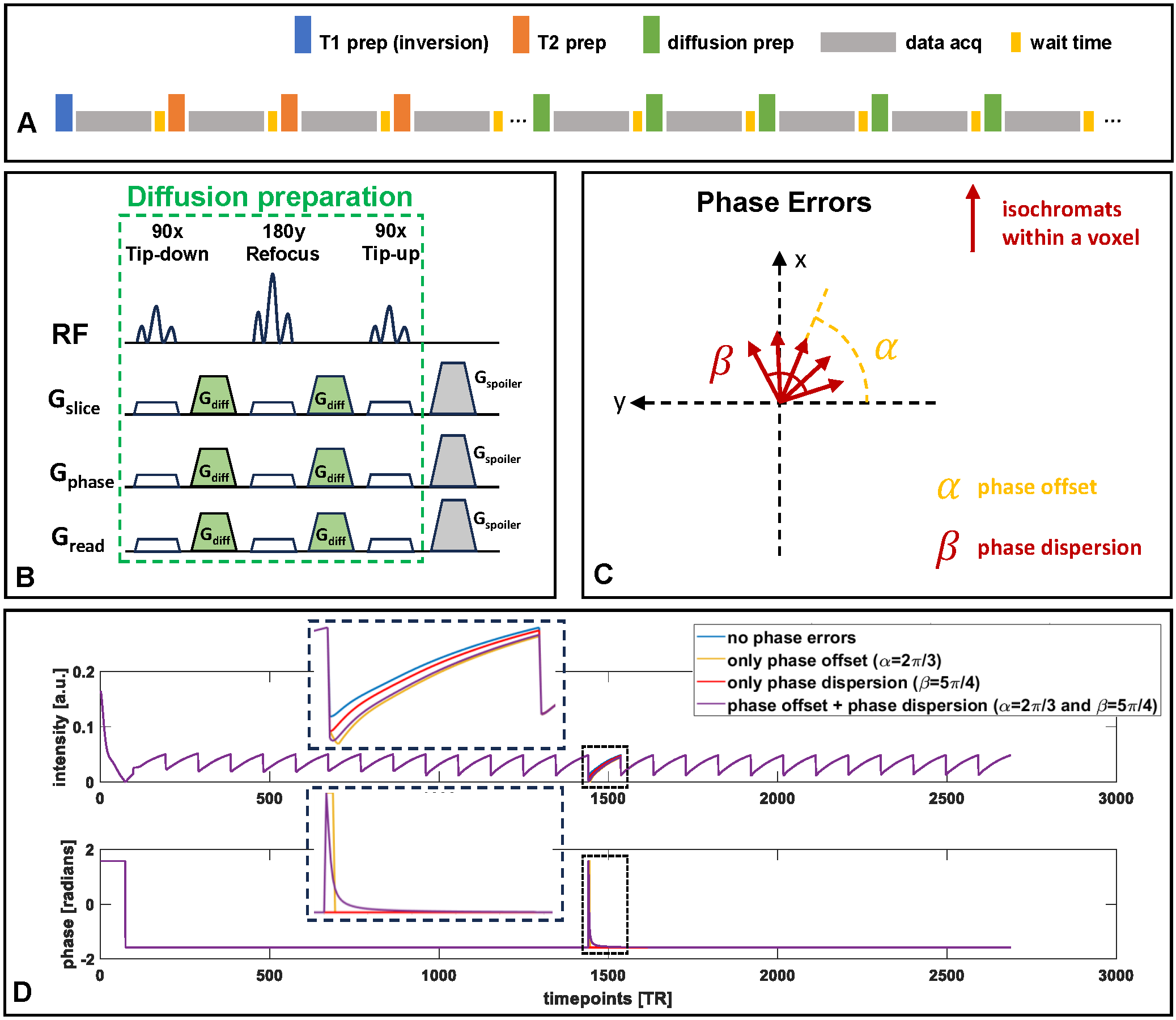
Figure 1. (A) mdMRF sequence consists of multiple segments, each staring with a preparation module, followed by data acquisition and ending with a wait time. (B) DP scheme used for diffusion encoding in mdMRF. (C) Phase offset and phase dispersion within a voxel are used to model the phase errors during diffusion preparation (before 90° tip-up pulse) for each diffusion-encoded segment. (D) Simulated mdMRF signal evolutions with and without phase errors. The phase errors cause signal attenuation and phase variation, with varying effects from phase offset and phase dispersion.
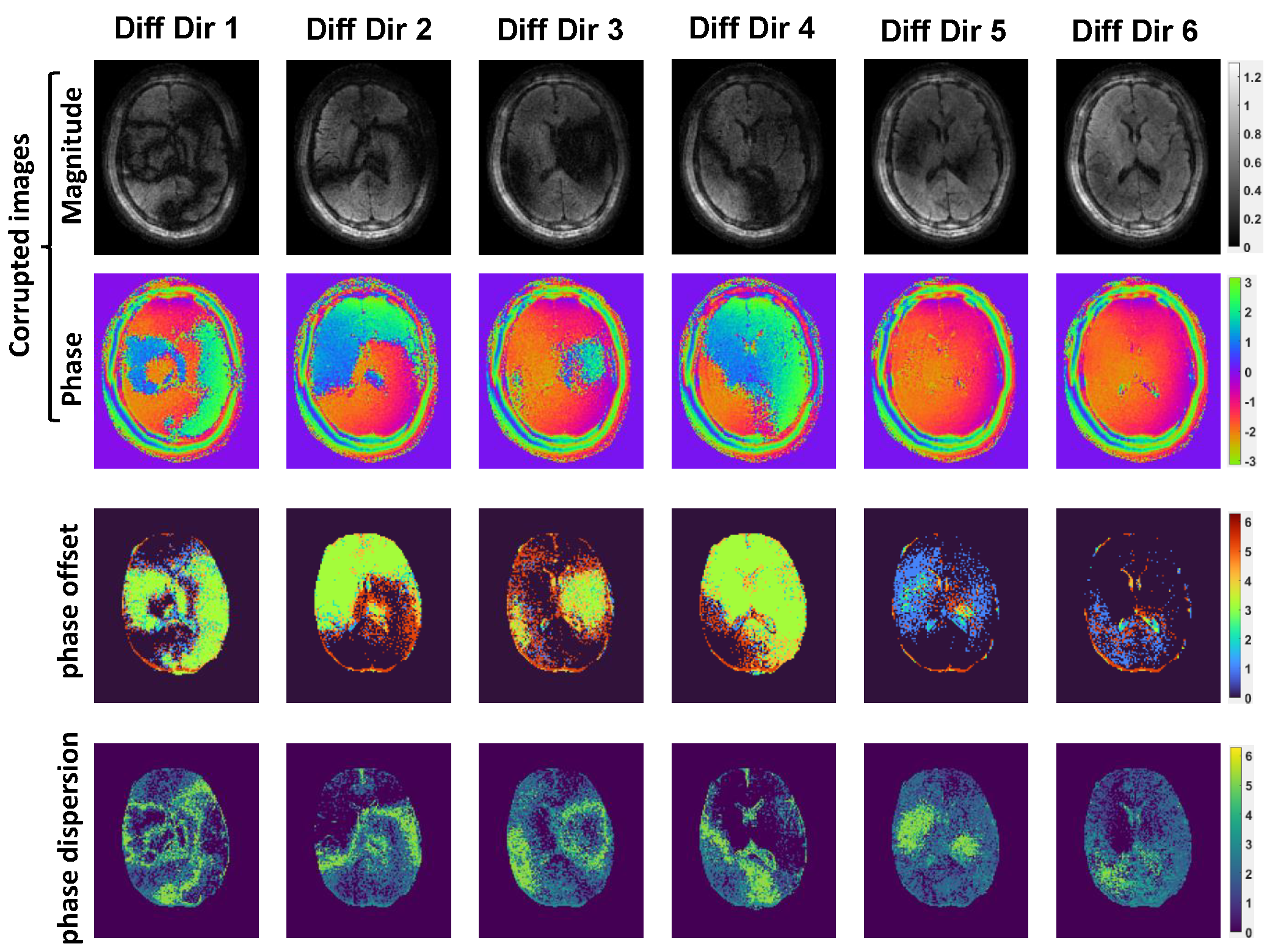
Figure 2. Reconstructed diffusion-weighted images that are affected by phase error effects including both magnitude (first row) and phase (second rows, unit: radians), the quantified phase offset maps (third row, unit: radians), and phase dispersion maps (fourth row, unit: radians) across all 6 diffusion encoding directions.
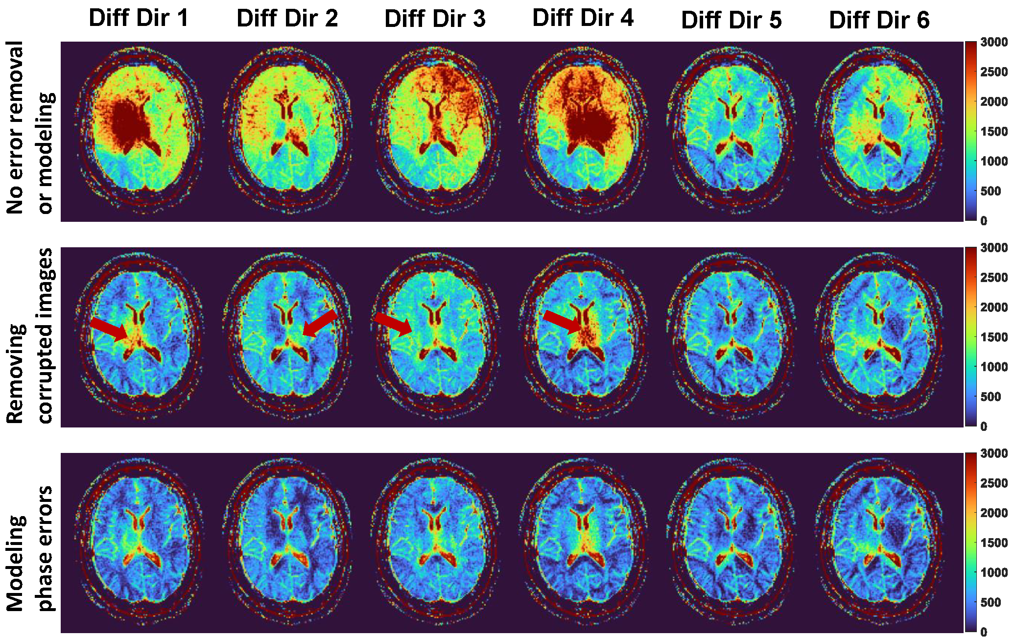
Figure 3. Comparison of ADC maps quantified using different methods. The ADC maps quantified without error removal or modeling suffer from severe shading artifacts (first row). By removing the corrupted segments, the artifacts can be significantly mitigated (second row). However, there are still some artifacts not removed, particularly around the ventricle anatomy (highlighted by red arrows). The proposed method can mitigate the artifacts more completely (third row). Color bar unit: 10-6 mm2/s.
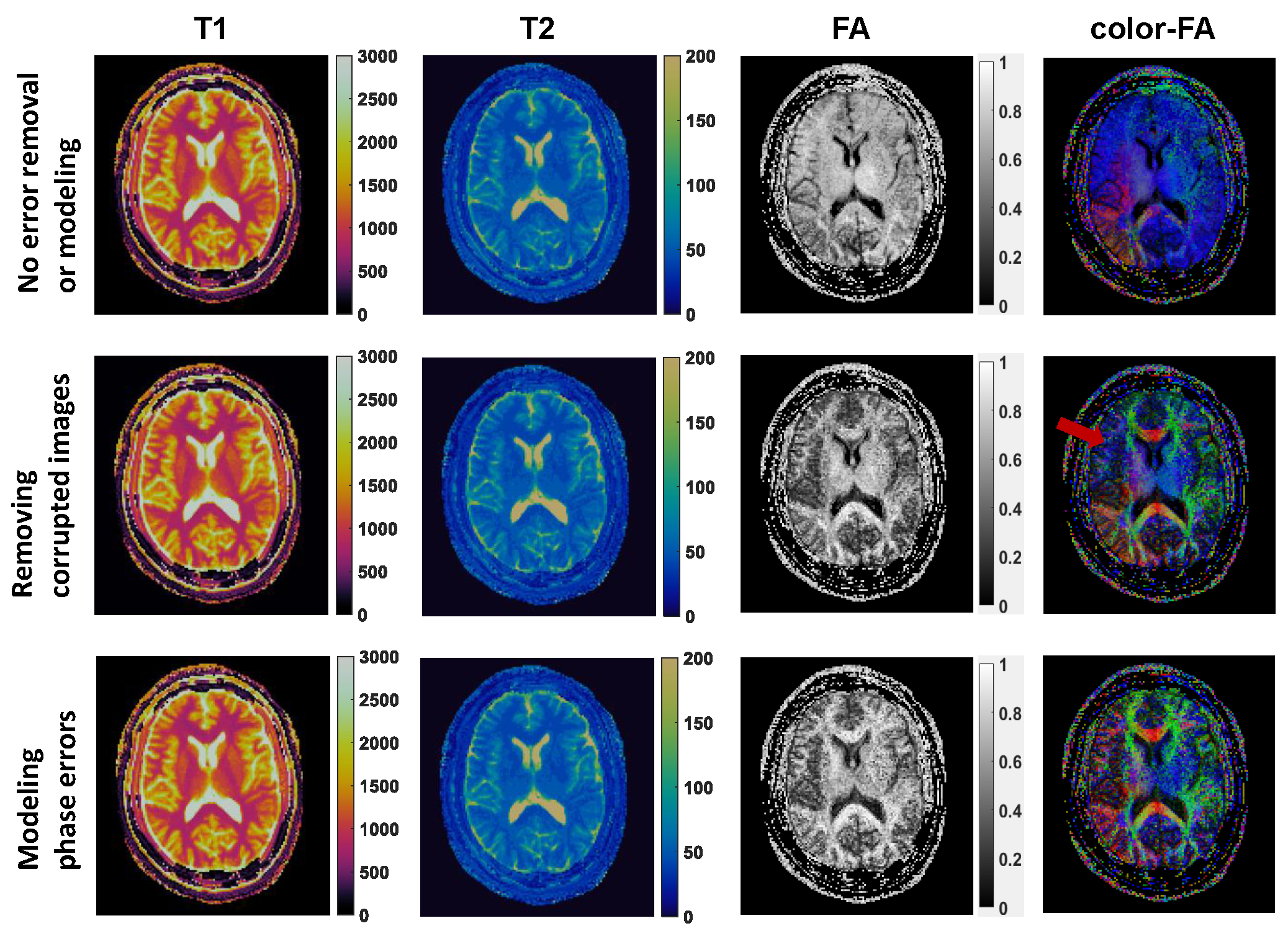
Figure 4. T1, T2, FA and colored FA maps from the first healthy subject scan. Without phase error removal or modeling, the FA and colored FA maps are totally corrupted (first row). With the corrupted segments excluded, the maps can be significantly improved, but still suffers from some artifacts (second row, highlighted by red arrows). The proposed method yields best diffusion parameter mapping (third row). T1 and T2 color bar unit: ms.
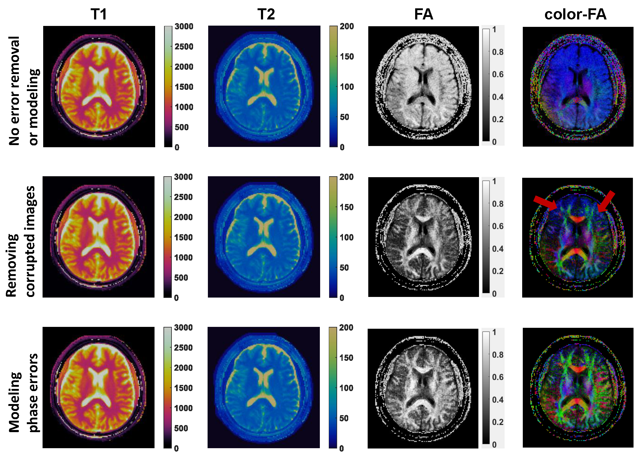
Figure 5. T1, T2, FA and colored FA maps from the second healthy subject scan. Without phase error removal or modeling, the FA and colored FA maps are totally corrupted (first row). With the corrupted segments excluded, the maps can be significantly improved, but still suffers from some artifacts (second row, highlighted by red arrows). The proposed method yields best diffusion parameter mapping (third row).