0621
Enhancing myocardial scar detection by combining fat-water separation with bright- and black-blood late gadolinium enhancement imaging1IHU LIRYC, Electrophysiology and Heart Modeling Institute, Université de Bordeaux – INSERM, Centre de Recherche Cardio-Thoracique de Bordeaux, Bordeaux, France, 2Department of Cardiothoracic Imaging, Hôpital Cardiologique du Haut-Lévêque, Bordeaux, France, 3Department of Diagnostic and Interventional Radiology, Lausanne University Hospital and University of Lausanne, Lausanne, Switzerland, 4Center for Biomedical Imaging (CIBM), Lausanne, Switzerland
Synopsis
Keywords: Myocardium, Fat
Motivation: Combined bright- and black-blood late gadolinium enhancement has shown potential for scar detection.However, accurate differentiation between scar and fat tissue in these images can pose a challenge.
Goal(s): To assess the SPOT-Dixon sequence, a joint bright- and black- blood methodology in combination with a two-point Dixon approach to improve myocardial scar detection and delineation.
Approach: The proposed sequence was tested in 13 patients with suspected cardiovascular diseases and the results were compared to those from reference sequences.
Results: The SPOT-Dixon sequence was able to reliably delineate scar tissue within the myocardium and differentiate it from fat in its proximity.
Impact: The proposed sequence combining the SPOT sequence and a two-point Dixon method gives reliable images of the myocardial scar and the fat tissue surrounding it, providing a valuable diagnostic advantage and potentially improving the accuracy of cardiovascular assessments.
Introduction
The combination of bright-blood (BR) and black-blood (BL) late gadolinium enhancement (LGE) techniques has demonstrated its efficacy in outlining myocardial scars in cardiac magnetic resonance imaging studies 1. Despite its success, accurately discriminating between scar and fat tissue in these images can be challenging, especially in patients with intramyocardial fat near the scar 2. On the other hand, the two-point Dixon method has been employed to create precise water- and fat-only images 3,4.In this proof-of-concept study, our primary aim is to assess the potential of incorporating a novel joint BR- and BL-LGE sequence with a two-point Dixon method to achieve a more distinct differentiation between myocardial scar tissue and fat.
Methods
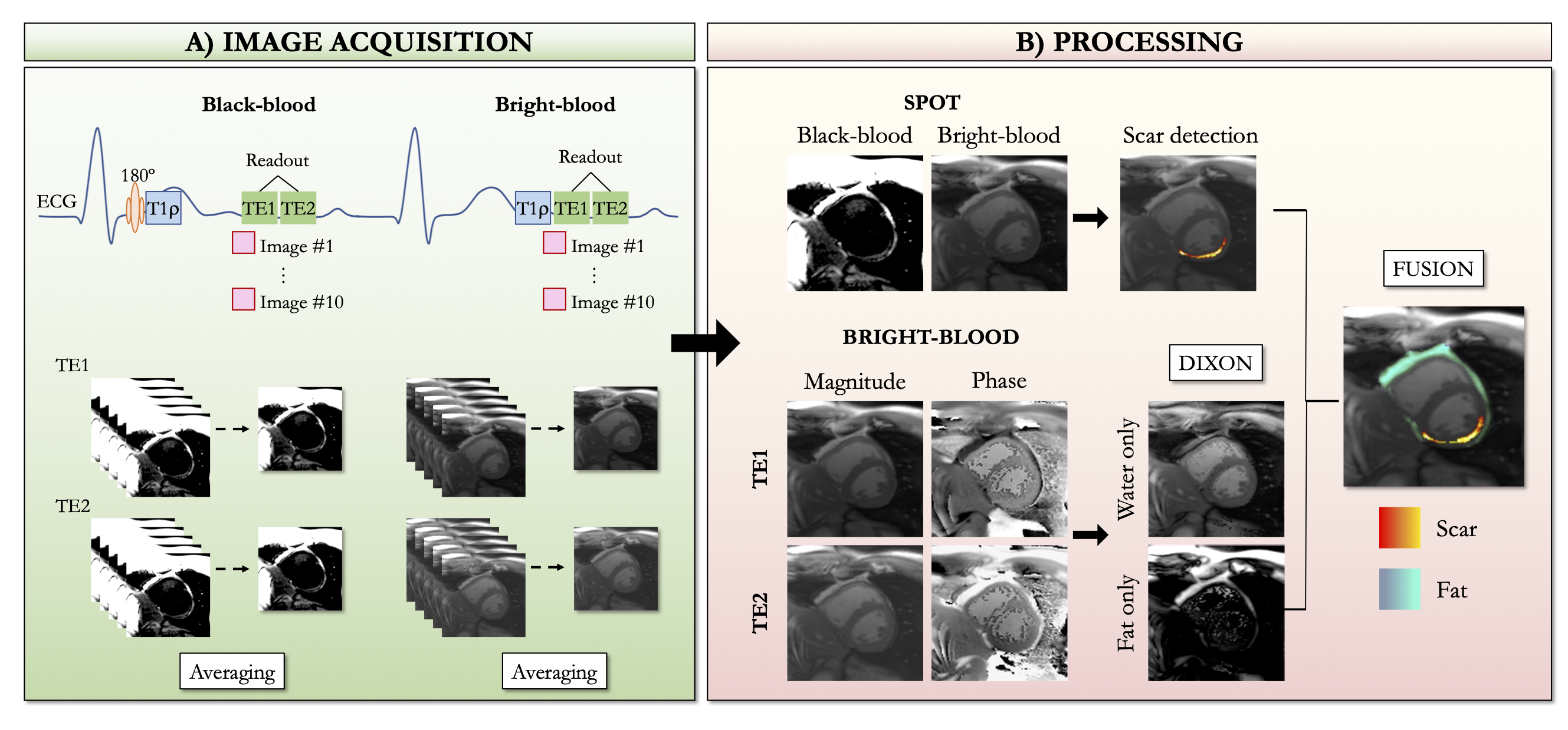
Acquisition: 13 patients (three women; mean age, 59±11.2 [SD] years; age range: 40−74 years) with suspected cardiovascular diseases were recruited for this study. The SPOT sequence was used, which simultaneously acquires both BR images, for anatomy visualization, and BL images, for scar visualization, by integrating inversion recovery with T1-rho pulses (Figure 1) 1. In order to apply a two-point Dixon method, whole-heart co-registered BR and BL SPOT images were obtained for each patient at two different echo times. Short-axis 2D whole-heart BR PSIR images were also acquired as reference 5.Imaging parameters: All images were collected on a 1.5T Siemens Aera system during a breath-hold. The imaging parameters were: four signal averages, 1.4mm x 1.4mm in-plane resolution, 8mm slice thickness, spin-lock frequency/duration=500Hz/27ms, FA=60°, GRAPPA x2, ~160ms acquisition window, TE1/TE2/TR=1.27/3.25/5.11ms, bandwidth=870Hz/pixel. A prior scout acquisition was used to determine the inversion time needed to null the myocardium signal (PSIR) and both the myocardial and the blood signals (SPOT).
Processing: A two-point Dixon method was implemented using the BR SPOT images in MATLAB (version R2023a, The MathWorks, Natick, Massachusetts, USA) (Figure 1). The B0-NICEbd algorithm was selected for fat-water separation due to its ability to generate reliable B0 and fat-water maps while effectively mitigating phase errors introduced during acquisition 6. This algorithm is based on the multi-point Dixon technique that incorporates magnitude information in B0 off-resonance mapping to perform a non-iterative correction of phase errors, known as B0-NICE 7. In addition to this, B0-NICEbd accommodates bipolar dual-echo acquisitions, with the goal of enhancing the robustness of fat-suppression in cardiac magnetic resonance imaging. Adjustments were made to the threshold selection for each patient, particularly because water-only pixels can exhibit notably high signal levels following the administration of contrast, causing the fat identification to fail. Once the images were acquired, fat and scar signals were superimposed with color layers onto the BR SPOT image to facilitate the most effective evaluation of fat content and the detection and localization of myocardial scar.
Image analysis: To determine relative contrasts between the myocardium, fat, scar, and the blood pool for each patient, we computed the difference in signal intensity (S1 - S2) between two of these tissues and divided it by S2. In the patient images, circular regions-of-interest with an area of 15.8 mm² were used to analyze all the tissues.
Results
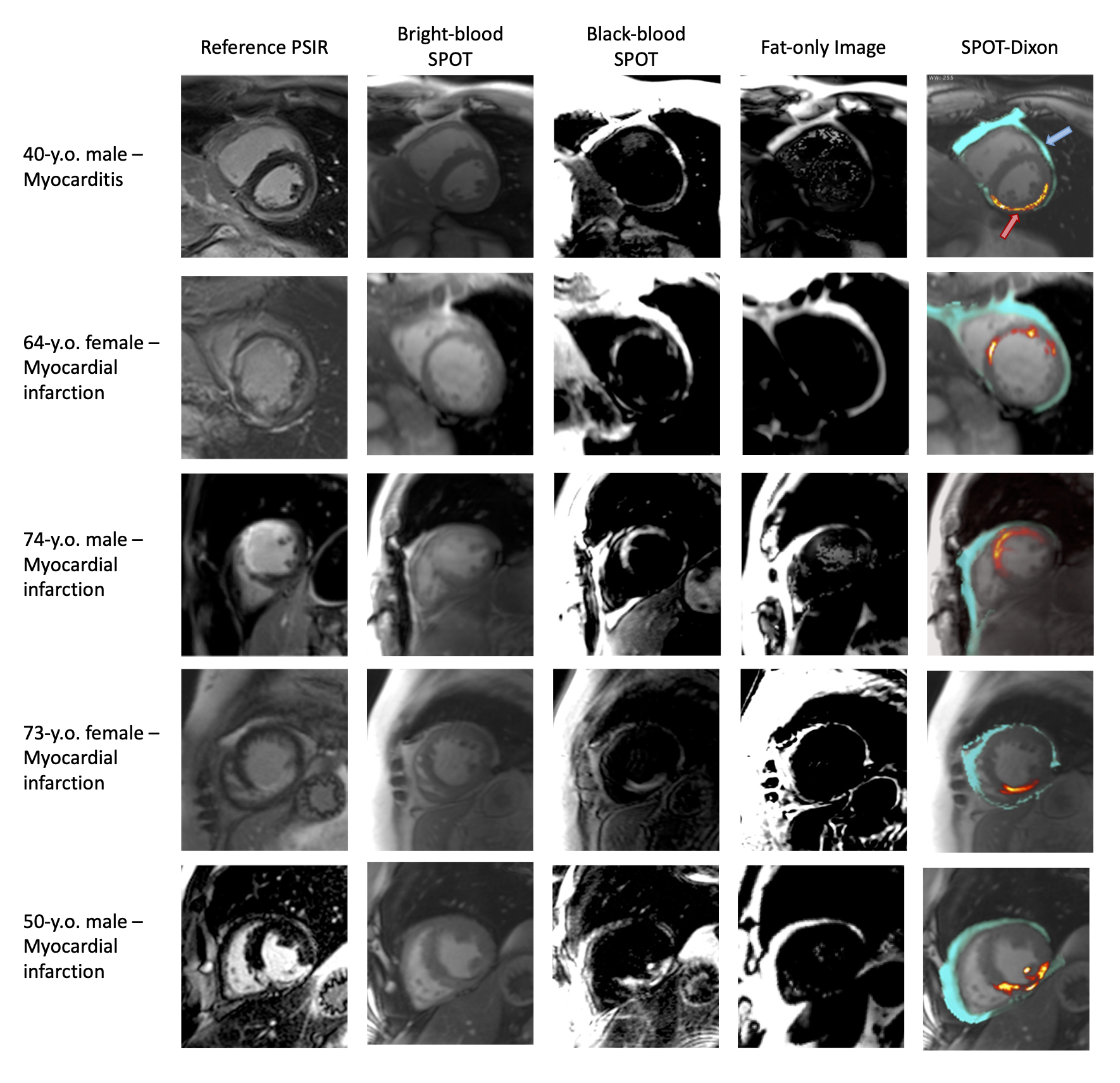
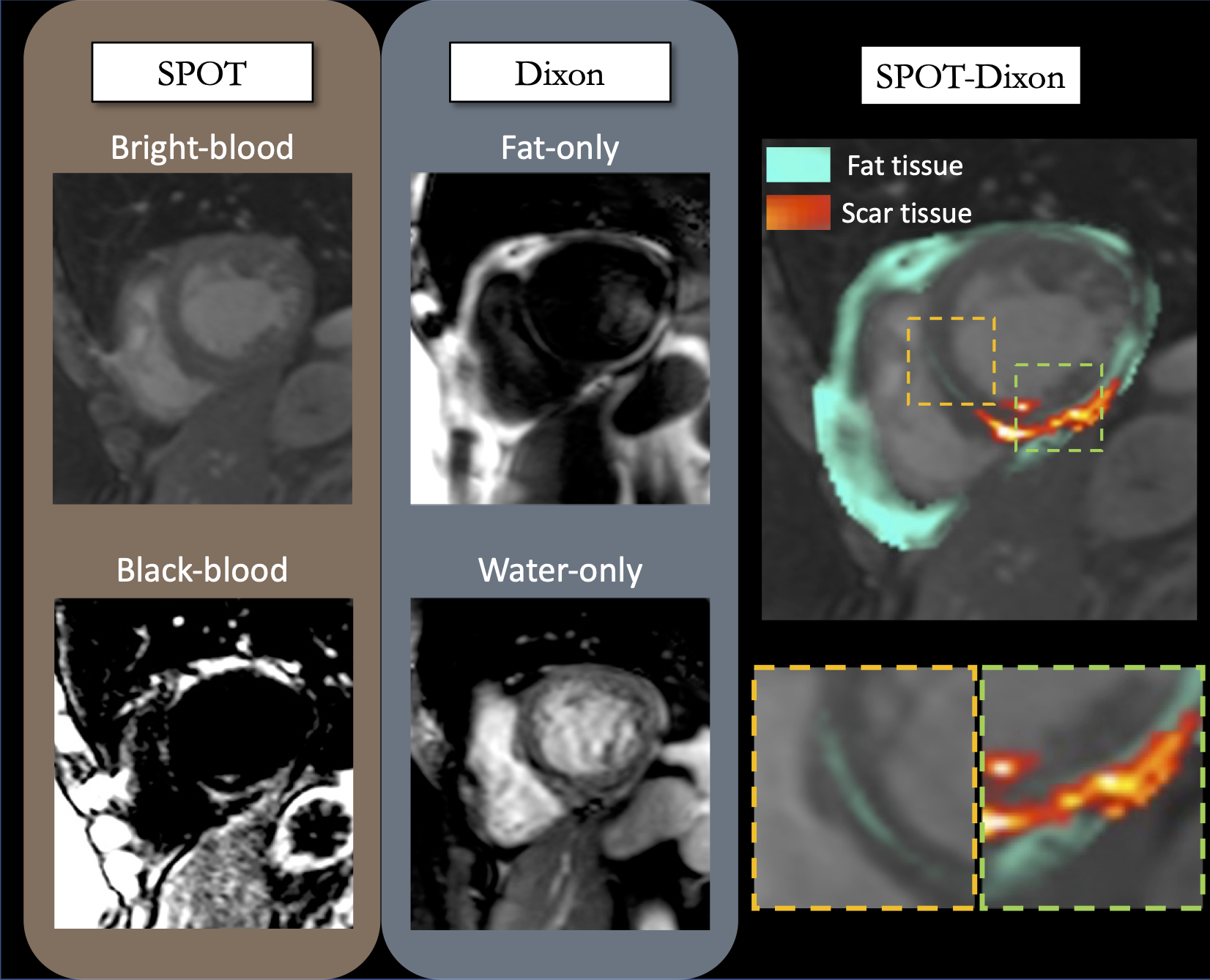
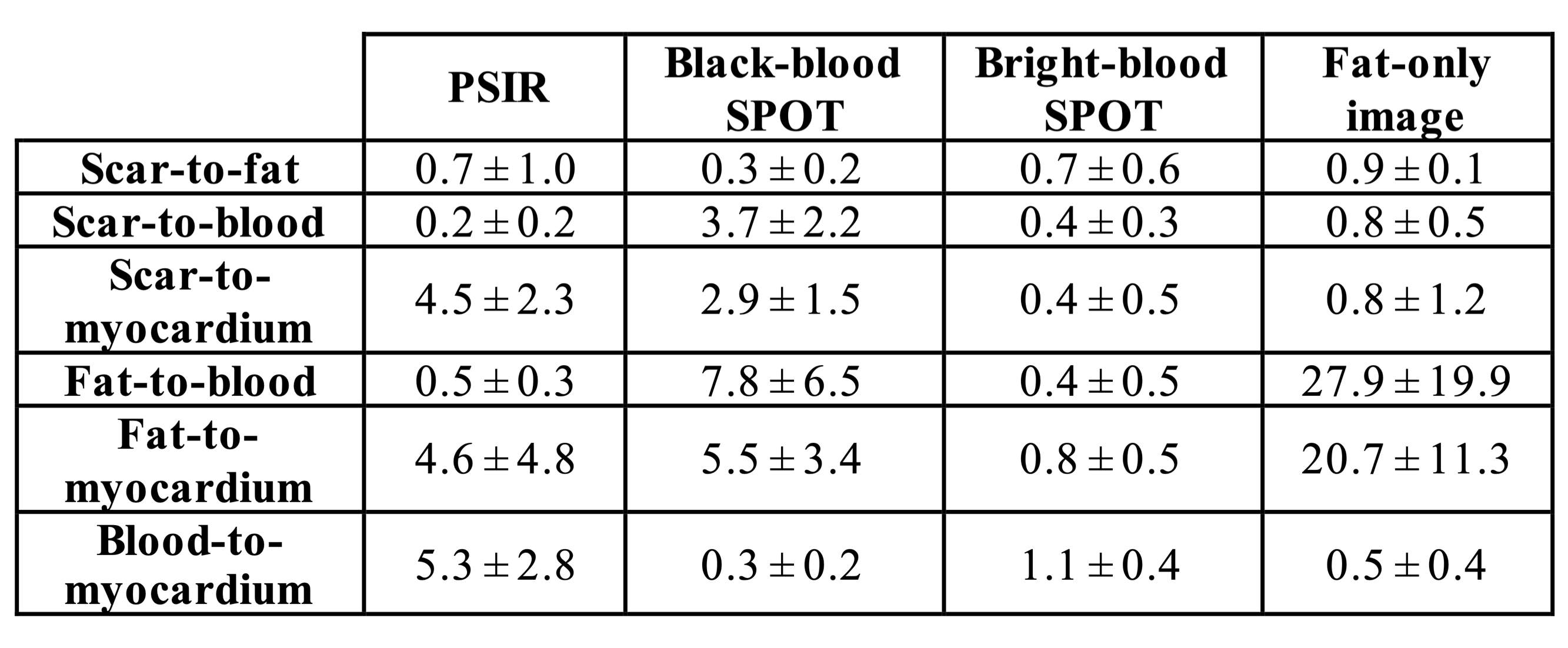
Our results showed that the mean scar-to-myocardium relative contrast was substantially higher in the BL SPOT images in comparison to the fat-only images (2.9±1.5 [SD] vs. 0.8±1.2). This emphasized the enhanced visibility of scars in the former images, while they were largely attenuated or entirely eliminated in the latter (Table 1). However, the BL images also presented considerable high contrasts between both fat tissue and the blood pool, and fat tissue and healthy myocardium, showcasing the potential challenge of distinguishing between them.The two-point Dixon method effectively achieved consistent water-fat separation within our patient cohort. This separation persisted across all patients, resulting in the suppression of blood, healthy myocardium, and scar tissue in the fat-only images. In combination with the SPOT sequence, this approach was able to enhance the visualization of myocardial scars by facilitating the differentiation between fat and scar tissue, as exemplified in Figure 2. Significantly, the SPOT-Dixon sequence successfully revealed myocardial fat in the septum of a patient diagnosed with left ventricular arrhythmogenic cardiomyopathy, where fibrofatty replacement was reported (Figure 3).
Conclusion
This proof-of-concept study aimed to demonstrate the capability of the SPOT sequence in conjunction with a two-point Dixon method to consistently outline myocardial scar tissue and distinguish it from adjacent fat tissue - thereby removing ambiguity. The results obtained emphasize the utility of the SPOT-Dixon sequence in highlighting myocardial scars while maintaining clear distinctions between fat, healthy myocardium, and blood, which could potentially provide a valuable diagnostic advantage.Acknowledgements
This work was supported by funding from the French National Research Agency under grant agreements Equipex MUSIC ANR-11-EQPX-0030, ANR-21-CE17-0034-01, Programme d’Investissements d’Avenir ANR-10-IAHU04-LIRYC, ANR-22-CPJ2-0009-01, and from the European Research Council (ERC) under the European Union’s Horizon 2020 research and innovation program (grant agreement N°101076351).References
- Bustin A, et al. Free-breathing joint bright- and black-blood cardiovascular magnetic resonance imaging for the improved visualization of ablation-related radiofrequency lesions in the left ventricle. EP Europace. 2022;24.
- Cannavale, Giuseppe et al. “Fatty Images of the Heart: Spectrum of Normal and Pathological Findings by Computed Tomography and Cardiac Magnetic Resonance Imaging.” BioMed research international. 2018, 5610347.
- Dixon WT. Simple proton spectroscopic imaging. Radiology. 1984 Oct;153(1):189-94.
- Kellman P, Hernando D, Shah S, et al. Multiecho Dixon fat and water separation method for detecting fibrofatty infiltration in the myocardium. Magn Reson Med. 2009 Jan;61(1):215-21.
- Kellman P, Arai AE, McVeigh ER, Aletras AH. Phase-sensitive inversion recovery for detecting myocardial infarction using gadolinium-delayed hyperenhancement. Magn Reson Med. 2002;47(2):372-383.
- Liu J, Peters DC, Drangova M. Method of B0 mapping with magnitude-based correction for bipolar two-point Dixon cardiac MRI. Magn Reson Med. 2017 Nov;78(5):1862-1869.
- Liu J, Drangova M. Method for B0 off-resonance mapping by non-iterative correction of phase-errors (B0-NICE). Magn Reson Med. 2015; 74:1177–1188.
Figures