0618
Regional fat distribution is associated with subclinical left atrial and left ventricular diastolic dysfunction in early adult obesity1West China Hospital of Sichuan University, Chengdu, China
Synopsis
Keywords: Heart Failure, Cardiovascular
Motivation: Whether obese subjects with no clinical signs or comorbidities have diastolic dysfunction is unclear.
Goal(s): We non-invasively assess diastolic function in adults with uncomplicated obesity and evaluate its association with fat distribution.
Approach: Left atrial (LA) and left ventricular (LV) strain and volume-time curve using cardiac magnetic resonance were compared.
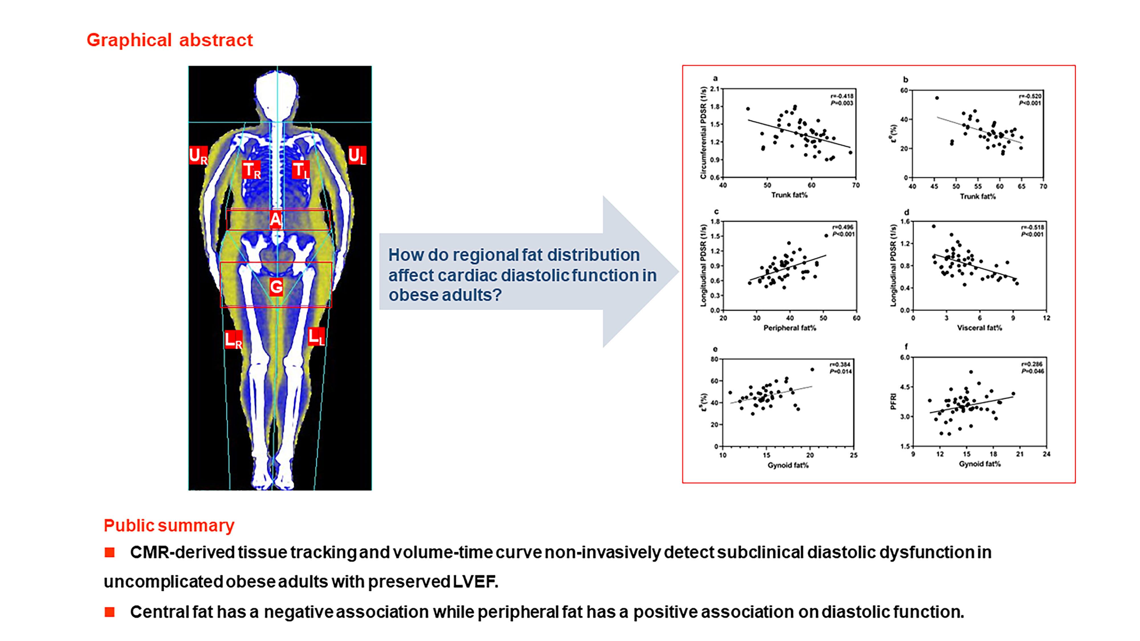
Results: The obese patients had impaired diastolic function, manifested as lower LV diastolic strain rates and peak filling rate index and declined LA reservoir and conduit function compared with controls. Central fat has a negative association while peripheral fat has a positive association on diastolic function.
Impact: CMR-derived feature tracking and volume-time curve non-invasively detected subclinical diastolic dysfunction in early adult obesity with preserved LVEF. This study also suggests that recognizing the role of different areas of fat on the heart may be beneficial for obese patients.
Methods: The study complied with the Declaration of Helsinki and was approved by the Institutional Review Board of our Hospital. Written informed consents were obtained from all participants. We prospectively recruited 49 mild-to-moderate obese subjects and 43 healthy volunteers between 18 and 60 years old. BMI was categorized into the three groups according to the Asia criteria: healthy weight (18.5-23.0 kg/m2), overweight (23.0-27.5kg/m2), and obesity (≥27.5kg/m2). Subjects were excluded if they had any of the following conditions: hypertension and diabetes; history of lipid-lowering, hypoglycemic, or antihypertensive drugs; history of cardiovascular diseases or cardiovascular procedures; major systemic diseases that may affect the myocardium, such as connective tissue disease; respiratory diseases that could affect heart, such as obstructive sleep apnea; renal failure; and any contraindication to CMR imaging. CMR examinations were performed with an 18-element surface coil on 3.0 T whole-body scanner (MAGNETOM Skyra, Siemens Medical Solutions, Erlangen, Germany). A balanced steady-state free precession sequence was used to acquire continuous cine images in the short-axis view and two- and four-chamber cine images in the long-axis view. The parameters are as follows: TR/TE=3.3/1.22 ms, flip angle=41°, slice thickness=8 mm, field of view=360×320 mm2, matrix size=208×166, and a temporal resolution=39.34 ms. CMR data was imported to commercially available software (CVI 42 version 5.11.3). LA strain [total, passive, and active strains (εs, εe, and εa) and peak positive, early and late negative strain rates (SRs, SRe, and SRa)], LV strain [peak diastolic/systolic strain rates (PDSR and PSSR)] and volume-time curve [peak filling rate/end‑diastolic volume (PFR/EDV) and peak ejection rate/end‑diastolic volume (PER/EDV)] were measured. DXA (Lunar iDXA, GE Medical Systems Lunar) was used to measure body fat distribution. All statistical analyses were performed using the SPSS (version 23). Continuous data between obesity and controls were compared using Student’s t-test and Whitney U-test. Pearson’s correlation and linear regression were used to estimate the correlation.
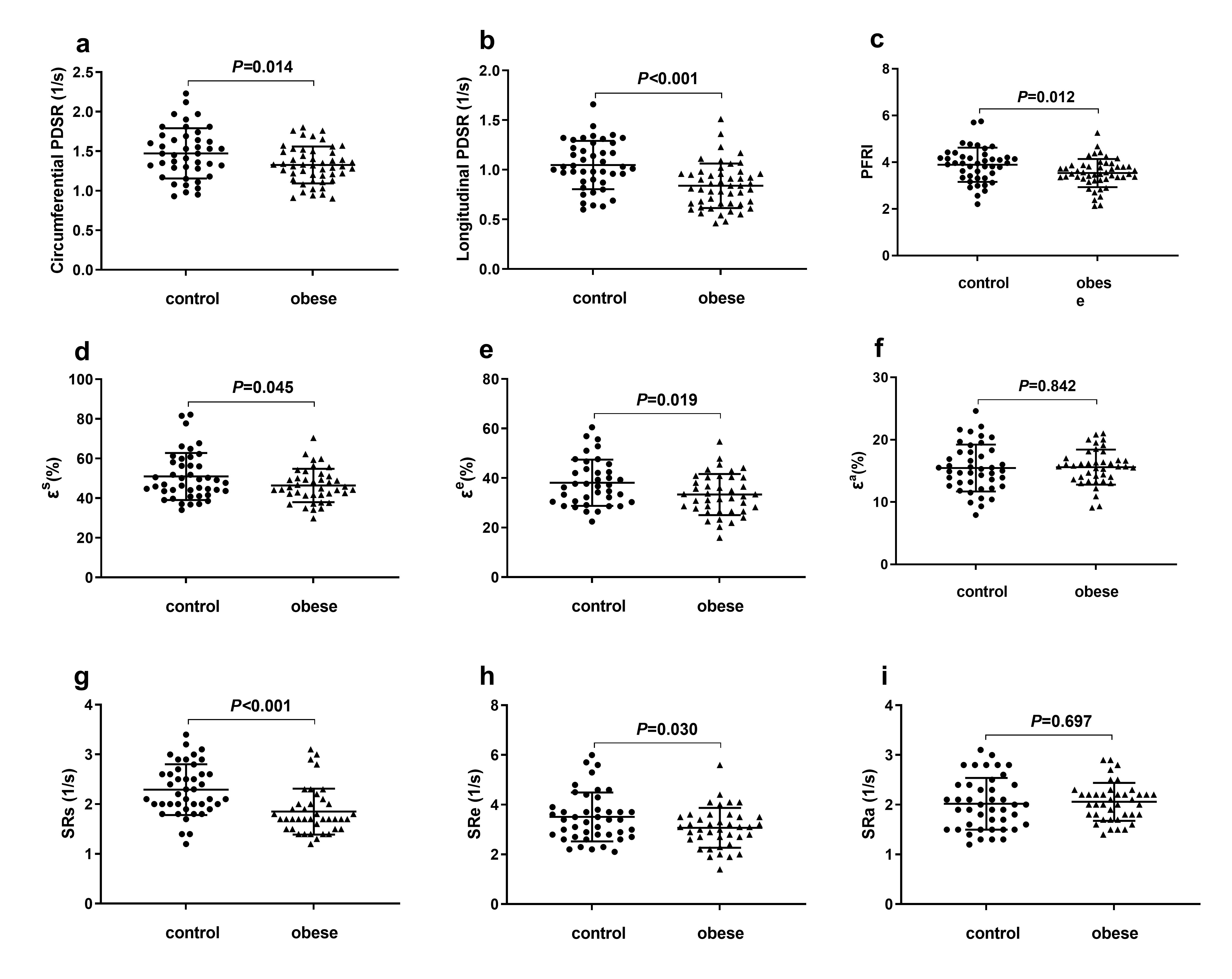
Results: The obese participants had impaired diastolic function, manifested as lower LV circumferential and longitudinal PDSR (1.3±0.2s-1 vs. 1.5±0.3s-1, 0.8±0.2s-1 vs. 1.1±0.2s-1, all P<0.05), LV PFR/EDV (3.5±0.6 s-1 vs. 3.9±0.7 s-1, P=0.012), and declined LA reservoir function [εs and SRs (46.4±8.4% vs. 51±12%, 1.9±0.5s-1 vs. 2.3±0.5s-1; all P<0.05)] and conduit function [εe and SRe (30.8±8.0% vs. 35.5±9.8%, 3.1±0.8s-1 vs. 3.5±1.0s-1; all P<0.05)] compared with controls. The LV systolic function parameters (PSSR and PER/EDV) and LA pumping function was not different between obesity and controls. Multivariable linear regression analysis demonstrated that trunk fat was independently associated with LA εe (β=-0.520, P=0.001; R2=0.270) and LV circumferential PDSR (β=−0.417, P=0.003; R2=0.174); visceral fat and peripheral fat were associated with LV longitudinal PDSR (β=−0.342, P=0.038; β=0.376, P=0.024; R2=0.389); gynoid fat was associated with LA εs (β=0.384, P=0.014; R2=0.148) and LV PFR/EDV (β=0.285, P=0.047; R2=0.081).
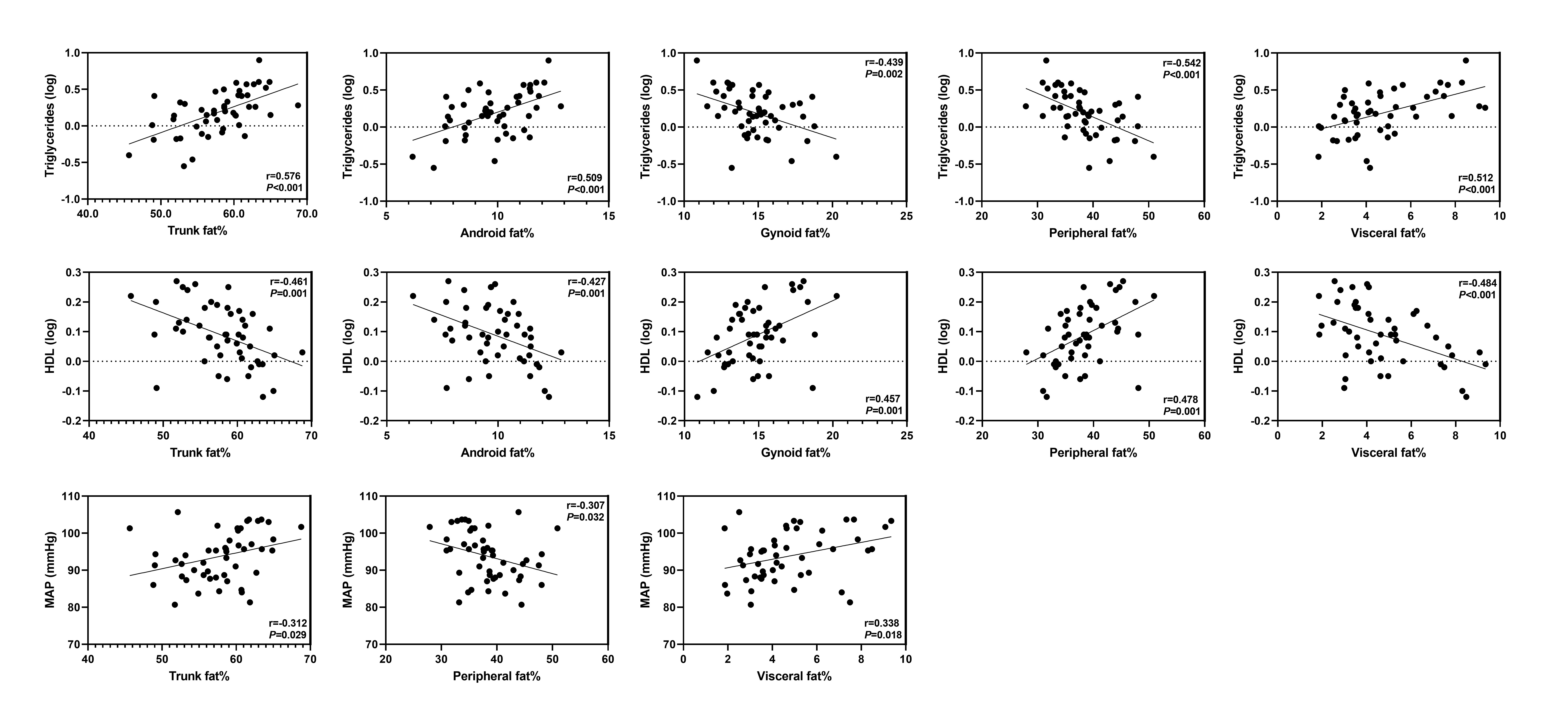
Discussion: In our study, subclinical diastolic dysfunction is found in obese adults with preserved LVEF. Earlier echocardiographic studies have shown both systolic and diastolic dysfunction in severe obesity. These findings may suggest that mild-moderate uncomplicated obesity initially develop diastolic dysfunction rather than systolic dysfunction. Our study also demonstrated that obese subjects had decreased LA reservoir and conduit function and preserved pump function compared with controls. Previous literature indicated that LA reservoir and conduit function were impaired in all grades of diastolic dysfunction, while pump function was increased in mild diastolic dysfunction and reduced as diastolic dysfunction progressed12, 13. The findings of LA function in our study may reveal mild diastolic dysfunction in these obese individuals. Moreover, our results showed that central fat distributions (visceral fat and trunk fat) had negative relationships while peripheral fat distributions (peripheral fat and gynoid fat) had positive relationships on diastolic function. The different effects of these fat distributions on metabolic factors (such as dyslipidemia), inflammatory cytokines, and adipokines may partially explain the discrepant effects of fat distributions on cardiac function.
Conclusion: CMR-derived feature tracking and volume-time curve non-invasively detected subclinical diastolic dysfunction in early adult obesity with preserved LVEF. Central fat has a negative association while peripheral fat has a positive association on diastolic function. This finding suggests that recognizing the role of different areas of fat on the heart may be beneficial for obese patients.
Acknowledgements
This work was supported by grants from the National Natural Science Foundation of China (No. 81601462), the Key Research & Development Project of Science and Technology of Sichuan Province (No. 2021YFS0142), the Science and Technology Department of Sichuan Province (No. 2019YFS0302), and the 1.3.5. Project for Disciplines of Excellence, West China Hospital, Sichuan University (No. ZYGD18017).References
1. Carbone S, Lavie CJ, Elagizi A, Arena R, Ventura HO. The Impact of Obesity in Heart Failure. Heart Fail Clin. Jan 2020;16(1):71-80.
2. Pandey A, LaMonte M, Klein L, et al. Relationship Between Physical Activity, Body Mass Index, and Risk of Heart Failure. Journal of the American College of Cardiology. Mar 7 2017;69(9):1129-1142.
3. Paulus WJ, Tschope C, Sanderson JE, et al. How to diagnose diastolic heart failure: a consensus statement on the diagnosis of heart failure with normal left ventricular ejection fraction by the Heart Failure and Echocardiography Associations of the European Society of Cardiology. Eur Heart J. Oct 2007;28(20):2539-2550.
4. Nagueh SF, Smiseth OA, Appleton CP, et al. Recommendations for the Evaluation of Left Ventricular Diastolic Function by Echocardiography: An Update from the American Society of Echocardiography and the European Association of Cardiovascular Imaging. Eur Heart J Cardiovasc Imaging. Dec 2016;17(12):1321-1360.
5. Claus P, Omar AMS, Pedrizzetti G, Sengupta PP, Nagel E. Tissue Tracking Technology for Assessing Cardiac Mechanics: Principles, Normal Values, and Clinical Applications. JACC Cardiovasc Imaging. Dec 2015;8(12):1444-1460.
6. Cameli M, Lisi M, Mondillo S, et al. Left atrial longitudinal strain by speckle tracking echocardiography correlates well with left ventricular filling pressures in patients with heart failure. Cardiovascular Ultrasound. Apr 21 2010;8.
7. Cameli M, Sparla S, Losito M, et al. Correlation of Left Atrial Strain and Doppler Measurements with Invasive Measurement of Left Ventricular End-Diastolic Pressure in Patients Stratified for Different Values of Ejection Fraction. Echocardiography. Mar 2016;33(3):398-405.
8. Petak S, Barbu CG, Yu EW, et al. The Official Positions of the International Society for Clinical Densitometry: body composition analysis reporting. J Clin Densitom. Oct-Dec 2013;16(4):508-519.
9. Vasan SK, Osmond C, Canoy D, et al. Comparison of regional fat measurements by dual-energy X-ray absorptiometry and conventional anthropometry and their association with markers of diabetes and cardiovascular disease risk. Int J Obes (Lond). Apr 2018;42(4):850-857.
10. Peppa M, Koliaki C, Hadjidakis DI, et al. Regional fat distribution and cardiometabolic risk in healthy postmenopausal women. Eur J Intern Med. Dec 2013;24(8):824-831.
11. Liu J, Li J, Yu J, et al. Regional Fat Distributions Are Associated With Subclinical Right Ventricular Dysfunction in Adults With Uncomplicated Obesity. Front Cardiovasc Med. 2022;9:814505.
12. Brecht A, Oertelt-Prigione S, Seeland U, et al. Left Atrial Function in Preclinical Diastolic Dysfunction: Two-Dimensional Speckle-Tracking Echocardiography-Derived Results from the BEFRI Trial. J Am Soc Echocardiogr. Aug 2016;29(8):750-758.
13. Guan Z, Zhang D, Huang R, Zhang F, Wang Q, Guo S. Association of left atrial myocardial function with left ventricular diastolic dysfunction in subjects with preserved systolic function: a strain rate imaging study. Clin Cardiol. Oct 2010;33(10):643-649.
Figures