0617
Interventional molecular MRI of early myocardial injury in a pig model of ischemia and reperfusion1Division of Medical Physics, Dept. of Diagnostic and Interventional Radiology, University Medical Center Freiburg, Faculty of Medicine, University of Freiburg, Freiburg, Germany, 2Dept. of Cardiology, University Medical Center Freiburg, Faculty of Medicine, University of Freiburg, Freiburg, Germany
Synopsis
Keywords: Myocardium, Heart
Motivation: Cardiac MRI has become the gold standard for non-invasive characterization of myocardial tissue. However, current MRI techniques only indirectly represent cardiac inflammation.
Goal(s): To assess if interventional molecular MRI allows for visualization of the cellular processes during the inflammatory response after myocardial infarction.
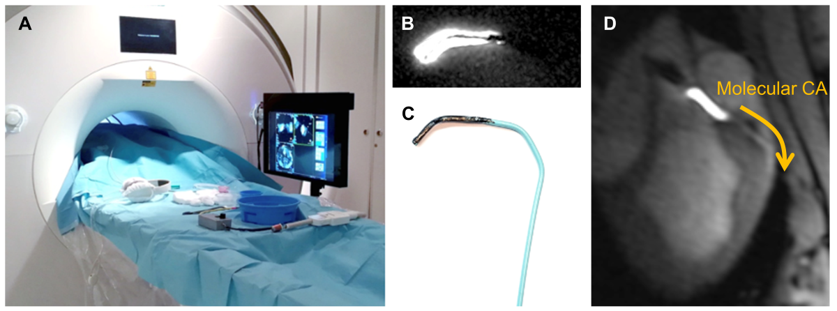
Approach: Iron-labeled P-selectin contrast agent was injected in pigs after 40 minutes of myocardial ischemia. To increase sensitivity, the contrast agent was injected into the coronary artery via an MR-guided intervention.
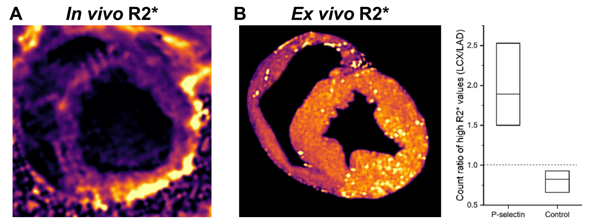
Results: Infarcted myocardial segments could be visualized using the iron-labeled contrast agent by increased R2* values both in vivo and ex vivo.
Impact: The intracoronary injection of molecular contrast agents using interventional MRI can add valuable information for preclinical studies of the early cellular processes after myocardial ischemia.
Background
Early reperfusion plays an essential role in preserving cardiac function after acute myocardial infarction (MI). Next to time of ischemia, inflammation is another driving force of wound healing. Assessment of inflammatory activity may therefore enable cardiovascular risk assessment after MI1. Even though cardiac MRI has become the gold standard for non-invasive characterization of myocardial tissue, current MRI techniques such as T1-, T2-, T2* mapping and LGE only indirectly represent cardiac inflammation. Molecular MRI with contrast agents that bind to specific antibodies allows for a direct visualization of the cellular processes during the inflammatory response after MI2–4. In this study, an iron-labeled P-selectin contrast agent is tested in a pig model for its potential to assess the early cardiac inflammatory response after ischemia/reperfusion. To increase the sensitivity, an intracoronary injection of the contrast agent is performed during an MR-guided coronary intervention5,6. The imaging results are compared to conventional cardiac tissue characterization methods.Methods
The interventional study was performed on 6 domestic landrace pigs (age: 3 months, body weight: 50-70 kg). The animals were anesthetized and mechanically ventilated during the experiments. Ischemia was induced under X-ray fluoroscopy with a balloon catheter that was inflated in the mid-segment of the left circumflex artery (LCX). After 40 min of ischemia, the balloon was deflated to establish reperfusion. Within 4 hours after reperfusion the animals were transferred to a clinical 3T MRI system (Siemens PrismaFit). First, functional imaging and relaxometric mapping (T1, T2, T2*) were performed. For T1-mapping, an inversion recovery bSSFP sequence was used, T2 mapping was performed with a T2-prepared FLASH sequence, and R2* maps were acquire with a multi-echo FLASH sequence.MR-guided coronary intervention was performed with a 6F active coronary catheter7 that was advanced via a femoral access. The catheter was maneuvered into the left coronary artery (LCA) under MR imaging with a real-time FLASH sequence. The successful intubation of the LCA was verified by a perfusion measurement during intracoronary injection of a small bolus of diluted Gd contrast agent. Next, a microparticles of iron oxide (MPIO)-based contrast agent was injected into the LCA via the catheter. In 3 pigs, an MPIO-labelled P-selectin antibody was injected, and whereas 3 control animals received unspecifically targeting IgG-MPIO.
T2* mapping was repeated after the coronary intervention, and LGE imaging was performed 15 min after intravenous injection of 2.5 mmol/kg Gd. The animals were euthanized immediately after the MR exam and the excised hearts were stored in PFA. Seven days after fixation T2* mapping was performed of the excised hearts with a 3D multi-echo FLASH dataset with 0.38 mm³ resolution.
Results
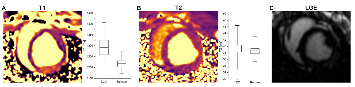
In vivo cardiac imaging showed no impaired wall motion and no late enhancement after 40 minutes of ischemia. In addition, no significant difference was seen in T2 between ischemic left ventricular segments (38.8 ± 2.7) ms and remote segments (38.4 ± 2.0) ms (p = 0.25), whereas T1 was significantly increased in segments with ischemia (1301 ± 53) ms over remote segments (1225 ± 28) ms (p < 0.05). Animals that received P-selectin targeting contrast agent showed increased R2* values in the ischemic segments compared to the control group. The measured R2* values were normalized to the values in remote segments. Ex vivo imaging supported the finding of higher amount of MPIOs in the ischemic segments in the P-selectin group. Here, the number of high R2* values in the ischemic segments was measured.Conclusion
This study shows that molecular imaging can be used to image and assess the early response after myocardial ischemia. The short occlusion of only 40 min lead to a mild injury so that conventional MRI techniques (LGE, T2-mapping) failed to detect the myocardial damage, whereas T1 mapping was more sensitive in detecting the ischemic myocardial segments. Thus, the proposed technique adds valuable insight into the cellular processes in the acute phase even after short myocardial ischemia. Local interventional administration of the molecular contrast agent through a catheter significantly enhanced the sensitivity – thus, MR-guided cardiac interventions may help to overcome the translational challenges associated with the larger amount of costly contrast agents in large animal studies as compared to small animal research of myocardial infarction with molecular MRI.Acknowledgements
This study is part of SFB1425, funded by the Deutsche Forschungsgemeinschaft (DFG, German Research Foundation #422681845). Grant support by the German Research Foundation (DFG) under RE 4876/1-1 is gratefully acknowledged.References
1. Nahrendorf, M., Pittet, M. J. & Swirski, F. K. Monocytes: Protagonists of Infarct Inflammation and Repair After Myocardial Infarction. Circulation 121, 2437–2445 (2010).
2. Jaffer, F. A., Libby, P. & Weissleder, R. Molecular Imaging of Cardiovascular Disease. Circulation 116, 1052–1061 (2007).
3. Lavin Plaza, B. et al. Molecular Imaging in Ischemic Heart Disease. Curr. Cardiovasc. Imaging Rep. 12, 31 (2019).
4. MacRitchie, N., Noonan, J., Guzik, T. J. & Maffia, P. Molecular imaging of cardiovascular inflammation. Br. J. Pharmacol. 178, 4216–4245 (2021).
5. Heidt, T. et al. Real-time magnetic resonance imaging – guided coronary intervention in a porcine model. Sci. Rep. 9, 8663 (2019).
6. Heidt, T. et al. Magnetic resonance imaging for pathobiological assessment and interventional treatment of the coronary arteries. Eur. Heart J. Suppl. 22, C46–C56 (2020).
7. Bock, M. et al. MR-guided intravascular procedures: Real-time parameter control and automated slice positioning with active tracking coils. J. Magn. Reson. Imaging 19, 580–589 (2004).
Figures