0613
Feasibility of water-fat separated free-running 3D cardiac cine imaging with a phase-sensitive approach1Radiology, Mayo Clinic, Rochester, MN, United States, 2Radiology, University Hospital and University of Lausanne, Lausanne, Switzerland, 3Center for Biomedical Imaging, Lausanne, Switzerland, 4MR R&D, Philips Healthcare, Rochester, MN, United States
Synopsis
Keywords: Myocardium, Heart, Cardiac CINE, free-running, water-fat separation, phase sensitive, fat suppression
Motivation: Water-fat separated (WFS) imaging can improve detection and characterization of various cardiovascular pathologies. We hypothesize that the signal phase at half the repetition time of balanced steady-state free precession (bSSFP) may be used for efficient WFS cine imaging.
Goal(s): To study the feasibility of WFS free-running 3D cardiac cine imaging using the phase-sensitive approach.
Approach: After reconstruction of 5D whole heart images, the global slowly varying phase was estimated and removed. Water and fat voxels were then identified according to the phase.
Results: Water and fat were sufficiently separated around the heart. A field map of off-resonance can partially mitigate peripheral water/fat swaps.
Impact: A phase sensitive approach for WFS only requires negligible computational cost and minor adjustment of repetition time. It might be implemented with a wide range of bSSFP cine imaging techniques for fat suppression or to provide complementary water/fat information.
Introduction
Free-running whole-heart 5D cardiac magnetic imaging (CMR)1, enabled by extra-dimensional golden-angle radial sparse parallel imaging (XD-GRASP)2, has the potential to radically simplify CMR workflow. Data are commonly acquired continuously with balanced steady-state free precession (bSSFP) for about 10 -15 minutes without the need for breath-holding and electrocardiogram (ECG) triggering. Three-dimensional cine images can then be reconstructed by exploiting sparsity along both cardiac motion and respiratory motion directions. Fat saturation pre-pulses or water excitation pulses are often used to suppress otherwise high fat signal. Both methods increase the specific absorption rate (SAR) and/or the scan time. Phase-sensitive water-fat separation3 was proposed previously for peripheral angiography utilizing a unique characteristic of bSSFP that the phase of the signal at TE = TR/2 is either 00 or 1800 depending on the product of off-resonance and the repetition time TR4. The goal of this study is to investigate the feasibility of fat suppression for free-running 3D cardiac cine imaging with this approach.Method
a. Data AcquisitionGolden-angle 3D radial bSSFP sequences5 based on a spiral phyllotaxis pattern6 were implemented on a 1.5T scanner (Ambition X, Philips Healthcare, Best, The Netherlands). Volunteer data were collected using a 28-channel torso coil. Each scan collected 5749 golden-angle-rotated interleaves. Each interleaf included 22 spokes and one superior-to-inferior (SI) navigator for respiratory motion detection. Cardiac time stamps were recorded by using a peripheral pulse unit. Data were acquired with the following scan parameters: field of view (FOV) = 220 mm3, resolution = 1.15 mm3, TE/TR=2.3/4.6 ms, flip angle = 55o. Acquisition time was 10 min 9 sec with SAR < 2.0W/kg and peripheral nerve stimulation (PNS) < 70%. A free-breathing Cartesian pre-scan was also performed to obtain a low-resolution off-resonance field map.
b. Reconstruction
Data were reconstructed using the MATLAB framework of the XD-GRASP2,7 for four respiratory and 11 cardiac phases. The basic steps of the proposed WFS are illustrated in figure 1. First, an initial constant phase of the whole image was estimated from the center of the imaging volume and removed from the whole image (phase correction). Subsequently, water and fat components were assigned according to the phase of each voxel as shown in figure 1, exploiting the fact that the phase of water and fat is close to 0o and 180o, respectively, where the off-resonance is in the range of [-110, 110] Hz. A global slowly varying phase was then estimated using the images obtained by complex subtraction of water and fat. This procedure was iterated to improve the phase correction. Finally, the obtained low-resolution off-resonance field map was used to correct any water/fat swaps resulting from absolute off-resonance higher than 110 Hz.
Results
An overall flat phase (represented by color) can be achieved after several iterations of phase estimation, as shown in figure 2. Most voxels can be correctly classified as water or fat as demonstrated with the images reconstructed from the whole data set (figures 2 and 3). Swaps of water and fat were observed outside the heart where the absolute off-resonance exceeds 110 Hz, which can be partially mitigated by an off-resonance field map (white arrows in figure 2) at a cost of obtaining a low-resolution off-resonance field map in approximately 20s. Water and fat were also successfully separated for 3D cine images as illustrated in the reformatted short axis and four chamber images for end systolic and diastolic phase (figure 4) as well as the time series for different short axis slices (figure 5) at the end of respiratory phase.Discussion
We demonstrate the feasibility of the proposed fat suppression approach for 3D cine imaging, although the reconstruction algorithm and parameters of XD-GRASP used in this study do not represent the state-of-the-art of the field. Shorter TR may be used to shorten the scan time with a trade-off of shifted water-fat out-of-phase bandwidth. For instance, the bandwidth would be [-135 85] Hz instead of [-110 110] Hz if the shortest TR=3.7 ms was used. Nevertheless, current scan time (10 min 9 sec) is much shorter than the approaches using fat saturation pre-pulses (15 min 36 sec) or water excitation pulses (13 min 33 sec) on our imaging platform. Partial volume effects as well as potential risk of misclassification of flow signal need to be carefully investigated. It might be feasible to combine this approach with fat saturation pre-pulses or water excitation to enhance fat suppression. This approach may also be applied with a wide range of bSSFP cine imaging techniques at negligible computational cost to provide complementary water/fat information.Acknowledgements
This work is supported in part by Philips Healthcare.References
1. Di Sopra L, Piccini D, Coppo S, Stuber M and Yerly J. An Automated Approach to Fully Self-Gated Free-Running Cardiac and Respiratory Motion-Resolved 5D Whole-Heart MR Imaging. Magn Reson Med 2019 Dec; 82(6):2118-2132
2. Feng L, Axel L, Chandarana H, Block KT, Sodickson DK, Otazo R. XD-GRASP: Golden-angle radial MRI with reconstruction of extra motion-state dimensions using compressed sensing. Magn Reson Med 2016;75:775–788
3. Hargreaves BA, Vasanawala SS, Nayak KS, Hu BS, Nishimura DG. Fat-suppressed steady-state free precession imaging using phase detection. Magn Reson Med 2003;50:210–213.
4. Scheffler K, Hennig J. Is TrueFISP a gradient-echo or a spin-echo sequence? Magn Reson Med 2003;49:395–397.
5. Coppo S, Piccini D, Bonanno G, Chaptinel J, Vincenti G, Feliciano H, van Heeswijk RB, Schwitter J, Stuber M. Free-running 4D whole- heart self-navigated golden angle MRI: Initial results. Magn Reson Med 2015;74:1306–1316.
6. D Piccini, A Littmann, S Nielles-Vallespin, and MO Zenge. Spiral phyllotaxis: the natural way to construct a 3D radial trajectory in MRI. Magn Reson Med 2011; 66 (4), 1049-1056
7. Feng L, Grimm R Block KT, Chandarana H, Kim S, Xu J, Axel L, Sodickson DK, Otazo R. Golden-angle radial sparse parallel MRI: Combination of compressed sensing, parallel imaging, and golden-angle radial sampling for fast and flexible dynamic volumetric MRI. Magn Reson Med. 2014 Sep;72(3):707-17.
Figures
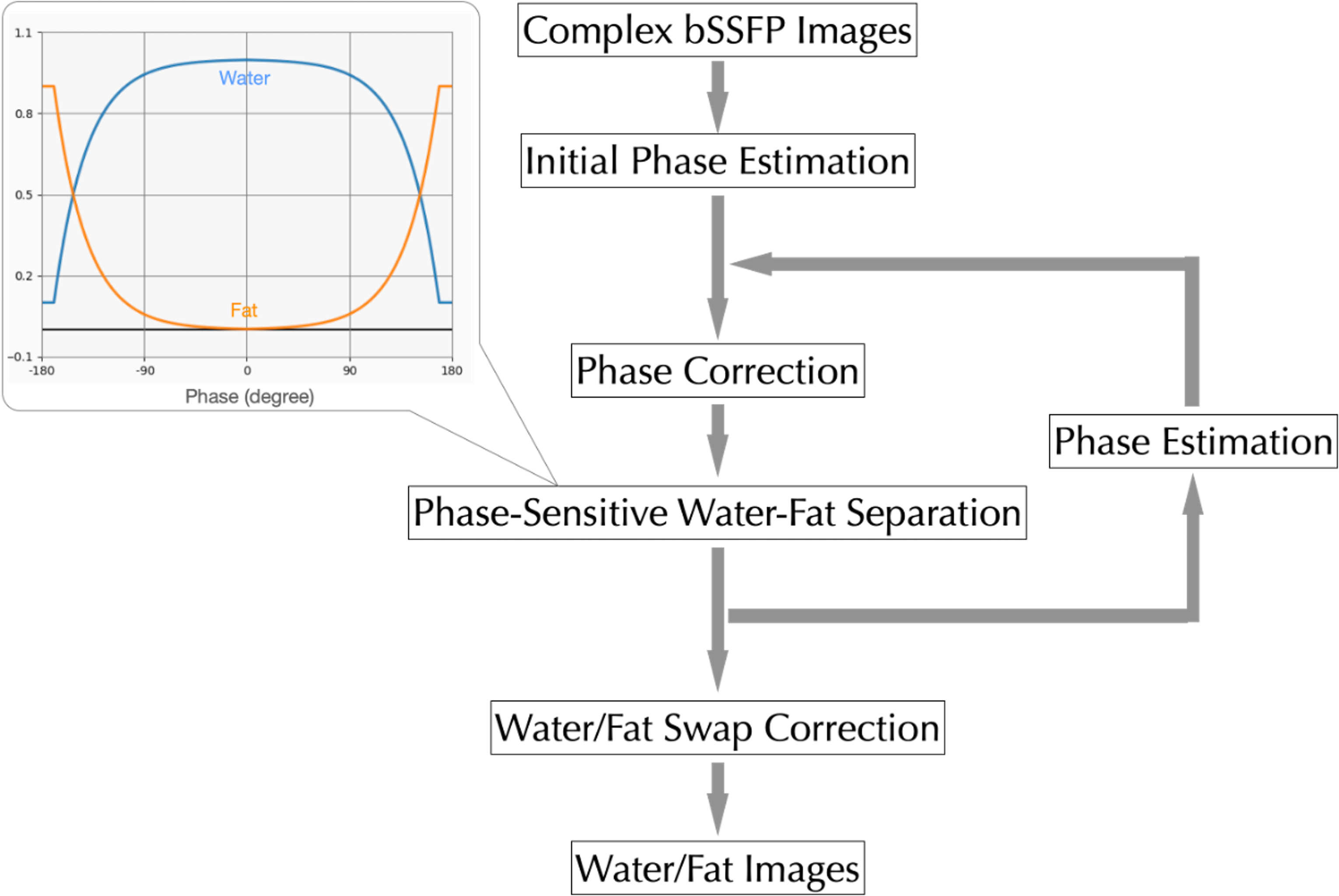
Figure 1 Flow chart of phase-sensitive water-fat separation for bSSFP images.
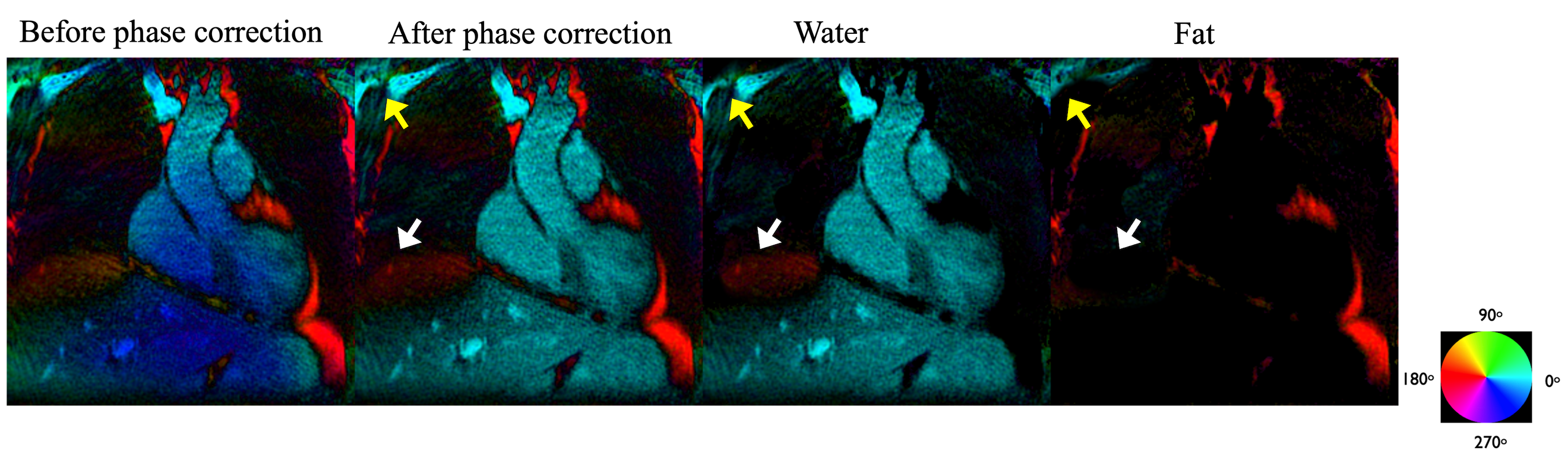
Figure 2 Demonstration of water-fat separation using phase information with whole data set of one volunteer. The phase of the coil combined image was estimated and corrected. After the phase correction, the phase of water and fat is close to 0o and 180o respectively, where the off-resonance is within [-110, 110] Hz. Outside this range, phase of water and fat can swap (pointed by the arrows). Off-resonance field map may be used to mitigate the swap of water and fat (the write arrows).
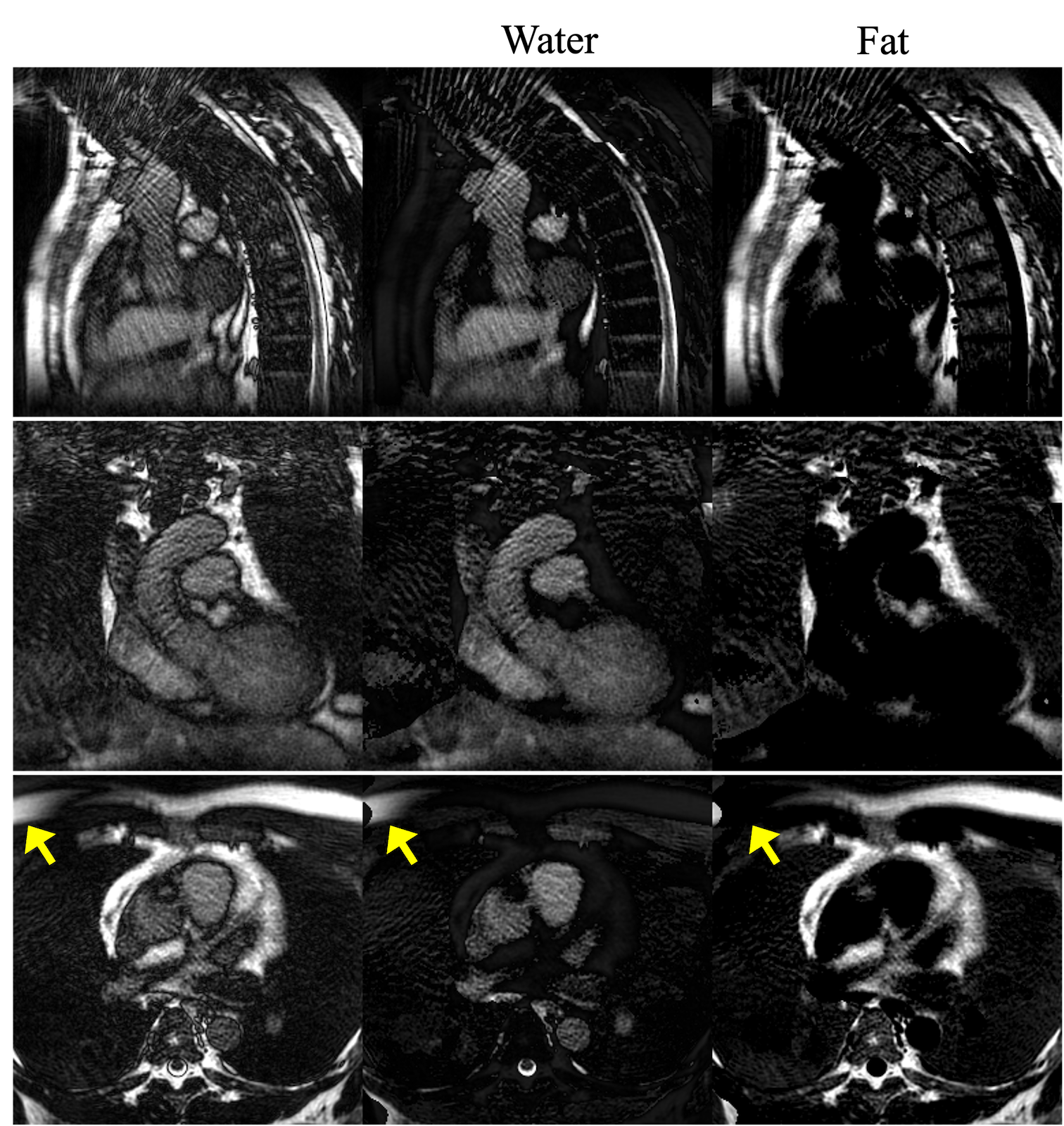
Figure 3 Water and Fat separation results using the whole data from another volunteer. Note the water/fat swap pointed by the arrows close to the banding artifacts.
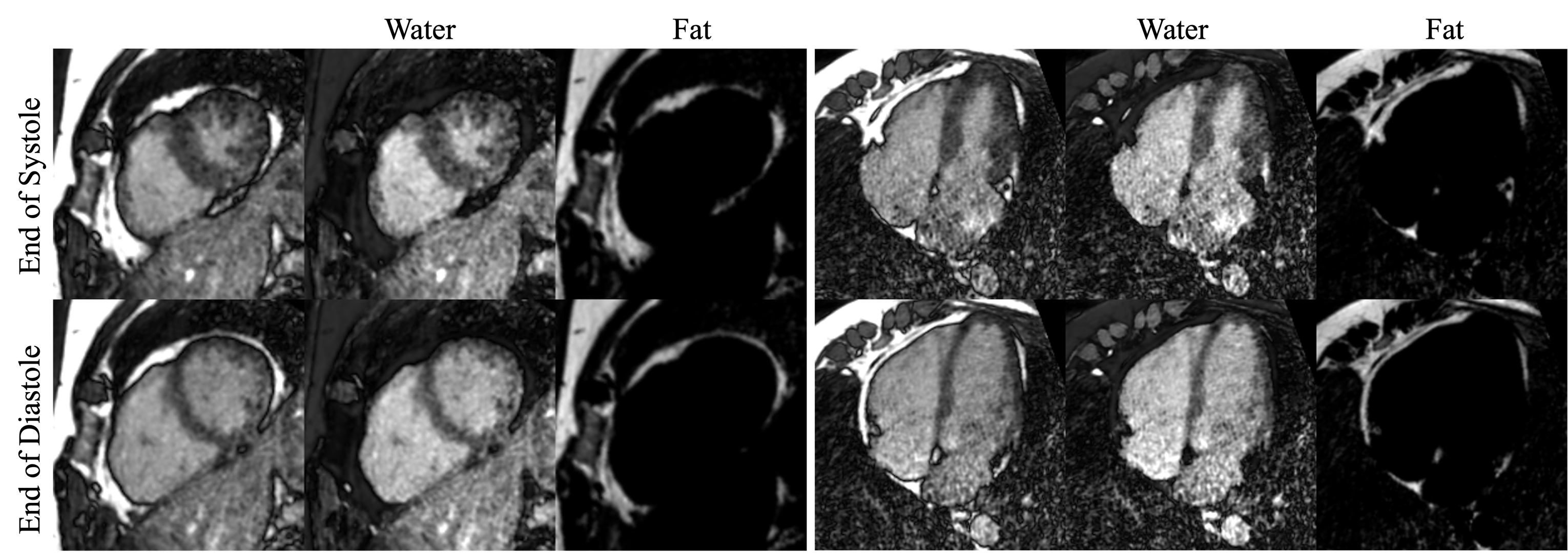
Figure 4 Images of short axis (left) and four chamber (right) at the end of expiration shown for two cardiac phases.
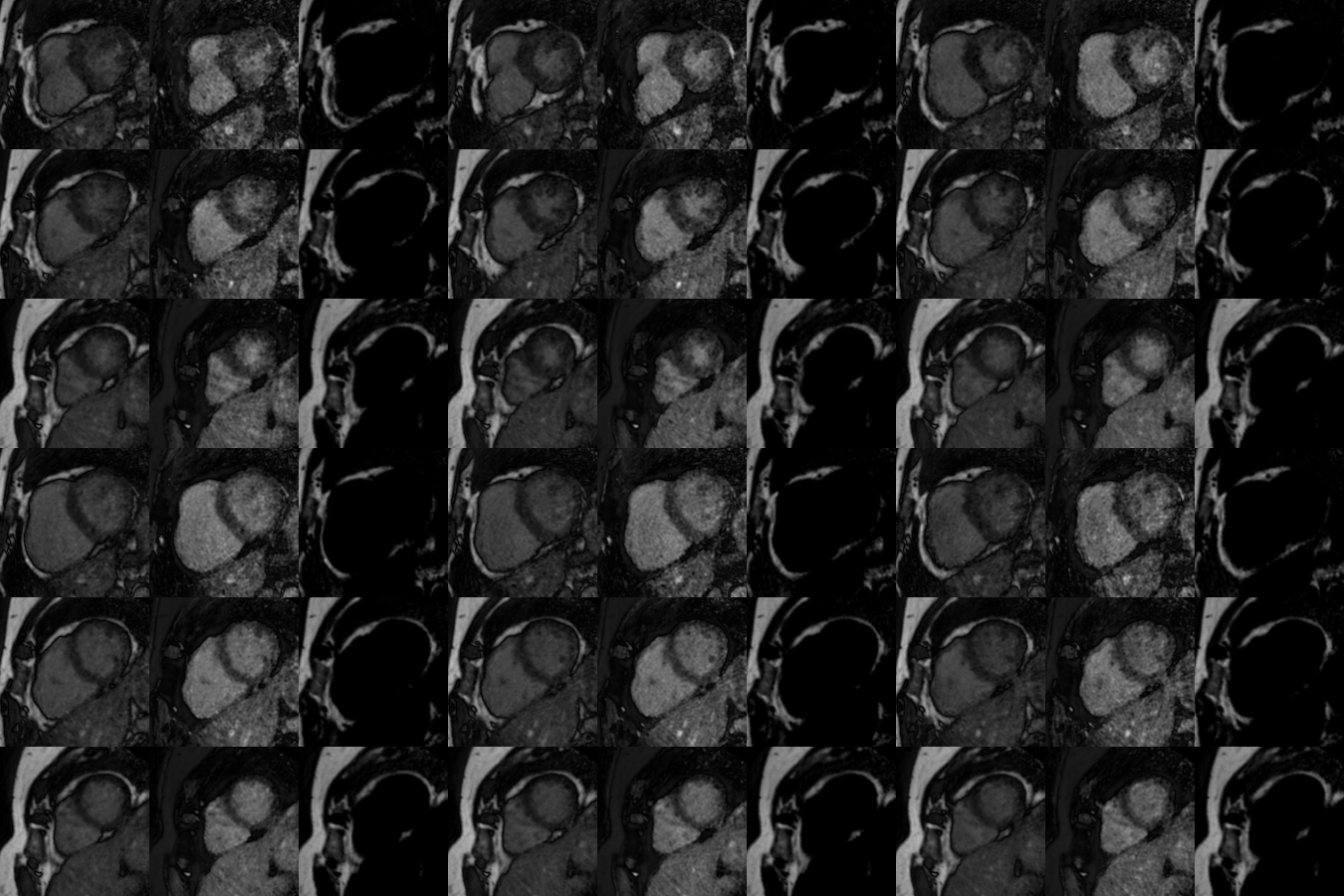
Figure 5 Short axis time series for different slices.