0608
3D Image Enhancement for High-Resolution ky-kz Accelerated 3D LGE CMR1Department of Medicine (Cardiovascular Division), Beth Israel Deaconess Medical Center and Harvard Medical School, Boston, MA, United States, 2Radiology, Beth Israel Deaconess Medical Center and Harvard Medical School, Boston, MA, United States
Synopsis
Keywords: Myocardium, Cardiovascular
Motivation: Prolonged scan times in high-resolution 3D late gadolinium enhancement (LGE) cardiovascular magnetic resonance (CMR) imaging hinders the full potential in clinical applications.
Goal(s): The primary goal of this study was to develop and evaluate a 3D enhanced-resolution for 2D (ky-kz) accelerated 3D LGE imaging.
Approach: A 3D generative adversarial network was implemented to enhance the spatial resolution of 2D-accelerated 3D LGE images.
Results: The proposed method at 6-fold acceleration (3-fold in ky and 2-fold in kz) maintained intricate scar details and improved image quality.
Impact: The improvement in acquisition speed by 2D acceleration may benefit patients presenting with heavy respiratory motion and may be less sensitive to contrast washout.
INTRODUCTION
Late gadolinium enhancement (LGE) cardiovascular magnetic resonance (CMR) is widely used for identifying and quantifying areas of myocardial fibrosis and scar. Although 2D LGE CMR is commonly used in clinical practice, 3D imaging improves SNR, spatial resolution, and volumetric coverage1. However, extended scan time required for high-resolution 3D LGE poses a challenge, especially in the presence of respiratory motion and its sensitivity to contrast washout and necessitates image acceleration techniques. Highly accelerated compressed-sensing techniques have been previously used to reduce scan time, albeit with prolonged reconstruction time2. Meanwhile, current deep learning (DL) models fail to leverage the full potential of 3D structures as they tend to simplify the problem by converting it into 2D slices3 and overlooks the intrinsic nature of 2D image acceleration in a 3D image acquisition. In this study, we sought to develop a 3D generative adversarial network for image enhancement to improve the spatial resolution of highly accelerated 3D LGE images acquired with low spatial resolution along phase and slice-encoding direction.METHODS
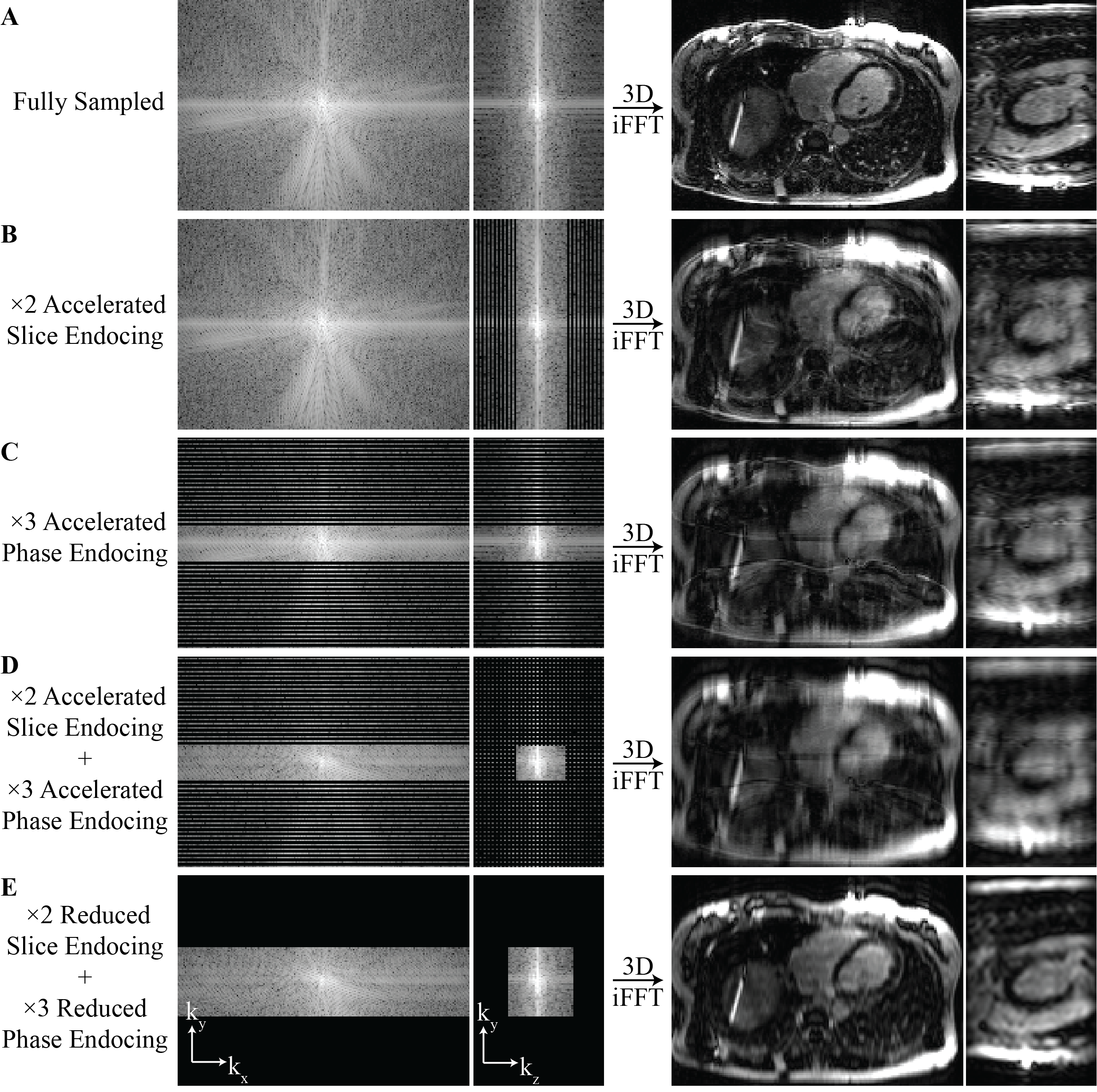
3D Resolution Enhancement: To accelerate a 3D Cartesian acquisition, in-plane, through-slice, or superimposition of two accelerations is generally used, leading to various aliasing patterns and challenges. The most common ways of accelerating a 3D Cartesian acquisition are depicted in Fig. 1B-D. Although individual in-plane (Fig. 1C) or through-slice (Fig. 1B) accelerations are easier to resolve, simultaneous aliasing in ky and kz dimensions when both accelerations are superimposed poses a greater challenge (Fig. 1D). Alternatively, the same amount of acceleration can be achieved by reducing the k-space dimension along ky and kz (Fig. 1E). In this work, 3D resolution enhancement aims to reconstruct 3D images from a single low-resolution 3D acquisition. Given their high-resolution counterparts as ground truth, a 3D generator is trained along with a 3D discriminator to minimize the following loss function:$$ L = \lambda_{pixel}L_{pixel} + \lambda_{VGG}L_{VGG} + \lambda_{FFT}L_{FFT} + \lambda_{GAN}L_{GAN},$$
where $$$\lambda$$$denotes the tradeoff parameters of different losses. Pixel loss is derived from Euclidean distance, visual geometry group (VGG) loss is derived from perceptual domain of VGG-19 network applied on axial views of the 3D volume, and fast Fourier transform (FFT) loss is derived from $$$\ell_1$$$ FFT to map images into a spatial frequency domain. Lastly, generative adversarial network (GAN) loss is derived from the enhanced super-resolution generative adversarial network (ESRGAN)4.
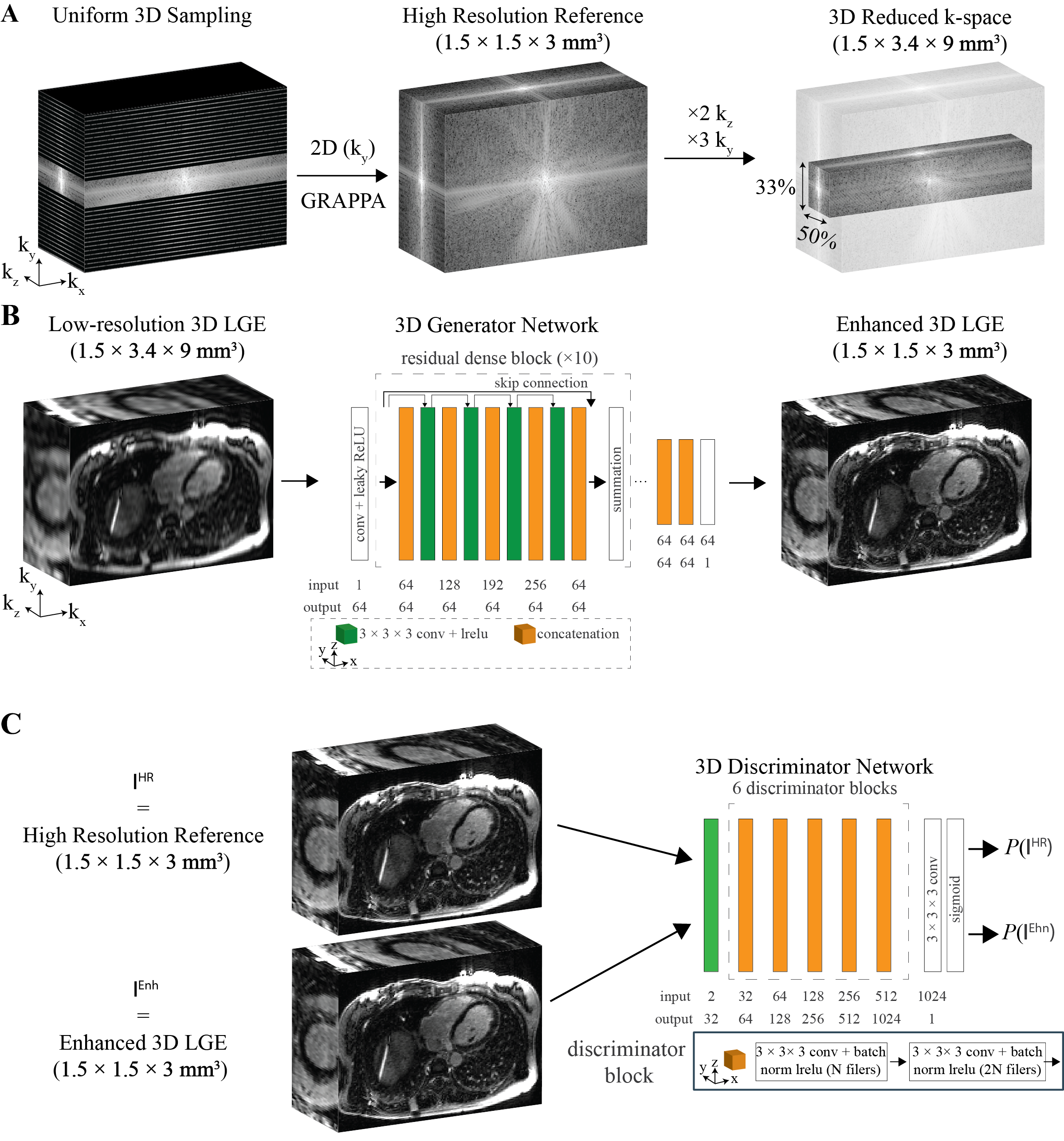
A 3D version of the recently proposed Resolution Enhancement Generative Adversarial Inline Neural Network (REGAIN)5 was used with the following hyperparameters: Adam optimizer with a LR=0.0001, $$$\beta_{1,2}$$$ = 0.9,0.999, $$$\lambda_{pixel,VGG,FFT,GAN}$$$=0.01,1,0.01,0.005. 3D REGAIN was trained using 64×64×32 randomly cropped image patches with batch size of 4 over 50 epochs. The 3D generator and discriminator networks are depicted in Fig. 2B-C.
The raw k-space data from 3D LGE images were extracted from 1348 patients (56 ± 16 years, 769 males) undergoing clinical CMR at 3T (MAGNETOM Vida Siemens Healthineers, Erlangen, Germany). A free-breathing ECG-triggered navigator-gated inversion-recovery sequence with GRE readout was used with the following imaging parameters: spatial resolution = 1-1.5 × 1-1.5 × 2-3 mm3, FOV = 300-420 × 380-420 × 100–120 mm3, flip angle = 15-40°, TR/TE = 2.7/1-1.24 msec, GRAPPA rate 1.8, and scan time of ~3min. Various 3D low-spatial resolution images were synthesized by discarding 44%-66% of outer phase-encoding (ky) lines along with 33%-44% of outer slice-encoding (kz) lines while maintaining the data in readout direction. A schematic of the training pipeline is depicted in Fig. 2A.
Testing was performed on 208 (57 ± 15 years, 126 males) patients that were unseen by the network. 6-fold 2D acceleration was performed by keeping 44% of the ky lines and 33% of the kz lines. Note that, compared to a three-minute conventional acquisition at 1.5 × 1.5 × 3 mm3, a combined 44% ky and 33% kz reduced k-space corresponds to a one-minute acquisition with 1.5 × 3.4 × 9 mm3 resolution. During testing, 3D patch averaging was performed using 80 × 80 × (#Slices) with 20% overlapping, which amounts to 8-12 patches per subject due to GPU memory limitations.
RESULTS
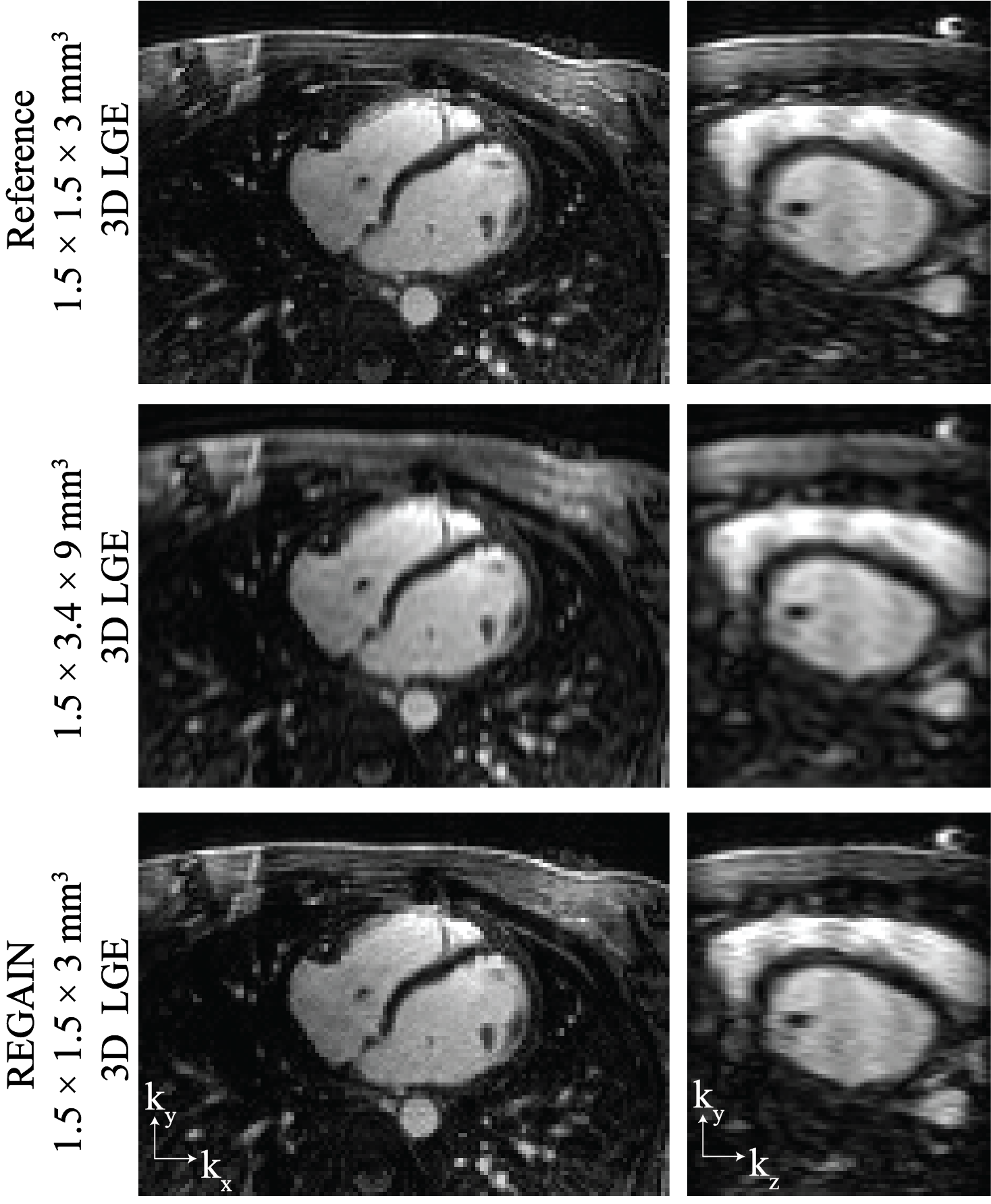
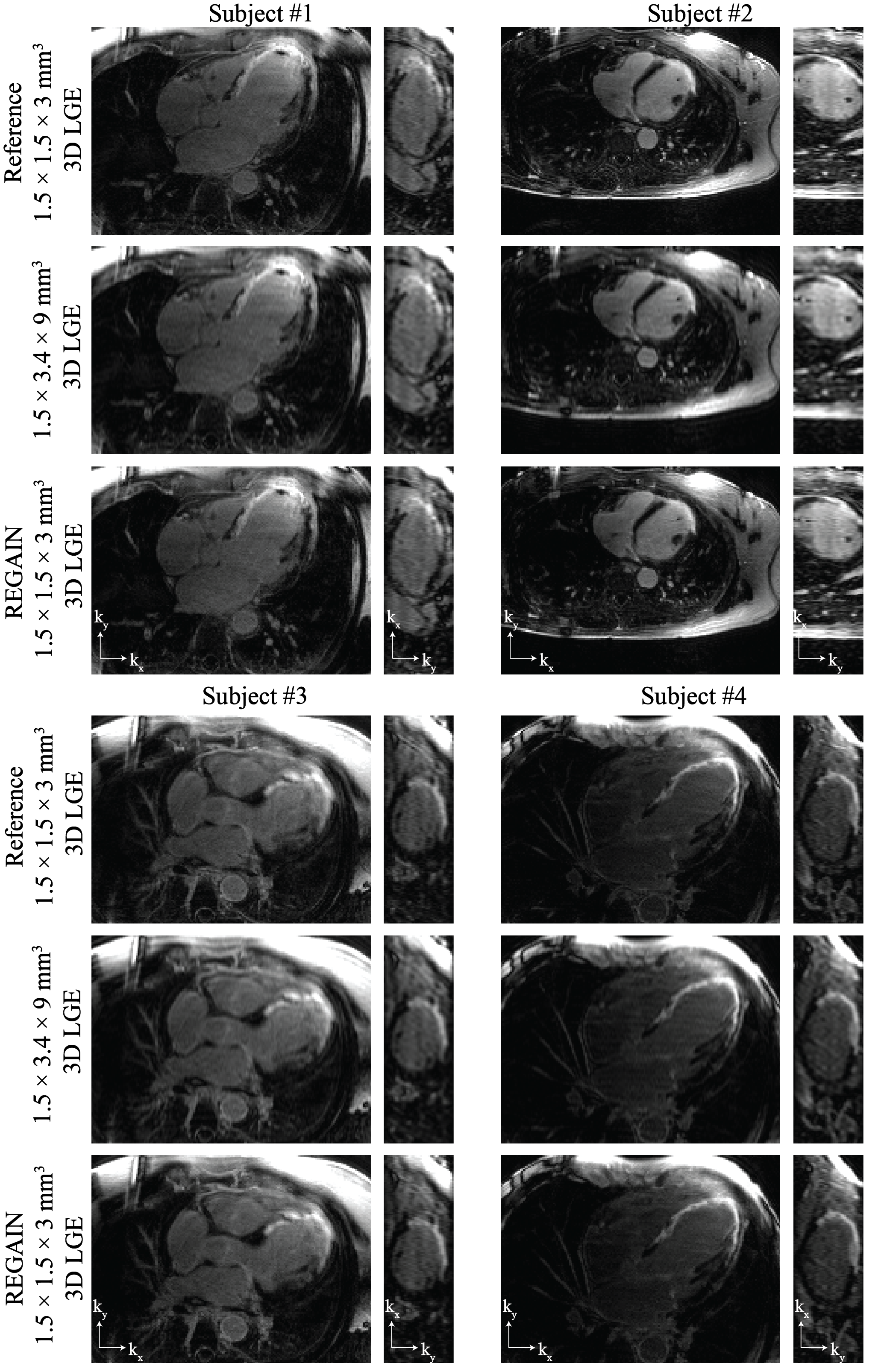
3D enhanced-resolution for 2D-accelerated 3D LGE visually improves image quality compared to low-resolution images while demonstrating a more accurate resemblance to high-resolution references especially in the ky-kz dimension, where the impact of substantial acceleration is most pronounced (Fig. 3). Example images in patients with myocardial scar are depicted in Fig. 4 where clean delineation of the scar is recovered with the 3D enhanced-resolution.CONCLUSION
We demonstrate the potential of 3D enhancement to improve image quality without generating additional artifacts for highly accelerated 3D LGE at 1.5 × 1.5 × 3 mm3 resolution.Acknowledgements
Funding: This study is supported by the National Institutes of Health.References
1) Toupin S, Pezel T, Bustin A, Cochet H. Whole-Heart High-Resolution Late Gadolinium Enhancement: Techniques and Clinical Applications. Journal of Magnetic Resonance Imaging 2022;55(4):967-87.
2) Basha TA, Akçakaya M, Liew C, Tsao CW, Delling FN, Addae G, et al. Clinical Performance of High-Resolution Late Gadolinium Enhancement Imaging With Compressed Sensing. Journal of Magnetic Resonance Imaging 2017;46(6):1829-38.
3) El-Rewaidy H, Neisius U, Mancio J, Kucukseymen S, Rodriguez J, Paskavitz A, et al. Deep complex convolutional network for fast reconstruction of 3D late gadolinium enhancement cardiac MRI. Nmr in Biomedicine 2020;33(7).
4) Wang XT, Yu K, Wu SX, Gu JJ, Liu YH, Dong C, et al. ESRGAN: Enhanced Super-Resolution Generative Adversarial Networks. 15th European Conference on Computer Vision (ECCV). 11133. Munich, GERMANY; 2018:63-79.
5) Yoon S, Nakamori S, Amyar A, Assana S, Cirillo J, Morales MA, et al. Accelerated Cardiac MRI Cine with Use of Resolution Enhancement Generative Adversarial Inline Neural Network. Radiology 2023;307(5).
Figures