0603
Neurite Beading Model of Acute Stroke from Tensor-Valued Diffusion Encoding Predicts Diffusion Time Effects with Oscillating Gradients1Biomedical Engineering, University of Alberta, Edmonton, AB, Canada, 2Radiology and Diagnostic Imaging, University of Alberta, Edmonton, AB, Canada, 3Neurosurgery, Medical College of Wisconsin, Milwaukee, WI, United States
Synopsis
Keywords: Simulation/Validation, Simulations
Motivation: Can two different diffusion experiments (tensor-valued encoding and oscillating-gradient-spin-echo) support the role of axon beading for diffusion restriction in acute ischemic stroke?
Goal(s): To evaluate whether tensor-valued diffusion encoding yields an axon beading model that predicts experimental ischemic changes of diffusivity measured with OGSE.
Approach: Tensor-valued and OGSE/PGSE diffusion MRI were measured in the same acute stroke patients. Monte Carlo simulations were used to assess the links between these two independent measurements.
Results: The tensor-valued derived beading model predictions were in general agreement with independent experiments of less diffusivity reduction with OGSE than PGSE within stroke lesions.
Impact: Novel diffusion MRI sequences such as tensor-valued encoding and oscillating-gradient-spin-echo are complementary methods that point to the same microstructural basis (i.e. beading and elevated intra-cellular volume fraction) for the clinically useful diffusion reduction in acute ischemic stroke.
Introduction
Neurite beading has been proposed as the underlying mechanism of lower mean diffusivity (MD) in acute ischemic stroke1. Diffusion time effects of tensor parameters like axial (AD) and radial diffusivity (RD) in human acute stroke have been shown by comparing oscillating-gradient- (OGSE) and pulsed-gradient-spin-echo (PGSE) diffusion MRI with changes that were consistent with Monte Carlo diffusion simulation of neurite beading2. Tensor-valued diffusion encoding using diffusion time ‘matched’ spherical (STE) and linear (LTE) tensor encoding gradient waveforms to disentangle orientation dispersion have shown reduced anisotropic kurtosis (MKA) and elevated isotropic kurtosis (MKI) in acute stroke3, also consistent with elevated intra-cellular volume fraction and beading4. The purpose here is to fit experimental measures of MKA, MKI and MD to tensor-valued Monte Carlo simulation of neurite beading, and subsequently test whether OGSE/PGSE diffusion simulation of the fitted model yields MD/AD/RD results consistent with experimental measures in the same acute stroke patients that have undergone both tensor-valued and OGSE/PGSE scans.Methods
Acute ischemic stroke patients (n=8) were 63±8 (51-74) years old, 7 males, NIH stroke scale score 8±5 (1-18), scanned 41±21 (6-71.5) hours after stroke onset, and with lesion volumes of 5±7 (0.02-18.1) cm3. Both tensor-valued diffusion encoding and OGSE(40 Hz)/PGSE were acquired with rapid single-shot spin-echo EPI protocols (3.5 min and 5 min, respectively) on a Siemens Prisma 3T (Table 1). Tensor-valued diffusion encoding yielded maps of MD, MKA and MKI5, while OGSE/PGSE yielded maps of MD, AD, and RD. Experimental values were extracted from regions-of-interest manually placed in the white matter of acute lesions and the contra-lateral hemisphere.Monte Carlo diffusion simulations6 of a beaded (ordered cylinders) impermeable surface axon model7 were performed independently for both the tensor-valued and OGSE/PGSE exact diffusion waveforms leading to predicted signal decay curves and values of MD, MKA and MKI (tensor-valued) and MD, AD, and RD (OGSE/PGSE). Geometries were generated for a range of radii (0.3-5 µm), beading amplitudes (0-0.8), and volume fractions (0.5-0.93) with the same intra- and extra-axonal intrinsic (i.e., free) diffusivities ranging from 1.7-2.2×10⁻³ mm2/s. Tensor-valued regression of experimental MD, MKA and MKI (contra and lesion) with simulation yielded best fit values of beading amplitude, volume fraction, axon radius and free diffusivity (contra and lesion). Note that the values of axon radius and free diffusivity were tied together between contra-lateral and lesion tissue. OGSE/PGSE values of MD, AD, and RD were then obtained from the best-fit beaded axon model for each patient, and these values compared with experimental OGSE/PGSE measures in the same patient using paired t-test. A complete flow chart is given in Figure 1.
Results
The tensor-valued measures of MD=(0.92±0.08×10⁻³ mm2/s contra, 0.55±0.08×10⁻³ mm2/s lesion), MKA=(0.84±0.27, 0.41±0.28), and MKI=(0.41±0.13, 0.85±0.38) in Figure 2A yielded the best-fit beaded axon models with beading amplitude=(0.15±0.08 contra, 0.46±0.10 lesion), intracellular volume fraction=(0.66±0.07, 0.82±0.06), radius=(3.7±0.4 µm), and free diffusivity=(1.95±0.10×10⁻³ mm2/s) in Figure 2B. Both in vivo experiment and OGSE/PGSE simulation of the best-fit tensor-valued beaded axon models show less OGSE MD reduction (-22% in vivo, -10% simulation) than PGSE MD reduction (-43%, -34%) in the lesion relative to contra-lateral tissue (Figure 3A). Similarly, OGSE lesion reductions in AD (-35% in vivo, -15% simulation) and RD (-14%, -2%) are less than PGSE reductions in AD (-48%, -31%) and RD (-38%, -44%), with both in vivo and simulation reporting greater OGSE/PGSE differences for RD (Figure 3B&C). Beaded axon model simulation and in vivo experiment also both yield MD, AD and RD increase from PGSE to OGSE which is greater in the stroke lesions than the contra-lateral white matter.Discussion
The tensor-valued derived beading models with greater intracellular volume fraction were in general agreement with independent experimental measurements of less diffusivity reduction with OGSE than PGSE in the ischemic lesions from the same acute stroke patients. Simulations were performed using coherent geometries (Figure 1C), but this will not generally be the case in vivo, and although tensor-valued diffusion MRI inherently accounts for orientation dispersion, OGSE/PGSE does not. Thus, OGSE/PGSE absolute differences between simulation and in vivo experiment are likely the result of orientation dispersion. However, simulated OGSE/PGSE parameters were very similar to experiment in a lesion within the ordered posterior limb of internal capsule (Figure 4). Using two different diffusion MRI experiments, this work supports the role of axon beading for diffusion restriction in acute ischemic stroke.Acknowledgements
Supported by the Heart and Stroke Foundation of Canada and China Scholarship Council.References
1. Budde MD, Frank JA. Neurite beading is sufficient to decrease the apparent diffusion coefficient after ischemic stroke. Proceedings of the National Academy of Sciences. 2010;107(32):14472-14477.
2. Baron CA, Kate M, Gioia L, et al. Reduction of diffusion-weighted imaging contrast of acute ischemic stroke at short diffusion times. Stroke. 2015;46(8):2136-2141.
3. Zhou M, Stobbe R, Szczepankiewicz F, Buck B, Beaulieu C. Exploring the Impact of Diffusion Time Difference on Tensor Valued Diffusion Encoding in Human Acute Ischemic Stroke. In Proceedings of ISMRM Diffusion Workshop 2022; Amsterdam, Netherlands.
4. Stobbe R, Budde M, Zhou M, Szczepankiewicz F, Beaulieu C. The Combination of B-Tensor Encoding & Diffusion Time Variation Enables Axon Volume Fraction & Beading Amplitude Mapping in Acute Stroke. In Proceedings of ISMRM Diffusion Workshop 2022; Amsterdam, Netherlands.
5. Szczepankiewicz F, van Westen D, Englund E, et al. The link between diffusion MRI and tumor heterogeneity: Mapping cell eccentricity and density by diffusional variance decomposition (DIVIDE). Neuroimage. 2016;142:522-532.
6. Cook PA, Bai Y, Hall MG, Nedjati-Gilani S, Seunarine KK, Alexander DC. Camino: Diffusion MRI reconstruction and processing. 2005.
7. Skinner NP, Kurpad SN, Schmit BD, Budde MD. Detection of acute nervous system injury with advanced diffusion‐weighted MRI: a simulation and sensitivity analysis. NMR in Biomedicine. 2015;28(11):1489-1506.
Figures
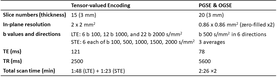
Table 1: Protocol parameters for tensor-valued encoding and PGSE/OGSE.
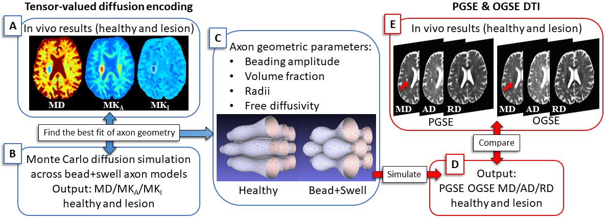
Figure 1. Experimentally measured tensor-valued MD, MKA, and MKI in contra-lateral and ischemic lesion (A) were compared to Monte Carlo simulations (B) leading to the best fit of axon geometric and free diffusivity parameters (C). PGSE- and OGSE-derived simulated signal and derived diffusion parameters were then calculated from the ‘best’ tensor-valued models (D) and compared to the in vivo PGSE/OGSE MD, AD, and RD parameters (E).
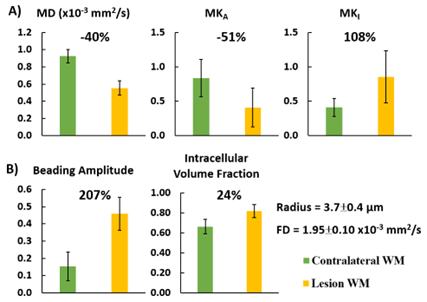
Figure 2. The alterations of the tensor-valued MD, MKA, and MKI in contralateral and lesion WM per patient were compared to Monte Carlo simulations with the same gradient waveforms to yield the beading amplitude and intracellular volume fraction of the best-fit beaded axon models over 8 acute stroke patients (mean ± SD). This also yielded cylinder radius and free diffusivity (FD) which were kept the same for contralateral and lesion WM in the model.
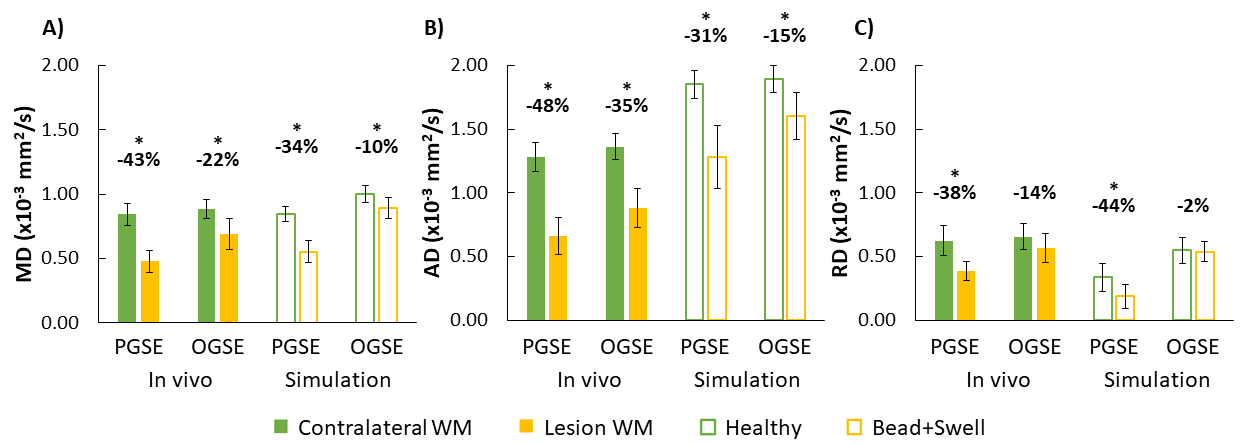
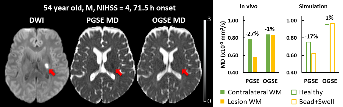
Figure 4. DWI, PGSE- and OGSE-derived MD maps show the low MD reversal of the lesion on OGSE (arrow). MD values in contralateral (filled-green) and lesion (filled-yellow) WM, as well as in simulated healthy (green border) and beading axons (yellow border) for PGSE and OGSE are plotted for the patient. The acute ischemic lesion located in the left posterior limb of internal capsule ‘disappears’ on the OGSE MD map, and notably this was also predicted by the best fit of the beading model from tensor-valued encoding (right plot).