0602
Coarse-graining with time-dependent diffusion reveals signatures of demyelination and axonal loss1Bernard and Irene Schwartz Center for Biomedical Imaging, Department of Radiology, New York University Grossman School of Medicine, New York, NY, United States, 2Center for Advanced Imaging Innovation and Research (CAI2R), Department of Radiology, New York University Grossman School of Medicine, New York, NY, United States, 3Athinoula A. Martinos Center for Biomedical Imaging, Department of Radiology, Massachusetts General Hospital, Boston, MA, United States, 4Harvard Medical School, Boston, MA, United States
Synopsis
Keywords: Simulation/Validation, Diffusion/other diffusion imaging techniques
Motivation: Time-dependent diffusion $$$D(t)$$$ is sensitive to brain microstructure. Using Monte Carlo (MC) simulations, $$$D(t)$$$ has been shown to provide information about structural changes caused by pathological conditions.
Goal(s): To establish the relation between the parameters of $$$D(t)$$$ and the extra-axonal space geometry.
Approach: We solve the Fick-Jacobs equation in the effective medium framework, connect $$$D(t)$$$ to correlations of density and local diffusivity, and validate with Monte-Carlo simulations.
Results: Time-dependence of $$$D(t)$$$ is quantitatively related to geometric characteristics of axonal packing, demyelination and axonal loss.
Impact: By coarse-graining the extra-axonal space, time-dependent diffusion explores the geometry relevant for demyelination and axonal loss, enabling quantifying axonal microstructure.
Introduction
The dependence on diffusion time $$$t$$$ of the diffusion coefficient $$$D(t)$$$ encodes information regarding brain microstructure1-9. By means of Monte Carlo (MC) simulations, $$$D(t)$$$ has been shown to identify structural changes caused by beading10,11, axon loss12-14, demyelination12-15, and inflammation16. For the extra-axonal space (EAS), $$$D(t)$$$ transverse to the axons has the form17:$$D(t)\simeq\;D_{\infty}+A\frac{\ln(t/t_c)}{t},\;t\,\gg\,t_c.\tag{1}$$
In this work, we relate $$$D(t)$$$ parameter $$$A$$$ to the extra-axonal space micro-geometry and its changes during axonal loss and demyelination.
Theory
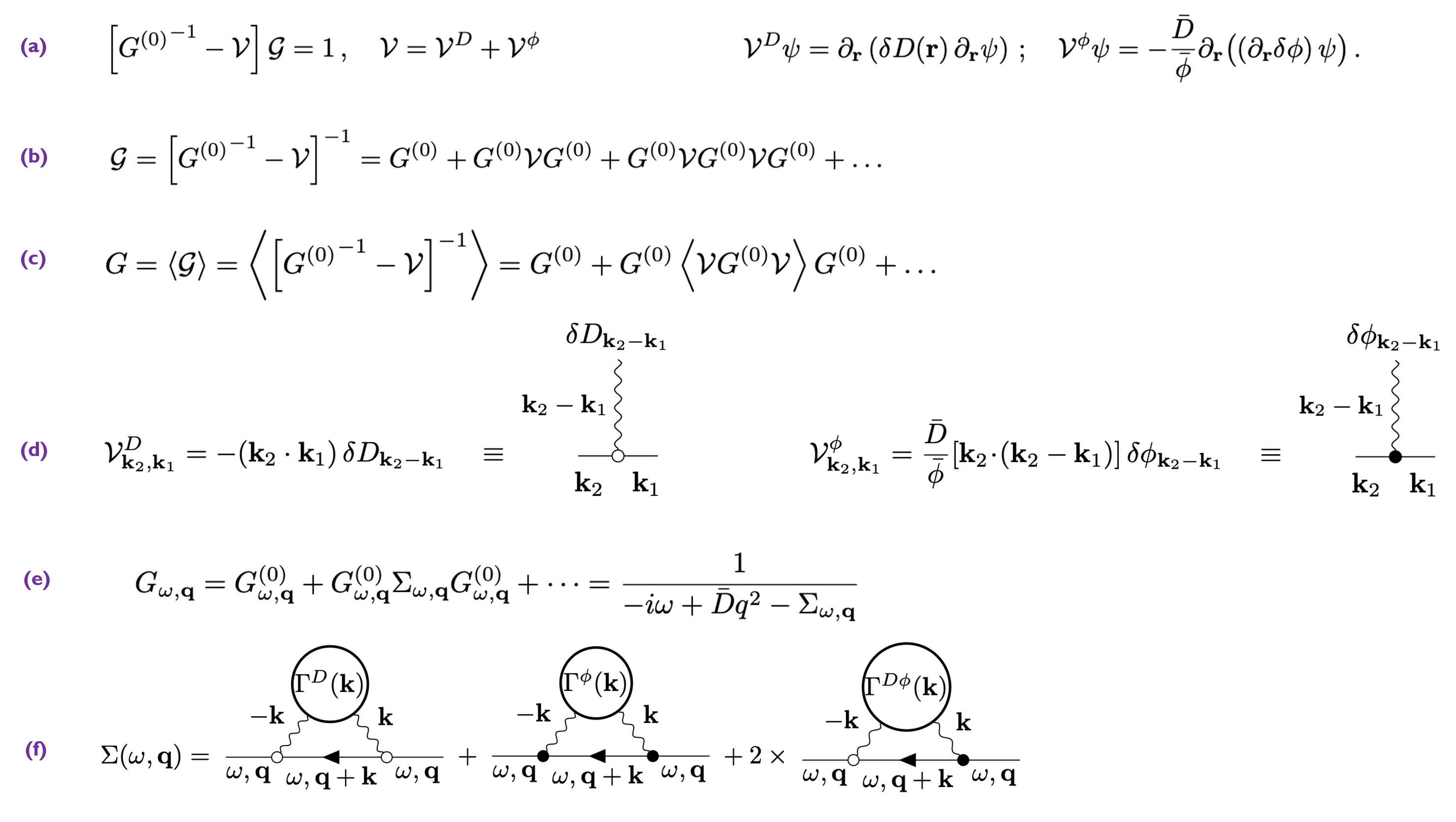
The ensemble averaging caused by the diffusion process irreversibly coarse-grains the heterogeneous structural properties of the medium8,9. However, it is possible to identify and extract from $$$D(t)$$$ structural information that survived this averaging. The $$$D(t)$$$ solution8,9,17 from the standard diffusion equation considers the locally varying diffusivity $$$D(\mathbf{r})$$$. However, it is important to also consider the locally varying EAS water fraction $$$\phi(\mathbf{r})$$$, since particle flux is governed by the gradient of concentration $$$\psi(\mathbf{r})/\phi(\mathbf{r})$$$ relative to the fraction of space the particles are free to move in, Fig.1a. This process is governed by the Fick-Jacobs (FJ) equation:$$\partial_t\psi(t,\mathbf{r})=\partial_\mathbf{r}\left[D(\mathbf{r})\phi(\mathbf{r})\partial_\mathbf{r}\left(\frac{\psi(t,\mathbf{r})}{\phi(\mathbf{r})}\right)\right].\tag{2}$$
Here, we treat stochastic quantities $$$D(\mathbf{r})$$$ and $$$\phi(\mathbf{r})$$$ as effective-medium parameters, i.e., already coarse-grained over a certain diffusion length $$$L(t)$$$. In particular, we calculate the local coarse-grained diffusivity $$$D(\mathbf{r})$$$ by emanating particles from each point $$$\mathbf{r}$$$ and computing local time-dependent cumulants, Fig.1b. Local coarse-grained EAS fraction $$$\phi(\mathbf{r})$$$ is obtained from the region with length $$$L(t)=\sqrt{4D(t)t}/2$$$ around $$$\mathbf{r}$$$, Fig.1b.
Generalizing9, at long enough $$$t$$$ the spatially varying components $$$\delta D(\mathbf{r})$$$ and $$$\delta \phi(\mathbf{r})$$$ become sufficiently small, such that Eq.(2) admits an asymptotically exact solution, which we find by developing the Feynman diagram technique for scattering off $$$\delta D(\mathbf{r})$$$ and $$$\delta \phi(\mathbf{r})$$$, Fig.2. Technically, scattering involves the three correlation functions:
$$\Gamma^{\phi}(\mathbf{r})=\langle\delta \phi(\mathbf{r_0}+\mathbf{r})\delta \phi(\mathbf{r_0})\rangle_{\mathbf{r_0}}\quad\Leftrightarrow\quad\Gamma^{\phi}(\mathbf{k})=\delta\phi(\mathbf{-k})\delta\phi(\mathbf{k})/V$$
$$\Gamma^{D}(\mathbf{r})=\langle\delta D(\mathbf{r_0}+\mathbf{r})\delta D(\mathbf{r_0})\rangle_{\mathbf{r_0}}\quad\Leftrightarrow\quad\Gamma^{D}(\mathbf{k})=\delta D(\mathbf{-k})\delta D(\mathbf{k})/V,\tag{3}$$
$$\Gamma^{D\phi}(\mathbf{r})=\langle\delta D(\mathbf{r_0}+\mathbf{r})\delta\phi(\mathbf{r_0})\rangle_{\mathbf{r_0}}\quad\Leftrightarrow\quad\Gamma^{D\phi}(\mathbf{k})=\delta D(\mathbf{-k})\delta \phi(\mathbf{k})/V.$$
While their large-k behavior gets suppressed with increasing diffusion time due to coarse-graining, their plateaus at $$$k\rightarrow0$$$ remain approximately intact as shown in Fig.1c. These plateaus in fact determine the asymptotically exact behavior of $$$D(t)$$$ in Eq.(1):
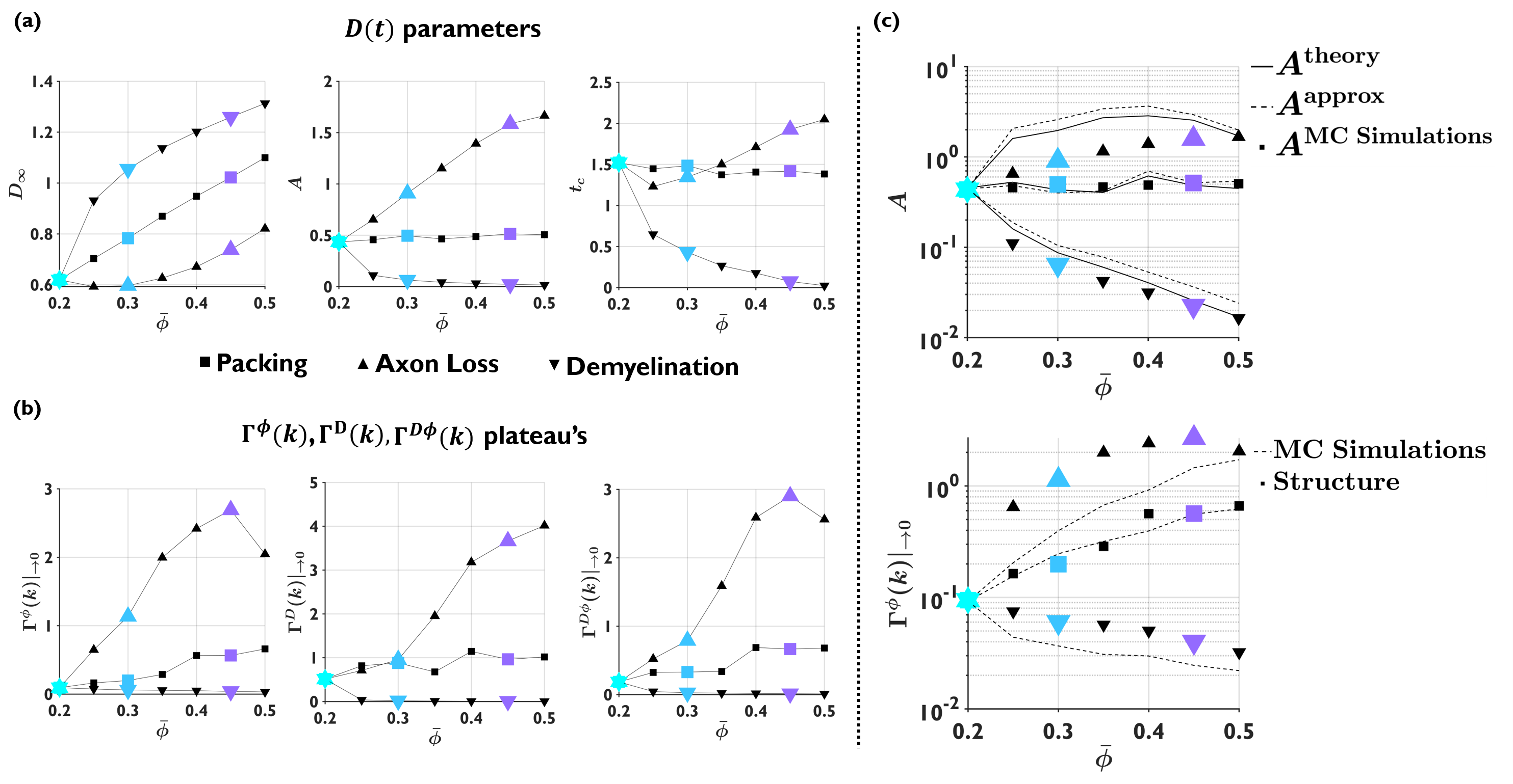
$$A^{\mathrm{theory}}=\frac{1}{8\pi}\left[\frac{\Gamma^{D}(k)|_{k\rightarrow0}}{D_\infty^2}+3\frac{\Gamma^{\phi}(k)|_{k\rightarrow0}}{\bar{\phi}^2}+2\frac{\Gamma^{D\phi}(k)|_{k\rightarrow0}}{D_\infty\bar{\phi}}\right],\tag{4}$$
where the first term has been previously found in17, while the other two terms describe the scattering off density fluctuations and the diffusivity-density fluctuations interference. From9, the approximation $$$\delta\;D(r)\approx\;C\delta\phi(r)$$$, with $$$C=\frac{\partial\;D_\infty}{\partial\bar{\phi}}$$$, can be used, leading to the approximation involving only EAS volume-fraction fluctuations
$$A^{\mathrm{approx}}=\frac{\Gamma^{\phi}(k)|_{\rightarrow0}}{8\pi D_\infty^2}\left[C^2+3\left(\frac{D_\infty}{\bar{\phi}}\right)^2+2C\left(\frac{D_\infty}{\bar{\phi}}\right)\right].\tag{5}$$
Methods
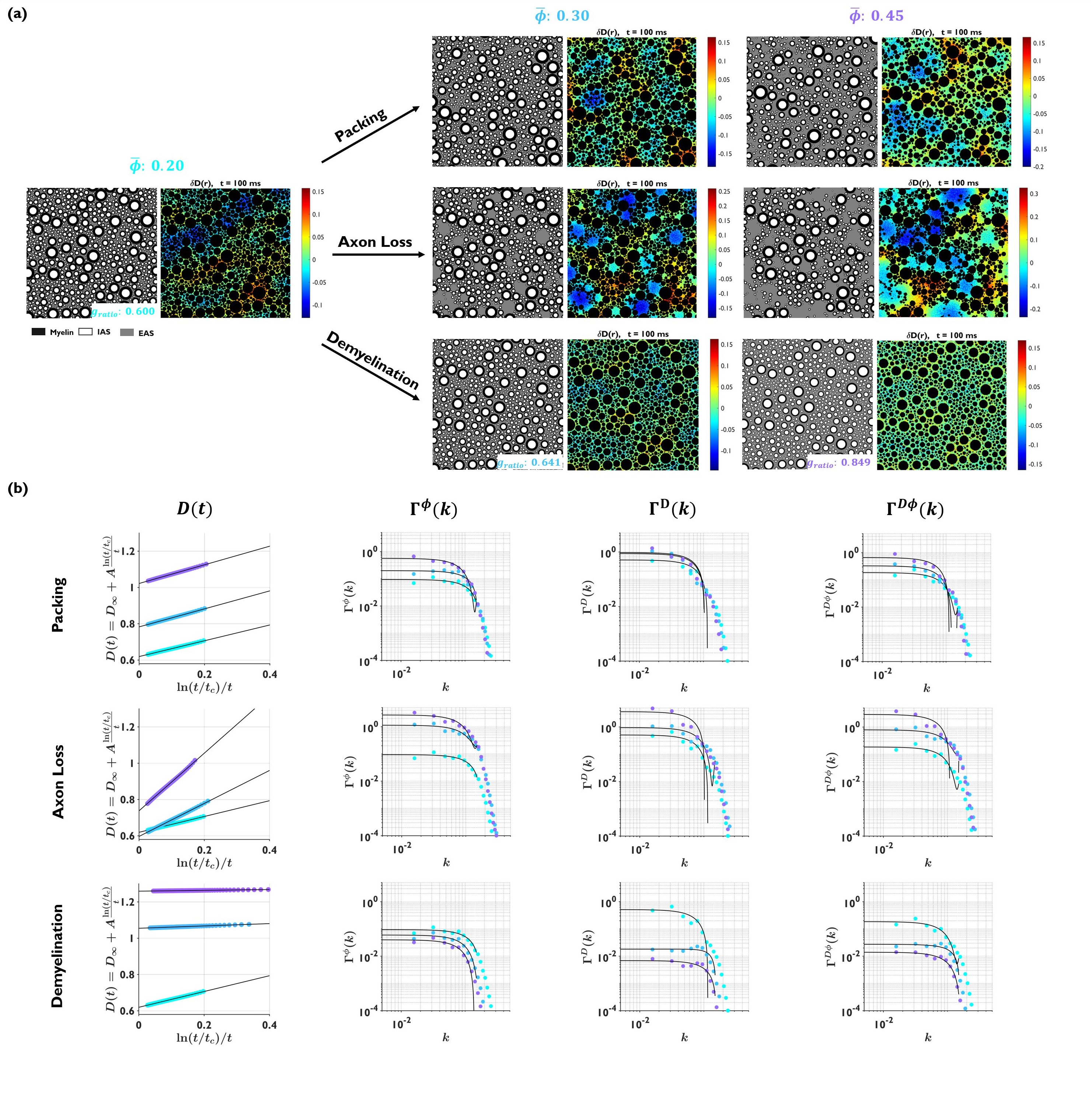
Substrate generation: Geometries of randomly packed disks (parallel cylinders in cross-section) with fractions from 0.5 to 0.8 (EAS fractions from 0.5 to 0.2) were generated18 starting from lowest disk density (packing in Fig. 3a). Disks diameters followed the distribution for axons in human corpus callosum19, and the number of disks varied from $$$3\times10^4$$$ for the higher density to $$$1.8\times10^4$$$ for the lower density, keeping the substrate size constant to $$$397\times397$$$ $$$\mu$$$m2.Models for axon loss and demyelination: from the generated disk packings at highest fraction 0.8, we simulate axon loss by randomly removing disks, yielding substrates with EAS from 0.2 to 0.5; and demyelination, by shrinking the outer disk diameter, yielding substrates with axon g-ratio from 0.60 to 0.75 (EAS from 0.2 to 0.5, assuming initial g-ratio20,21 of 0.60).
Monte Carlo simulations in EAS22: MC simulations on each substrates of disks were performed with $$$1\times10^3$$$ random walkers per EAS pixel in a 1444 lattice. Time step $$$4.15\times10^{-6}$$$ ms (step length 5.8 nm). $$$D_0=D(t)|_{t=0}=2$$$ ms/mm2 and total diffusion time of 175 ms.
Results
Locally varying $$$\delta D(\mathbf{r})$$$ and $$$\delta\phi(\mathbf{r})$$$ at increasing diffusion time, are shown in Fig.1b. Coarse-graining affects high-frequencies while low $$$k$$$ plateaus remain approximately intact (Fig.1c). Examples of the substrates generated for packing, axon loss and demyelination are shown in Fig.3a. Conditions with equal $$$\bar{\phi}$$$ show structural differences reflected in $$$\delta D(\mathbf{r})$$$ at $$$t=100$$$ ms. This differences are also captured by $$$D(t)$$$, $$$\Gamma^\phi(k)$$$, $$$\Gamma^D(k)$$$, $$$\Gamma^{D\phi}(k)$$$ as shown in Fig.3b. The dependence of the structural and time-dependent parameters of interest on EAS fraction $$$\bar{\phi}$$$ is shown in Fig.4a and Fig.4b, respectively. As previously shown10-12, these parameters can differentiate between pathological conditions. Fig.4c shows good agreement between $$$A^{\mathrm{theory}}$$$, Eq.(4), and $$$A$$$ estimated from MC simulations, $$$A^{\mathrm{approx}}$$$, Eq.(5), also shows good agreement. Using $$$A^{\mathrm{approx}}$$$ we can predict $$$\Gamma^\phi(k)$$$ only using $$$D(t)$$$ parameters obtained from MC simulations.Conclusions
In this work, we considered Fick-Jacobs equation for extra-axonal space and derived an asymptotically exact analytical expression for the time-dependent tail of $$$D(t)$$$. We observed good agreement between theory and MC simulations for disk (axon) packings in a wide range of densities, as well as for demyelination and axon loss.Acknowledgements
Research was supported by the National Institute of Neurological Disorders and Stroke of the NIH under awards R01 NS088040 and R21 NS081230, and by the Hirschl foundation, and was performed at the Center of Advanced Imaging Innovation and Research (CAI2R, www.cai2r.net), a Biomedical Technology Resource Center supported by NIBIB with the award P41 EB017183. H.H.L. was funded by the Office of the Director of the NIH under award DP5 OD031854.References
1. MA Horsfield, GJ Barker, WI McDonald. Self-diffusion in CNS tissue by volume-selective proton NMR. Magn Reson Med. 1994;31:637-644.
2. Els Fieremans, Lauren M Burcaw, Hong-Hsi Lee, Gregory Lemberskiy, Jelle Veraart, Dmitry S Novikov. In vivo observation and biophysical interpretation of time-dependent diffusion in human white matter. Neuroimage. 2016;129:414-427.
3. Hong-Hsi Lee, Antonios Papaioannou, Sung-Lyoung Kim, Dmitry S Novikov, Els Fieremans. In vivo observation and biophysical interpretation of time-dependent diffusion in human cortical gray matter. Neuroimage. 2020;222:117054
5. CA Baron, C Beaulieu. Oscillating gradient spin-echo (OGSE) diffusion tensor imaging of the human brain. Magn Reson Med. 2014;72:726-736.
6. Manisha Aggarwal, Matthew D Smith, Peter A Calabresi. Diffusion-time dependence of diffusional kurtosis in the mouse brain. Magn Reson Med. 2020;84(3):1564-1578.
7. Ileana O. Jelescu, Alexandre de Skowronski, Marco Palombo, Dmitry S. Novikov. Neurite Exchange Imaging (NEXI): A minimal model of diffusion in gray matter with inter-compartment water exchange. Neuroimage. 2022; 256:119277.
8. Dmitry S Novikov, Valerij G Kiselev. Effective medium theory of a diffusion-weighted signal. NMR Biomed. 2010;23(7):682-97.
9. Dmitry S Novikov, Jens H Jensen, Joseph A Helpern, Els Fieremans. Revealing mesoscopic structural universality with diffusion. Proc Natl Acad Sci USA. 2014;111(14):5088-93.3.
10. Hong-Hsi Lee, Antonios Papaioannou, Sung-Lyoung Kim, Dmitry S Novikov, Els Fieremans. A time-dependent diffusion MRI signature of axon caliber variations and beading. Commun Biol. 2020;3(1):354.
11. Ali Abdollahzadeh, Ricardo Coronado-Leija, Subah Mehrin, Hong-Hsi Lee, Alejandra Sierra, Els Fieremans, and Dmitry S. Novikov. Characterization of white matter myelinated and unmyelinated axons from diffusion MRI perspective. Proc. ISMRM 4322, 2023.
12. E. Fieremans, J. H. Jensen, J. Helpern, S. Kim, R. Grossman, M. Inglese, D. Novikov. Diffusion distinguishes between axonal loss and demyelination in brain white matter. Proc. Intl. Soc. Mag. Reson. Med. 2012.
13. D. S. Novikov, E. Fieremans. Relating extracellular diffusivity to cell size distribution and packing density as applied to white matter. Proc. Intl. Soc. Mag. Reson. Med. 2012
14. Ricardo Coronado-Leija, Hong-Hsi Lee, Els Fieremans, and Dmitry S. Novikov. Characterizing time-dependent diffusion in the extra-axonal space of white matter for axon loss and demyelination. Proc. ISMRM 3402, 2021.
15. Ginsburger Kévin, Poupon Fabrice, Beaujoin Justine, Estournet Delphine, Matuschke Felix, Mangin Jean-François, Axer Markus, Poupon Cyril. Improving the Realism of White Matter Numerical Phantoms: A Step toward a Better Understanding of the Influence of Structural Disorders in Diff!usion MRI. Frontiers in Physics. 2018;6.
16. André Döring, Maryam Afzali, Elena Kleban, Roland Kreis, and Derek K Jones. Realistic simulations of diffusion MR spectroscopy: The effect of glial cell swelling on non-Gaussian and anomalous diffusion. Proc. ISMRM 3401, 2021.
17. Lauren M Burcaw, Els Fieremans, Dmitry S Novikov. Mesoscopic structure of neuronal tracts from time-dependent diffusion. Neuroimage. 2015;114:18-37.
18. Aleksandar Donev, Salvatore Torquato, and Frank H. Stillinger. Neighbor list collision-driven molecular dynamics simulation for nonspherical hard particles. I. Algorithmic details. Journal of Computational Physics 2005;202(2).
19. F Aboitiz, A B Scheibel, R S Fisher, E Zaidel. Fiber composition of the human corpus callosum. Brain Res. 1992;598(1-2):143-53.
22. Hong-Hsi Lee, Katarina Yaros, Jelle Veraart, Jasmine L Pathan, Feng-Xia Liang, Sungheon G Kim, Dmitry S Novikov, Els Fieremans. Along-axon diameter variation and axonal orientation dispersion revealed with 3D electron microscopy: implications for quantifying brain white matter microstructure with histology and diffusion MRI. Brain Struct Funct. 2019;224(4):1469-1488.
21. Ali Abdollahzadeh, Ilya Belevich, Eija Jokitalo, Jussi Tohka, Alejandra Sierra. Automated 3D Axonal Morphometry of White Matter. Sci Rep. 2019 Apr 15;9(1):6084.
22. Hong-Hsi Lee, Els Fieremans, Dmitry S Novikov. Realistic Microstructure Simulator (RMS): Monte Carlo simulations of diffusion in three-dimensional cell segmentations of microscopy images. J Neurosci Methods. 2021;350:109018.
Figures
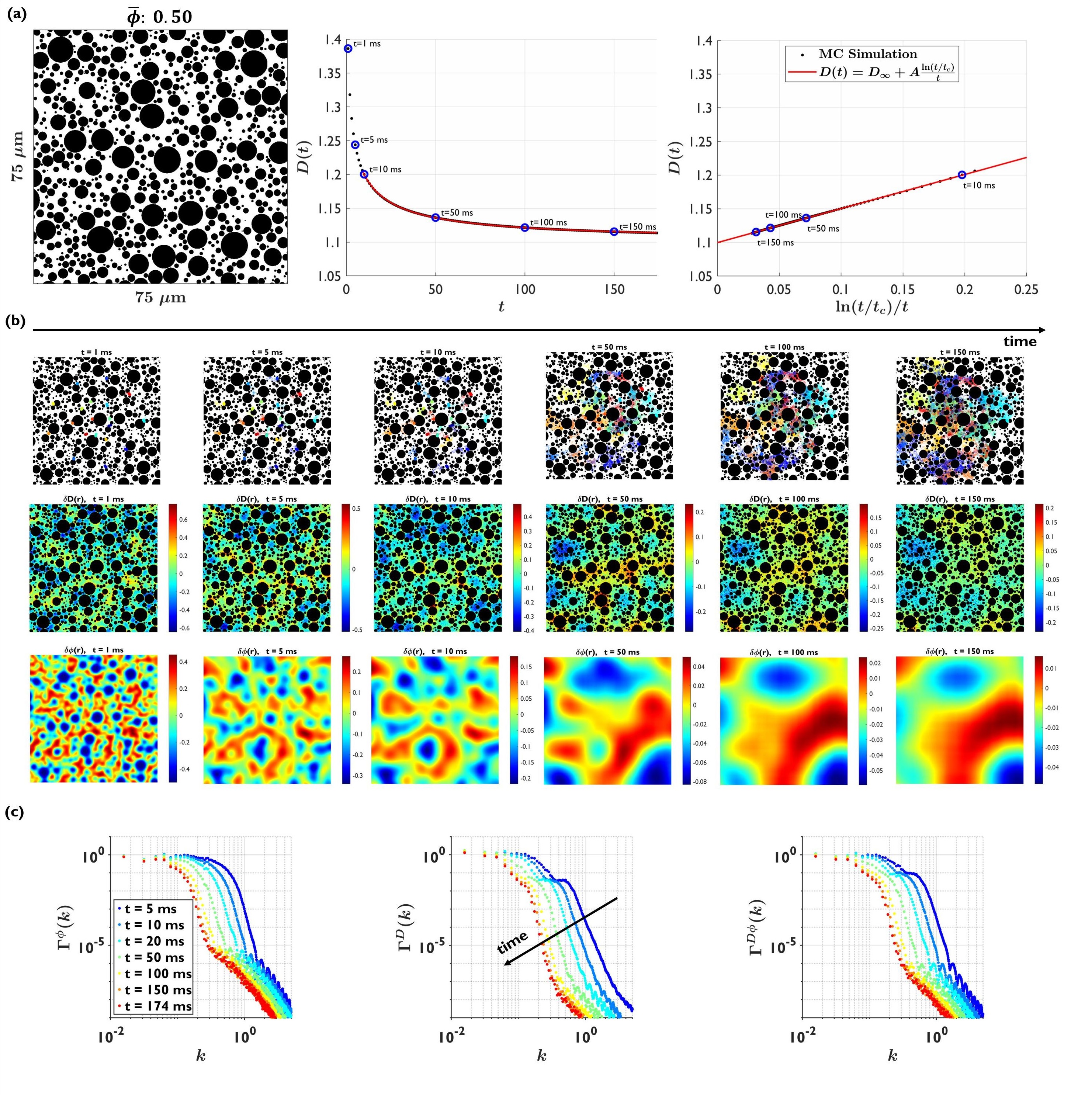
Fig.1. (a) $$$D(t)$$$ follows Eq.(1). (b) Top: diffusion process showing the local environment explored by 19 particles (different colors) at increased time. Middle: local diffusivity $$$\delta D(\mathbf{r})$$$ at increased time. Bottom: local density $$$\delta\phi(\mathbf{r})$$$ at increased time. The length scale of the fluctuations is similar than for $$$\delta D(\mathbf{r})$$$. (c) The coarse-graining caused by the increased diffusion time, only affects the high frequency part of the $$$\Gamma(k)$$$'s, while the low $$$k$$$ plateau remains approximately intact.