0601
Tuned Exchange Imaging (TEXI) – A modified Filter-Exchange Imaging pulse sequence for applications with thin slices and restricted diffusion1Department of Diagnostic Radiology, Lund University, Lund, Sweden, 2Danish Research Centre for Magnetic Resonance, Centre for Functional and Diagnostic Imaging and Research, Copenhagen University Hospital - Amager and Hvidovre, Copenhagen, Denmark, 3Department of Medical Radiation Physics, Lund University, Lund, Sweden, 4MR Section, DTU Health Tech, Technical University of Denmark, Lyngby, Denmark, 5Department of Radiology, Brigham and Women's Hospital, Harvard Medical School, Boston, MA, United States, 6Department of Clinical Sciences Lund, Radiology, Lund University, Lund, Sweden
Synopsis
Keywords: Diffusion Acquisition, Diffusion/other diffusion imaging techniques, exchange, restricted diffusion
Motivation: Thin slices in filter-exchange imaging lead to biased exchange rates, as thin slices require strong crushers. No current approach accounts for crushers in the presence of restricted diffusion.
Goal(s): We set to address the bias in FEXI due to the influence of strong crushers and restricted diffusion.
Approach: Tuned exchange imaging (TEXI) relies on gauging exchange and restriction weighting. We modify FEXI to ensure constant restriction weighting also with strong crushers. The accuracy of exchange mapping was evaluated using Monte Carlo simulations.
Results: TEXI yields consistent exchange rates independent of slice thickness and restriction size even if strong crushers are used.
Impact: TEXI could be useful to maximize exchange sensitivity and specificity with thin slices and in the presence of restricted diffusion.
INTRODUCTION
Filter exchange imaging (FEXI) is a double diffusion encoding (DDE) sequence sensitive to exchange between sites with different apparent diffusivities1-2 increasingly used in research3-11. In FEXI, the filter diffusion encoding block is followed by the detection block at varying mixing times to map the apparent exchange rate (AXR). Using long mixing times enhances the sensitivity to exchange4,12-14, but requires a stimulated echo sequence with crusher gradients. The amplitudes of crushers are inversely proportional to the slice thickness, which for thinner slices leads to significant diffusion weighting and biased exchange rate estimates13,10,11.For Gaussian diffusion, the crusher-induced bias can be corrected13,10,11, but this approach likely fails in the presence of time-dependent diffusion15-17, as the diffusion-time of the crusher-related diffusion encoding increases with the mixing time. The confounding effects of restricted diffusion in FEXI have been discussed18,6,19 but they have not yet been resolved. The effects of crushers in the presence of restricted-diffusion need to be considered particularly when long mixing times are used in DDE20-22.
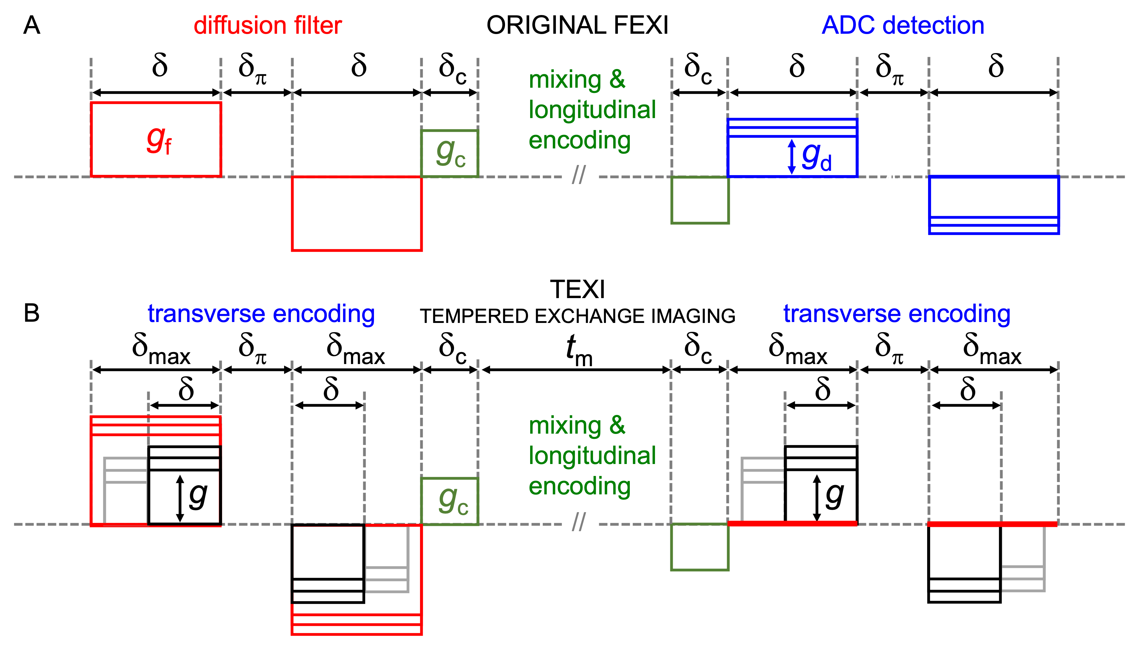
This work addresses the bias in FEXI due to the influence of strong crushers and restricted diffusion. We introduce a modified FEXI protocol, based on two principles: (1) the effects of the crushers are included in the forward exchange model based on Ning’s cumulant expansion approximation12,14 and (2) the timing parameters of diffusion encoding gradients in the filter and encoding blocks are adjusted to retain the same level of restriction encoding independent of the mixing times, resulting in the tuned exchange imaging (TEXI) protocol.
METHODS
Sensitivity to exchange for arbitrary gradient waveforms can be analyzed based on Ning’s cumulant expansion12, where the first and second cumulants are due to mean diffusivity $$$⟨D⟩$$$ and diffusion variance $$$V_D=⟨D^2 ⟩-⟨D⟩^2$$$. Signal attenuation is given by$$\ln(S⁄S_0 )≈-b⟨D⟩+\frac{b^2}{2} h(k) \cdot V_D,$$ where $$$k$$$ is the exchange rate constant and $$$h(k)$$$ is the exchange-weighting function depending on the dephasing waveform $$$q(t)$$$.
Sensitivity to restricted diffusion for arbitrary gradient waveforms can be analyzed in the frequency domain23-25. Using the Gaussian phase approximation and the low-frequency approximation for the diffusion spectrum $$$D(\omega)$$$ yields signal as
$$ \ln(S⁄S_0) ≈ -b⋅V_\omega⋅G_R,$$
where $$$G_R$$$ is the geometry factor related to restriction size26, $$$b$$$ is diffusion weighting and $$$V_\omega$$$ is the spectral variance,
$$V_\omega = \frac{\gamma^2}{b} \int_0^T g^2 (t) dt.$$
Tuned exchange imaging (TEXI) sequence design considers the triple diffusion encoding (TDE) in Figure 1B. The total b-value and restriction weighting are given by the contributions from the transverse and longitudinal encoding blocks as
$$b=b_T+b_L=\overbrace{q^2\delta'}^\text{tranverse enc.} +\overbrace{q_c^2 t'_m}^\text{longitudinal enc.}$$
and
$$b V_\omega=b_T V_T+b_L V_L=\overbrace{4\frac{q^2}{\delta}}^\text{tranverse enc.} +\overbrace{2\frac{q_c^2}{\delta_c}}^\text{longitudinal enc.}$$
where $$$t'_m=t_m+2\delta_c/3$$$ and $$$\delta'=\delta+(3\delta_\pi)/2.$$$ To maintain a constant restriction weighting, we need to reduce the transverse b-value as the longitudinal b-value increases with $$$t_m$$$, and we need to keep the ratio $$$\frac{q^2}{\delta}$$$constant as we do so.
Monte-Carlo (MC) simulations14 with resolution of 0.2 µs, D0 = 2x10⁻⁹ m2/s and 5x10⁶ particles were performed in substrates of equally sized and hexagonally packed spheres and cylinders (50/50 packing, diameters 1-11 µm) with the true exchange rates 0-20 s-1.
RESULTS & DISCUSSION
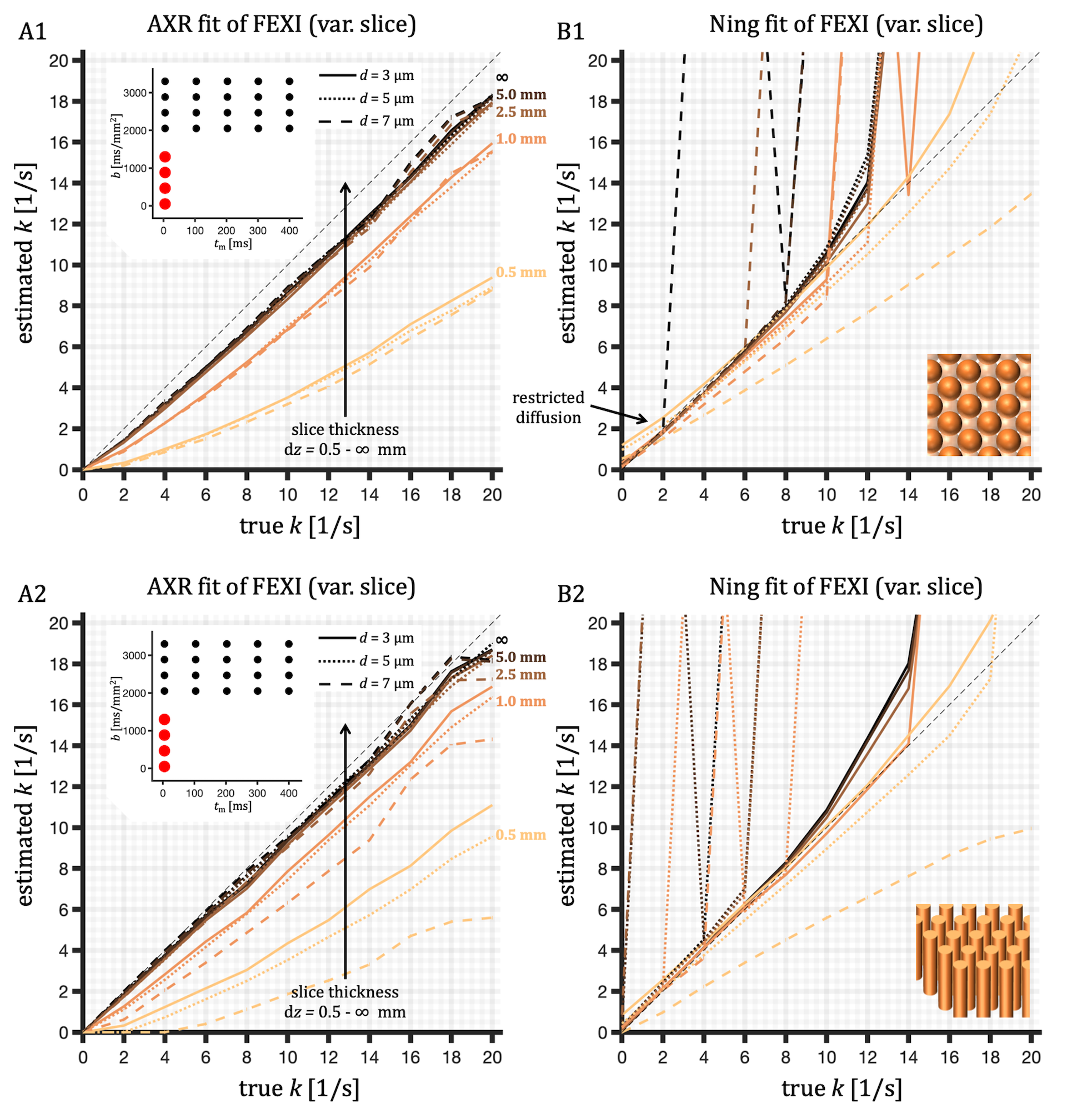
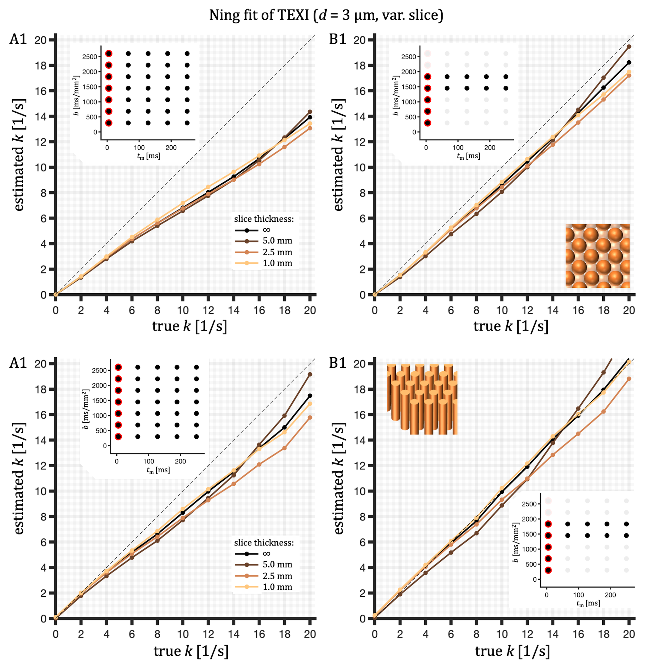
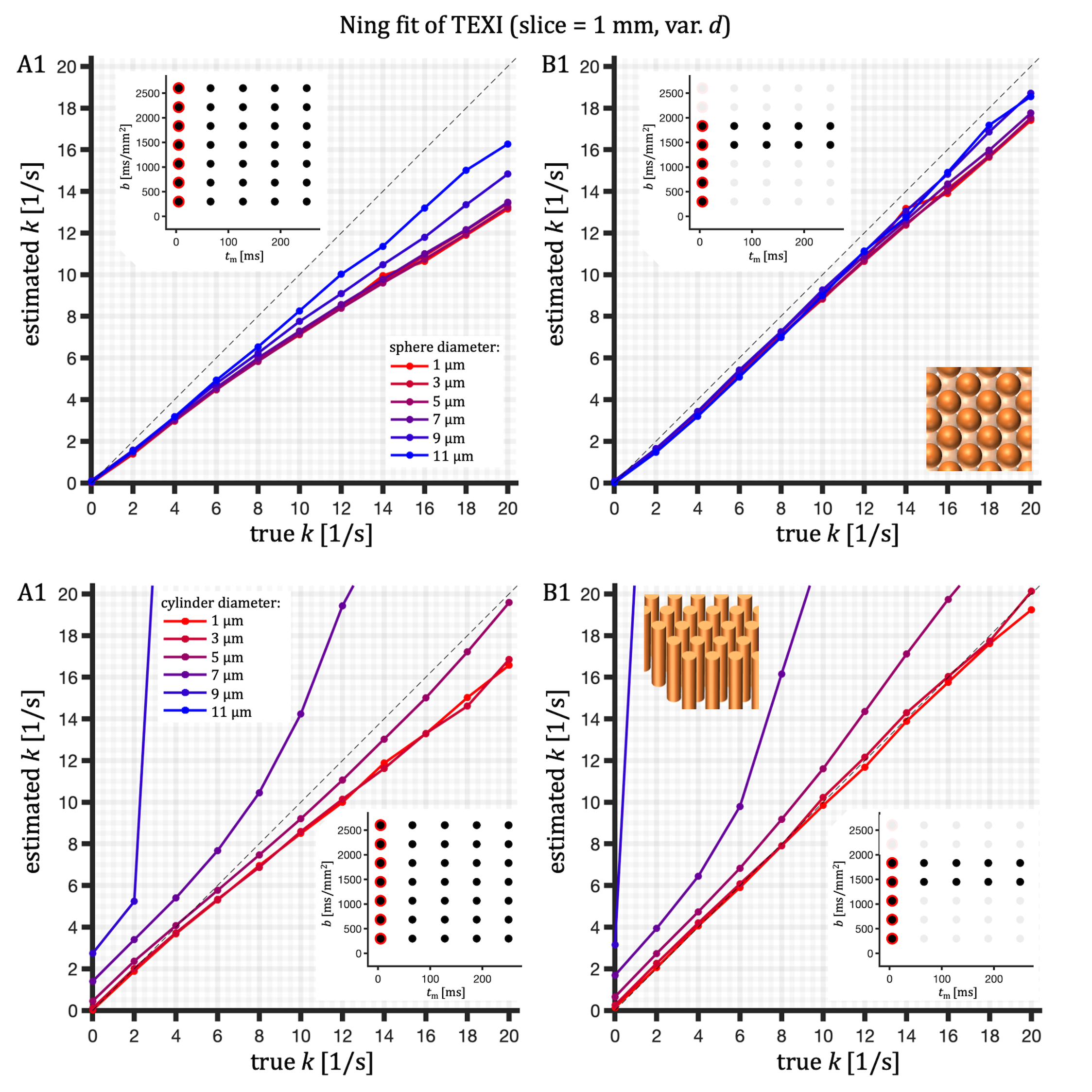
The Ning model could mitigate some of the crusher-induced bias in the original AXR fit of FEXI, but the exchange rate estimation was unreliable for faster exchange rates and larger sizes (Figure 2). Application of the Ning model in combination with the new TEXI protocol addresses both sources of bias. TEXI yields consistent exchange rates independent of slice thickness (Figure 3) and restriction size (Figure 4) even if strong crushers are used. The results for cylinders showed similar trends as for spheres, but with further reduced accuracy for larger diameters. In contrast to spheres, the dependence on diameter is stronger in the case of cylinders, where overestimated exchange rates were observed for larger diameters (Figure 4).TEXI relies on two approximations: the Ning exchange model12,14 and the spectral variance $$$V_\omega$$$26. This work addressed only accuracy and used simple MC simulations. Accuracy may be affected in more realistic substrates with hindered time-dependent diffusion15-17. Precision considerations need to be included in future investigations. As an alternative for mapping exchange with high imaging resolution and thin slices, super-resolution techniques may be considered27,28.
CONCLUSIONS
TEXI can address the bias in FEXI due to strong crushers and restricted diffusion. While the exchange rate is estimated based on the Ning model, the transverse encoding blocks are adjusted to retain constant restriction encoding independent of the mixing time, yielding consistent exchange rates independent of slice thickness and restriction size. TEXI could be useful for mapping exchange in combination with thin imaging slices and in the presence of restricted diffusion.Acknowledgements
This research was funded by: VR (Swedish Research Council) (grant number 2020–04549), NIH (National Institutes of Health) (grant numbers R01NS125781, R01MH074794 and P41EB015902) and
ERC (European
Research Council) under the European Union’s Horizon 2020 research and innovation programme
(grant number 804746).
References
1. Lasič S, Nilsson M, Lätt J, Ståhlberg F, Topgaard D: Apparent exchange rate mapping with diffusion MRI. Magn Reson Med 2011; 66:356–365.
2. Nilsson M, Lätt J, van Westen D, et al.: Noninvasive mapping of water diffusional exchange in the human brain using filter-exchange imaging. Magn Reson Med 2013; 69:1572–80.
3. Schilling F, Ros S, Hu D-E, et al.: MRI measurements of reporter-mediated increases in transmembrane water exchange enable detection of a gene reporter. Nat Biotechnol 2016; 35:75–80.
4. Lampinen B, Szczepankiewicz F, van Westen D, et al.: Optimal experimental design for filter exchange imaging : apparent exchange rate measurements in the healthy brain and in intracranial tumors. Magn Reson Med 2017; 77:1104–1114.
5. Tian X, Li H, Jiang X, Xie J, Gore JC, Xu J: Evaluation and comparison of diffusion MR methods for measuring apparent transcytolemmal water exchange rate constant. J Magn Reson 2017; 275:29–37.
6. Ludwig D, Laun FB, Ladd ME, Bachert P, Kuder TA: Apparent exchange rate imaging: On its applicability and the connection to the real exchange rate. Magn Reson Med 2021; 86:677–692.
7. Dickie BR, Parker GJM, Parkes LM: Measuring water exchange across the blood-brain barrier using MRI. Prog Nucl Magn Reson Spectrosc 2020; 116:19–39.
8. Bai R, Li Z, Sun C, Hsu Y, Liang H, Basser P: Feasibility of Filter-exchange Imaging (FEXI) in Measuring Different Exchange Processes in Human Brain. Neuroimage 2020:117039.
9. Li Z, Pang Z, Cheng J, et al.: The direction-dependence of apparent water exchange rate in human white matter. Neuroimage 2022; 247:118831.
10. Ohene Y, Harris WJ, Powell E, et al.: Filter exchange imaging with crusher gradient modelling detects increased blood – brain barrier water permeability in response to mild lung infection. Fluids Barriers CNS 2023; 20:1–18.
11. Powell E, Ohene Y, Battiston M, Dickie B, Parkes LM, Parker GJ: Blood-brain barrier water exchange measurements using FEXI: impact of modelling paradigm and relaxation time effects. Magn Reson Med 2023; 90:1–17.
12. Ning L, Nilsson M, Lasič S, Westin C-F, Rathi Y: Cumulant expansions for measuring water exchange using diffusion MRI. J Chem Phys 2018; 148.
13. Lasič S, Lundell H, Topgaard D, Dyrby TB: Effects of imaging gradients in sequences with varying longitudinal storage time—Case of diffusion exchange imaging. Magn Reson Med 2018; 79:2228–2235.
14. Chakwizira A, Westin CF, Brabec J, et al.: Diffusion MRI with pulsed and free gradient waveforms: Effects of restricted diffusion and exchange. NMR Biomed 2023; 36:e4827.
15. Novikov DS, Fieremans E, Jensen JH, Helpern J a: Random walk with barriers. Nat Phys 2011; 7:508–514.
16. Novikov DS, Jensen JH, Helpern JA, Fieremans E: Revealing mesoscopic structural universality with diffusion. PNAS 2014; 111:5088–93.
17. Novikov DS, Fieremans E, Jespersen SN, Kiselev VG, Mri D, Bird TA: Quantifying brain microstructure with diffusion MRI : Theory and parameter estimation. NMR Biomed 2018; e3998:1–53.
18. Ulloa P, Methot V, Koch MA: Experimental validation of a bias in apparent exchange rate measurement. Curr Dir Biomed Eng 2017; 3:529–532.
19. Li C, Fieremans E, Novikov DS, Ge Y, Zhang J: Measuring water exchange on a preclinical MRI system using filter exchange and diffusion time dependent kurtosis imaging. Magn Reonance Med 2023; 89:1441–1455.
20. Henriques RN, Jespersen SN, Shemesh N: Correlation tensor magnetic resonance imaging. Neuroimage 2020; 211(February):116605.
21. Chakwizira A, Szczepankiewicz F, Nilsson M: Diffusional exchange versus microscopic kurtosis from CTI: two conflicting interpretations of the same data. arXiv 2023.
22. Chakwizira A, Westin CF, Brabec J, et al.: Diffusion MRI with pulsed and free gradient waveforms: Effects of restricted diffusion and exchange. In Proc Intl Soc Magn Reson Med 31; 2023:0687.
23. Stepišnik J: Time-dependent self-diffusion by NMR spin-echo. Phys B 1993; 183:343–350.
24. Challaghan P, Stepišnik J: Frequency-Domain Analysis of Spin Motion Using Modulated-Gradient NMR. J Magn Reson A 1995; 117:118–122.
25. Stepišnik J: Validity limits of Gaussian approximation in cumulant expansion for diffusion attenuation of spin echo. Phys B 1999; 270:110–117.
26. Nilsson M, Lasič S, Drobnjak I, Topgaard D, Westin CF: Resolution limit of cylinder diameter estimation by diffusion MRI: The impact of gradient waveform and orientation dispersion. NMR Biomed 2017; 30:1–13.
27. Vis G, Nilsson M, Westin CF, Szczepankiewicz F: Accuracy and precision in super-resolution MRI: Enabling spherical tensor diffusion encoding at ultra-high b-values and high resolution. Neuroimage 2021; 245.
28. Spotorno N, Strandberg O, Vis G, Stomrud E, Nilsson M, Hansson O: Measures of cortical microstructure are linked to amyloid pathology in Alzheimer’s disease. Brain 2023; 146:1602–1614.
Figures