0600
How does dMRI signal evolve during diffusion encoding: theoretical analysis and numerical simulations for Gaussian diffusion1Center for Biomedical Imaging Research, Department of Biomedical Engineering, School of Medicine, Tsinghua University, Beijing, China, 2Center for Nano and Micro Mechanics, Department of Engineering Mechanics, Tsinghua University, Beijing, China, 3Institute of Imaging Science, Vanderbilt University Medical Center, Nashville, TN, United States, 4Department of Radiology and Radiological Sciences, Vanderbilt University Medical Center, Nashville, TN, United States, 5Department of Biomedical Engineering, Vanderbilt University, Nashville, TN, United States, 6Department of Physics and Astronomy, Vanderbilt University, Nashville, TN, United States
Synopsis
Keywords: Microstructure, Microstructure, dMRI signal analysis
Motivation: Until now, most attention has been focused on the final dMRI signals acquired, ignoring the signal evolution during diffusion encoding. However, the methods, which incorporated water exchange between compartments into modelling to extract more comprehensive tissue information, need to consider the signal evolution within the different compartments, as described in Karger model.
Goal(s): Figure out the dMRI signal evolution in the simplest case: Gaussian diffusion.
Approach: Theoretical analysis, Monte-Carlo and finite difference simulations.
Results: Signal-evolution curves provided by analytical expressions and numerical simulations are consistent. An “observation-size” effect emerges, the signal-evolution curve depends on the spatial size of the observation area.
Impact: Clarifying the actual dMRI signal evolution during diffusion encoding will inspire us to revisit the theoretical framework of Karger model. The results show that it is necessary to revise the current Karger-model-based methods for the “observation-size” effect.
Introduction
Calculations on dMRI signals sampled under different diffusion forms (Gaussian, restricted, and other no-Gaussian diffusion) are well established for a given acquisition sequence1,2. However, the signal evolution during diffusion encoding has always been neglected and the actual evolution curve is unknown. Recently, more and more studies attempted to establish biophysical models of in-vivo tissues by incorporating water exchange between compartments, to obtain more accurate and comprehensive information3. Karger model4 provides the most used framework for processing water exchange, which involves the dMRI signal evolutions within different compartments. Typically, only rough estimations can be provided for signal evolutions and the actual evolution curve is unknown.In this work, we first derived the analytical expression of dMRI signal evolution for the simplest case: one-dimensional Gaussian diffusion, and then performed Monte-Carlo5 and finite difference simulations6 to validate our theoretical results. The signal-evolution curves obtained from analytical expressions and numerical simulations were consistent. Furthermore, we found that the actual evolution curves were highly dependent on the spatial size of the observation area, i.e., an “observation-size” effect. The above findings show that the classic Karger-model-based framework may be problematic because it ignores this “observation-size” effect.
Theory
If numerous molecules are released from a single point $$$x_0$$$, as shown in Fig.1 (a), the spin phase of an individual molecule is $$$\Phi(t)=\gamma \int_0^t g\left(t_1\right) x\left(t_1\right) d t_1$$$ at any moment $$$t$$$. The total signal is the sum of magnetizations of all molecules released from $$$x_0$$$ and can be computed as:$$S(t)=S_0\langle\exp (-\boldsymbol{i} \Phi(t))\rangle \text { and } \ln (S)=\sum_{n=1}^{\infty} \frac{(\boldsymbol{i})^n}{n !}\left\langle\Phi^n\right\rangle_c \tag{1}$$
For Gaussian diffusion (n = 1, 2) with diffusivity $$$D$$$, we can derive that:
$$S(t)=S_0 \cdot \exp \left(-\gamma^2 D t G(t)^2+2 \gamma^2 D G(t) \int_0^t G\left(t_1\right) d t_1-\gamma^2 D \int_0^t G\left(t_1\right)^2 d t_1\right) \cdot \exp \left(\boldsymbol{i} \gamma x_0 G(t)\right) \tag{2}$$
Note that $$$G(t)=\int_0^t g\left(t_1\right) d t_1$$$ is usually unequal to 0 during diffusion encoding, as opposed to the published expressions.
If molecules are released from a region of finite width (-L, L), as shown in Fig.1 (b), the total signal is obtained by integrating over the spatial position $$$x$$$, and then:
$$|S(t)|=S_0 \cdot \exp \left(-\gamma^2 D t G(t)^2+2 \gamma^2 D G(t) \int_0^t G\left(t_1\right) d t_1-\gamma^2 D \int_0^t G\left(t_1\right)^2 d t_1\right) \cdot \frac{\sin (\gamma L G(t))}{\gamma L G(t)} \tag{3}$$
In practice, acquired signals come from a limited voxel while the region of molecular release is infinite, as shown in Fig.1 (c). Based on Eq. (3), we can obtain the expression for signal evolution in this case:
$$|S(t)|=S_0 \cdot \exp \left(-\gamma^2 D \int_0^t G\left(t_1\right)^2 d t_1\right) \cdot \frac{\sin (\gamma L G(t))}{\gamma L G(t)} \tag{4}$$
For simplicity, we termed the above three cases: “one-point-release-infinite-measurement”, “finite-release-infinite-measurement” and “infinite-release-finite-measurement”.
Method
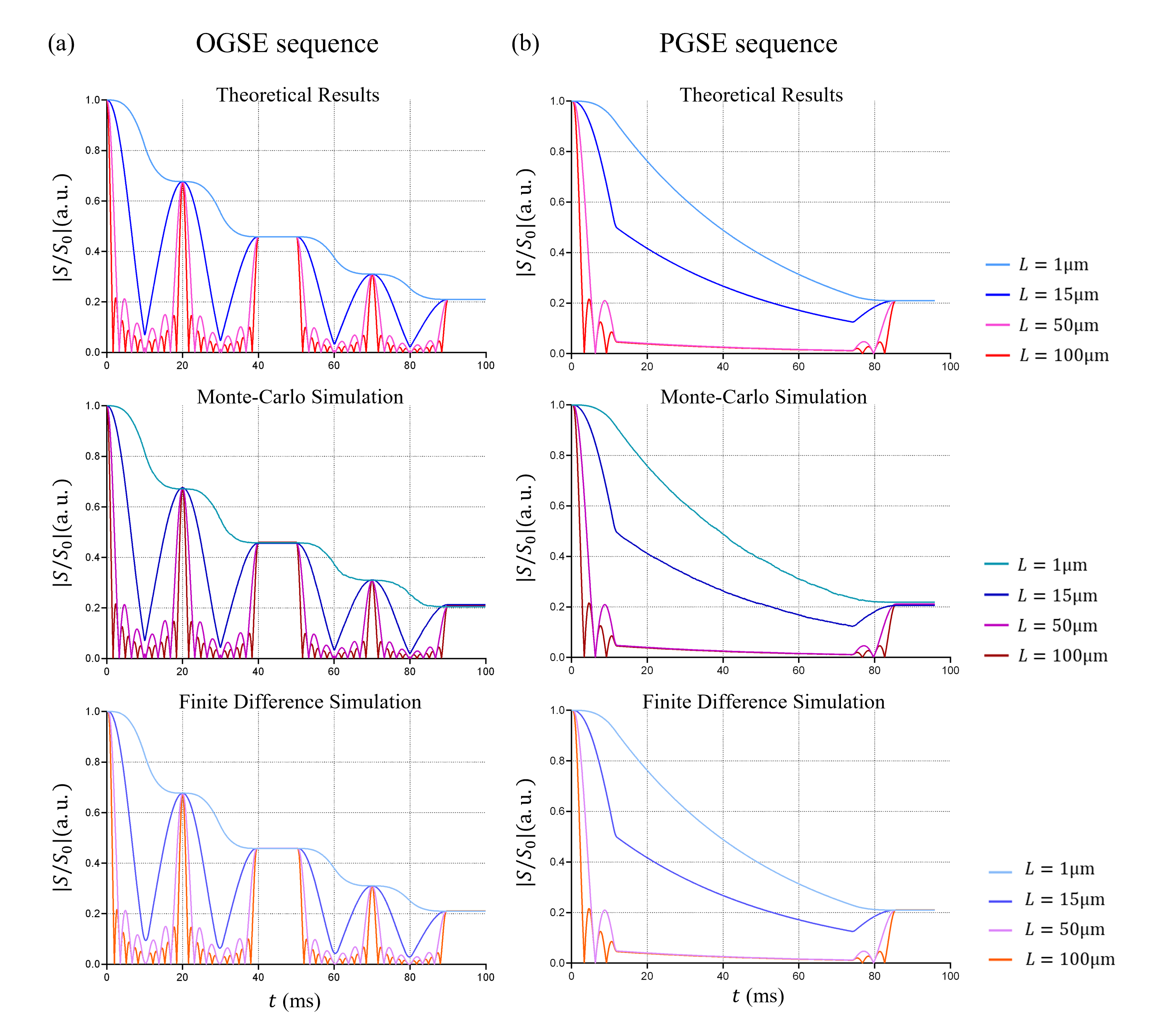
Monte-Carlo and finite difference simulations were performed to validate the theoretical results. We set different boundary conditions to suit the three cases: free boundary condition for “one-point-release-infinite-measurement” and “finite-release-infinite-measurement”, periodic boundary condition for “infinite-release-finite-measurement”. Note that the finite difference method was only used to simulate the signals for “infinite-release-finite-measurement”, due to its limitation. OGSE and PGSE were tested with the parameters shown in Table 1. The Gaussian diffusivity $$$D$$$ was set as 1.56 μm2/ms.Results and Discussion
As shown in Figs 2-4, the signal-evolution curves obtained from the analytical expressions and numerical simulations are consistent in three cases. Obviously, the evolution curves vary with different spatial sizes but converge to the same value when $$$G(t)=0$$$. Note that the signal evolution corresponding to larger L shows periodicity during $$$G(t) \neq 0$$$, and the time period or signal amplitude becomes shorter or smaller as L increases, contributing from the term $$$\sin (\gamma L G(t)) / \gamma L G(t)$$$. If the spatial size L reaches the actual spatial resolution ~1mm, we speculate that the entire evolution curve converges to 0 during $$$G(t) \neq 0$$$ and the dMRI signal is non-zero only at $$$G(t)=0$$$. We call this phenomenon the “observation-size” effect in dMRI signal evolution. Karger model has not considered this effect and its theoretical framework is independent of spatial size. But this work points out that once signal evolution is involved, the impact of the spatial-size-dependent signal is non-negligible for $$$G(t) \neq 0$$$. Therefore, Karger model may be problematic and needs to be revised.Conclusion
In this work, we systematically investigate the dMRI signal evolution for one-dimensional Gaussian diffusion. The analytical expressions were derived and numerical simulations were performed to validate the theoretical results. We found that the “observation-size” effect of the signal evolution will appear at $$$G(t) \neq 0$$$ during diffusion encoding. This suggests that the current Karger model and related methods are imperfect because they ignored this effect.Acknowledgements
No acknowledgement found.References
1. Stejskal EO, Tanner JE. Spin diffusion measurements: spin echoes in the presence of a time‐dependent field gradient. The journal of chemical physics 1965;42(1):288-292.2.
2. Jiang X, Li H, Xie J, Zhao P, Gore JC, Xu J. Quantification of cell size using temporal diffusion spectroscopy. Magnetic resonance in medicine 2016;75(3):1076-1085.3.
3. Jiang X, Devan SP, Xie J, Gore JC, Xu J. Improving MR cell size imaging by inclusion of transcytolemmal water exchange. NMR in Biomedicine 2022;35(12):e4799.4.
4. Karger J, Pfeifer H, Heink W. Principles and Application of Self-Diffusion Measurements by Nuclear Magnetic Resonance. In: Waugh JS, editor. Advances in Magnetic and Optical Resonance. Volume 12: Academic Press; 1988. p 1-89.5.
5. Hall MG, Alexander DC. Convergence and parameter choice for Monte-Carlo simulations of diffusion MRI. IEEE transactions on medical imaging 2009;28(9):1354-1364.6.
6. Xu J, Does MD, Gore JC. Numerical study of water diffusion in biological tissues using an improved finite difference method. Physics in Medicine & Biology 2007;52(7):N111.
Figures