0593
An anisotropic capillary based phantom for validation of diffusion-relaxation models1Department of Chemistry, University of Bergen, Bergen, Norway, 2Department of Health and Functioning, Western Norway University of Applied Sciences, Bergen, Norway
Synopsis
Keywords: Simulation/Validation, Phantoms
Motivation: We aim to create a universal procedure for experimental verification of diffusion models using model systems based on glass capillaries.
Goal(s): Spatially ordered glass capillaries mimics the characteristic geometry of white matter and are ideal for performing a 'stress test' of various diffusion models. We aim to verify this through diffusion and relaxation measurements at varying spatial directions and time scales.
Approach: Combined diffusion- and relaxation-weighted MRI based measurements verify the geometry of the glass capillaries at different spatial scales.
Results: The glass capillary phantom was established as a ground truth model for modelling of white matter at different diffusion and relaxation regimes.
Impact: Glass capillaries present a unique object with simple physical and geometrical features mimicking white matter. We tested a glass capillary phantom in terms of a diffusion-relaxation model using conventional sequences and provide a simple theoretical interpretation of experimental results.
Introduction
Conventional diffusion MRI (dMRI) techniques almost exhausts its potential in order to provide an accurate picture of underlying microstructure in living tissue1. Complexity of the tissue modelling, and relevant experimental implementations motivate researchers to search for dMRI alternatives2. Recent achievements in in vivo diffusion-relaxation MRI opens up new opportunities in our understanding of the brain organisation3. Nevertheless, new methods demand comprehensive validation, in particular, probing the limits of the new models and/or techniques. A good example of an object with known structure and boundless manipulations is an artificial phantom4. Glass capillaries present a unique chance to build up such an object with clear physical features mimicking white matter, in particular, internal/external water pools, susceptibility variations and strong anisotropy5. In the present study, we built and tested a glass capillary phantom in terms of a simple diffusion-relaxation MRI model. We aim to obtain diffusion-relaxation data using conventional pulse gradient sequences and provide a simple theoretical interpretation of experimental results.Methods
Fused silica capillaries with an internal diameter equals to 20±2 µm and with an external diameter of 90±6 µm were accurately cut into short pieces and manually placed in a 3 mm o.d. NMR tube. The NMR tube was filled with distilled water and placed in vacuum for approximately 5 hours to remove microscopic air bubbles. Subsequently, the tube was sealed up and placed in a 10 mm o.d. NMR tube filled with fluorinated oil. All measurements were performed at 11.7 T Bruker Ascend 500 MHz vertical wide bore spectrometer using a commercial Bruker Biospin MicWB40 micro imaging probe with triple gradients (max strength of 1.5 T/m in each direction) and Paravision v6.0.1 software. Parametric T2-maps were obtained using a multi-echo pulse sequence with TR = 2000 ms, TE = [4.3, 8.6, …, 86] ms. Parametric diffusion-maps were obtained using a diffusion-weighted spin echo sequence with TE/TR = 30/2000 ms, diffusion time = 20 ms, and 6 b-values between 0.1 and 1 ms/µm2. The capillaries are aligned along the z-axis and diffusion measurements were performed orthogonally along x, y, and z. All images were obtained with an in-plane resolution of 19x19 µm2 and a slice thickness of 1 mm along the z-axis. In addition, 'spectroscopic' spin echo diffusion measurements (i. e. with no spatial encoding) were performed with diffusion time = 25 ms, TE's between 30 and 130 ms, and 16 b-values between 0.05 and 4 ms/µm2. A corresponding 'spectroscopic' measurement of T2 was performed using a CPMG sequence with an inter-echo spacing of 2.5 ms. All numerical estimations have been performed with help of Matlab and Julia6 v1.6.7 in JupyterLab7.Results
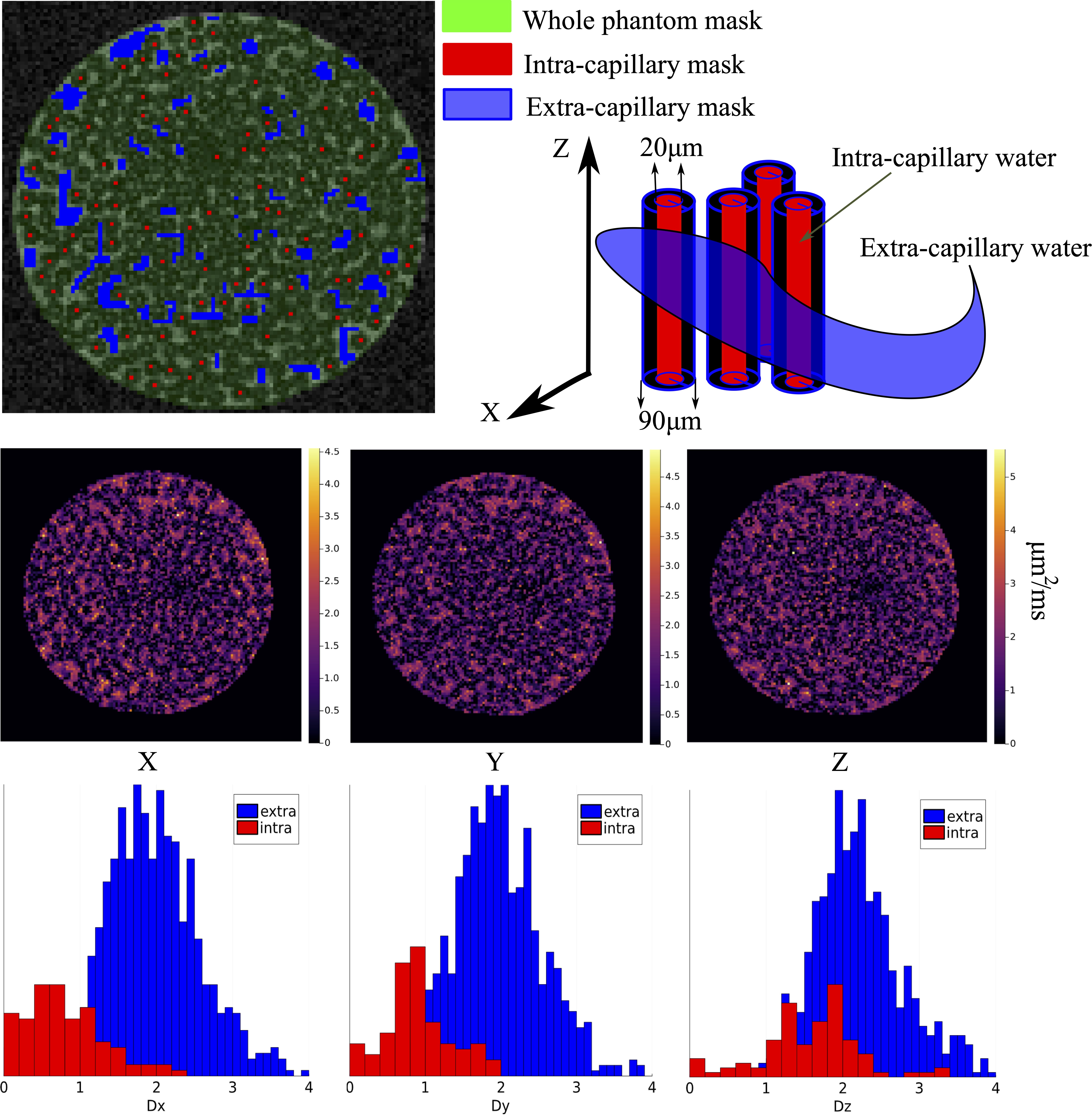
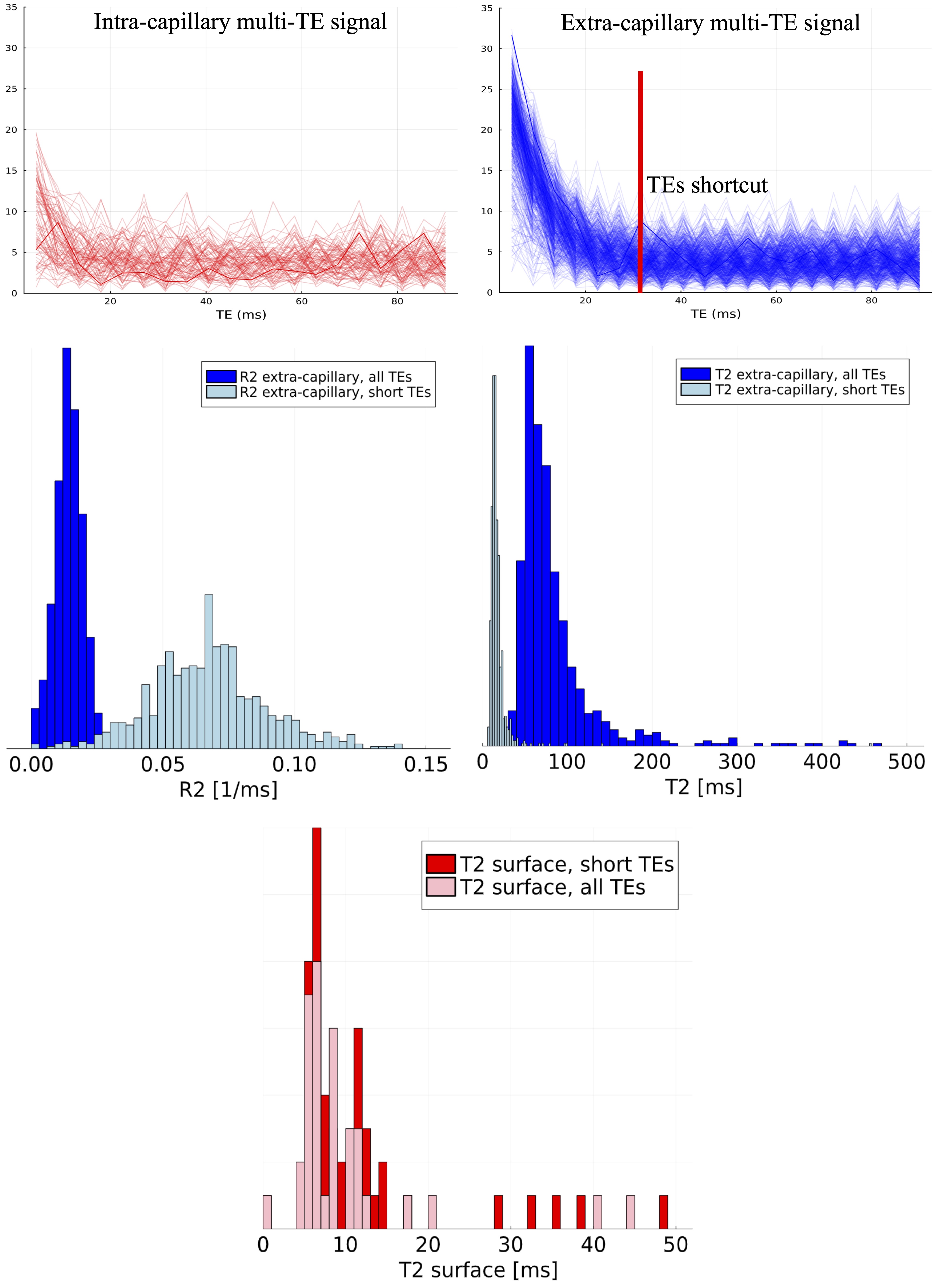
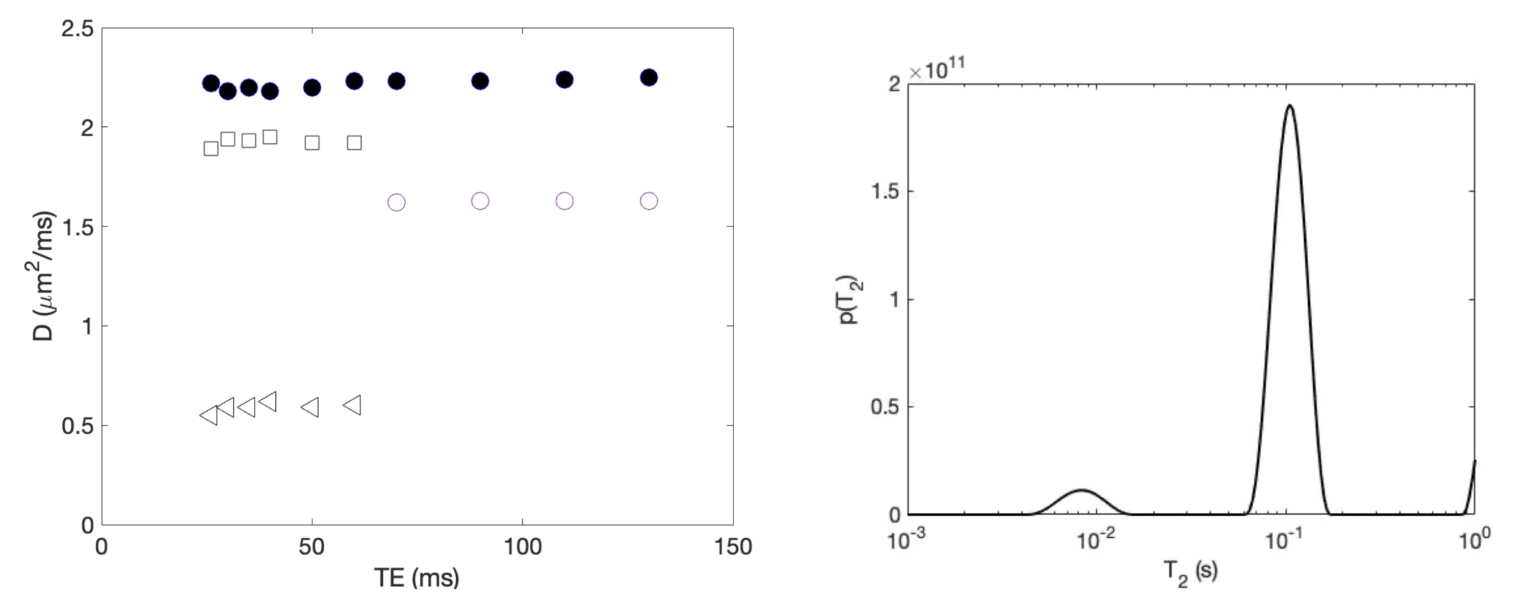
The structure of the capillary phantom is presented in Fig. 1 as a cross section MR-image. In order to evaluate the diffusion coefficients for water, we masked internal and external voxels. The histograms in Fig. 1 represent distributions of diffusion coefficients from the different masking and along the three orthogonal axis. Along the z-axis the intra- and extra-capillary distributions are strongly overlapping, while along the x- and y-axis the two distributions of diffusion coefficients are more separated, revealing the anisotropy of the system and two different water fractions/pools. In Fig. 2 the relaxation time distributions for the same masks estimated from the multi-echo measurements are presented. The intra- and extra-capillary T2-distributions can clearly be distinguished and further verify the two different fractions/pools.Diffusion coefficients at varying TE's together with T2-distributions, both obtained from the 'spectroscopic' measurements are presented in Fig. 3. These data, obtained at a greater dynamic range of b-values and at shorter inter-echo spacings, verify the validity of the parametric images presented above.
Discussion
Model systems with a clear structure and ability to determine the most important diffusion-relaxation features are in high demand. The constructed glass capillary model allowed us to create a simple and feature-rich model consisting of the most relevant parameters: two water pools, different internal/external diffusion-relaxation coefficients, capillary diameters with the known size, known field inhomogeneity. Our preliminary results demonstrated that these features could be estimated and compared with background truth, potentially allowing one to adapt various dMRI approaches for in vivo measurements.Conclusion
The anisotropic diffusion-relaxation phantom described in this study offers an excellent basis for the verification of theoretical models of white matter.Acknowledgements
The equipment used in this study is part of NNP- Norwegian NMR Platform.References
- D. Novikov, V. Kiselev, S. Jespersen, On modeling. Magn. Reson. Med. 2018; 79:3172-3193.
- J. T. Rosenberg, S. C. Grant, D. Topgaard, Nonparametric 5D D-R distribution imaging with single-shot EPI at 21.1 T: Initial results for in vivo rat brain. J. Magn. Reson. 2022; 341:107256.
- B. Lampinen, F. Szczepankiewicz, J. Lätt, L. Knutsson, J. Mårtensson, I. M. Björkman-Burtscher, D. van Westen, P. C. Sundgren, F. Ståhlberg, M. Nilsson, Probing brain tissue microstructure with MRI: principles, challenges, and the role of multidimensional diffusion-relaxation encoding. Neuroimage 2023; 282:120338.
- E. Fieremans, H-H. Lee, Physical and numerical phantoms for the validation of brain microstructural MRI: A cookbook. Neuroimage 2018; 182:39-61
- S. Vellmer, D. Edelhoff, D. Suter, I. Maximov, Anisotropic diffusion phantoms based on microcapillaries. J. Magn. Reson. 2017; 279:1-10.
- J. Bezanson, A. Edelman, S. Karpinski, V. B. Shah, Julia: A Fresh Approach to Numerical Computing. SIAM Rev. 2017; 59:65-98.
- https://jupyterlab.readthedocs.io/en/stable/index.html
Figures