0591
Tuneable Digital Phantoms for Grey Matter Modelling1CUBRIC, Cardiff University, Cardiff, United Kingdom, 2Blue Brain Project, École polytechnique fédérale de Lausanne, Lausanne, Switzerland, 3Cuntz lab, Ernst Strüngmann Institute (ESI) for Neuroscience in Cooperation with Max Planck Society, Frankfurt, Germany
Synopsis
Keywords: Simulation/Validation, Simulations
Motivation: There are several generators of white matter phantoms that have been developed in recent years and have significant potential in developing and validating diffusion MRI techniques through computational simulation. However, no such generator has been proposed for grey matter phantoms.
Goal(s): We aim to provide a means of generating grey matter phantoms compatible with diffusion MRI simulations.
Approach: Combing the network growth presented in the Contextual Fibre Growth (ConFiG) algorithm with the generative method of topological neuro synthesis (TNS), to create non intersecting morphologically realistic cellular structures.
Results: We can show that our algorithm can generate non-intersecting realistic voxels of grey matter.
Impact: We have developed a highly versatile algorithm, ConCeG, which can generate realistic digital phantoms of GM. These phantoms are ready to be incorporated into dMRI simulators, such as Camino, DiSimPy and MCDS for testing and validating diffusion MRI techniques
Introduction
IntroductionNumerical phantoms play an indispensable role in advancing and validating magnetic resonance imaging (MRI) techniques[1]. This is particularly the case of diffusion MRI (dMRI), where the need arises for generating adjustable and microstructurally accurate representations of complex biological tissues. While considerable effort has been dedicated to developing numerical phantoms for brain white matter (WM)[2-4], the same tuneable phantoms capable of replicating the complexity of grey matter (GM) microstructure are lacking.
In this work, we introduce Contextual Cellular Growth (ConCeG) as a solution to synthesize morphologically accurate GM tissue substrates, employing principles similar to the Contextual Fibre Growth (ConFiG) approach applied to WM[2].
Methods
To create realistic GM phantoms, it is essential to define the key morphological features required to faithfully replicate neural cells, including neurons and glia[5]. We categorized these attributes into three groups: structural, topological, and shape (Fig.1). Real neural cell reconstructions from open-access datasets such as neuromorpho.org[6] and the Allen brain atlas[7] were used to estimate distributions of these morphological features within the GM of the healthy adult mouse brain, utilising the TREES toolbox in MATLAB[8].ConCeG
ConCeG is the algorithm introduced to synthesize realistic GM phantoms and it is implemented in Matlab. It takes as inputs: voxel size, the number of nodes, predefined morphological parameters (structural, topological, and shape, see Fig.1), and layer-specific density profiles for each cell type. The output consists of cells formatted in SWC[9] format.
Similar to ConFiG[2], ConCeG relies on a network of connected nodes. The algorithm starts by distributing soma nodes within specified dimensions, following the user-defined cell density with respect to cortical depth. Additional nodes are randomly placed within the voxel space, creating a connected network for cellular projections. This network serves as a guide for cellular projections to navigate, considering biologically informed cost functions. The projections extend toward attractor points efficiently while avoiding nodes already occupied by existing projections or nodes that would lead to unrealistic projection shrinkage. Fig.2 illustrates the growth process.
ConCeG facilitates branching by employing the topological neuron synthesis approach[10]. As a projection grows, its path length increases, and the probability of branching is determined in relation to the birth lengths of remaining bars in the barcode. The branching probability increases as the path length approaches the birth length of another bar and satisfy the probability of branching given the current branch order. Newly initiated branches select angles from the angle distribution and pick attractor points that satisfies this angle in relation to the parent branch. Cellular growth occurs contextually, with the maximum node radius for nodes in the network updated at each growth step.
Results
Fig. 3 demonstrates that ConCeG can recreate cellular structures accurately when cells are grown individually, preserving key morphological characteristics' distributions (see table in Fig.3).Fig. 4 shows that the same degree of accuracy can be achieved when cells are grown contextually. Finally, in Fig. 5 we illustrate ConCeG's flexibility and capabilities by synthesizing a column of the mouse visual cortex based on density profiles from the Allen brain atlas[11] and[12, 13].
Discussion
Here, we have developed a highly versatile approach, ConCeG, which leverages real cellular data to create realistic digital phantoms of brain GM. These phantoms are ready to be incorporated into dMRI simulators, such as Camino[14], DiSimPy[15] and MCDS[16]The current implementation of ConCeG enables the generation of large voxels within reasonable computational time (e.g. ~5 hours to generate a 100x100x1200 µm3 voxel using a single CPU-thread). However, further developments are necessary to achieve dense cellular packing. In fact, the densest voxel we were able to generate thus far has a 31% intracellular volume fraction. Future work will focus on increasing ConCeG ability to achieve denser cellular packings (e.g. 60-70%), implementing strategies such as post-growth optimization similar to ConFiG[2]and MEDUSA[4].
Conclusion
Here we introduced ConCeG: a significant contribution to the field of neuroimaging and computational biology, enabling more accurate and insightful simulations of complex neural tissues.Acknowledgements
UKRIReferences
1. Fieremans, E. and H.H. Lee, Physical and numerical phantoms for the validation of brain microstructural MRI: A cookbook. Neuroimage, 2018. 182: p. 39-61.
2. Callaghan, R., et al., ConFiG: Contextual Fibre Growth to generate realistic axonal packing for diffusion MRI simulation. Neuroimage, 2020. 220: p. 117107.
3. Villarreal-Haro, J.L., et al., CACTUS: a computational framework for generating realistic white matter microstructure substrates. Front Neuroinform, 2023. 17: p. 1208073.
4. Ginsburger, K., et al., MEDUSA: A GPU-based tool to create realistic phantoms of the brain microstructure using tiny spheres. Neuroimage, 2019. 193: p. 10-24.
5. Zisis, E., et al., Digital Reconstruction of the Neuro-Glia-Vascular Architecture. Cereb Cortex, 2021. 31(12): p. 5686-5703.
6. Akram, M.A., et al., An open repository for single-cell reconstructions of the brain forest. Sci Data, 2018. 5: p. 180006.
7. Atlas, D.M.B., Allen brain atlas. 2006.
8. Cuntz, H., et al., One rule to grow them all: a general theory of neuronal branching and its practical application. PLoS Comput Biol, 2010. 6(8).
9. Cannon, R.C., et al., An on-line archive of reconstructed hippocampal neurons. Journal of neuroscience methods, 1998. 84(1-2): p. 49-54.
10. Kanari, L., et al., Computational synthesis of cortical dendritic morphologies. Cell Rep, 2022. 39(1): p. 110586.
11. Gouwens, N.W., et al., Classification of electrophysiological and morphological neuron types in the mouse visual cortex. Nat Neurosci, 2019. 22(7): p. 1182-1195.
12. Keller, D., C. Ero, and H. Markram, Cell Densities in the Mouse Brain: A Systematic Review. Front Neuroanat, 2018. 12: p. 83.
13. Tsai, P.S., et al., Correlations of neuronal and microvascular densities in murine cortex revealed by direct counting and colocalization of nuclei and vessels. J Neurosci, 2009. 29(46): p. 14553-70.
14. Alexander, D., et al. Camino: Open-source diffusion-MRI reconstruction and processing. in Proceedings of the 14th Scientific Meeting of the International Society for Magnetic Resonance in Medicine (ISMRM), Seattle, WA, USA. 2006.
15. Kerkelä, L., et al., Disimpy: A massively parallel Monte Carlo simulator for generating diffusion-weighted MRI data in Python. Journal of Open Source Software, 2020. 5(52).
16. Rafael-Patino, J., et al., Robust Monte-Carlo Simulations in Diffusion-MRI: Effect of the Substrate Complexity and Parameter Choice on the Reproducibility of Results. Front Neuroinform, 2020. 14: p. 8.
17. Kanari, L., et al., A Topological Representation of Branching Neuronal Morphologies. Neuroinformatics, 2018. 16(1): p. 3-13.
Figures
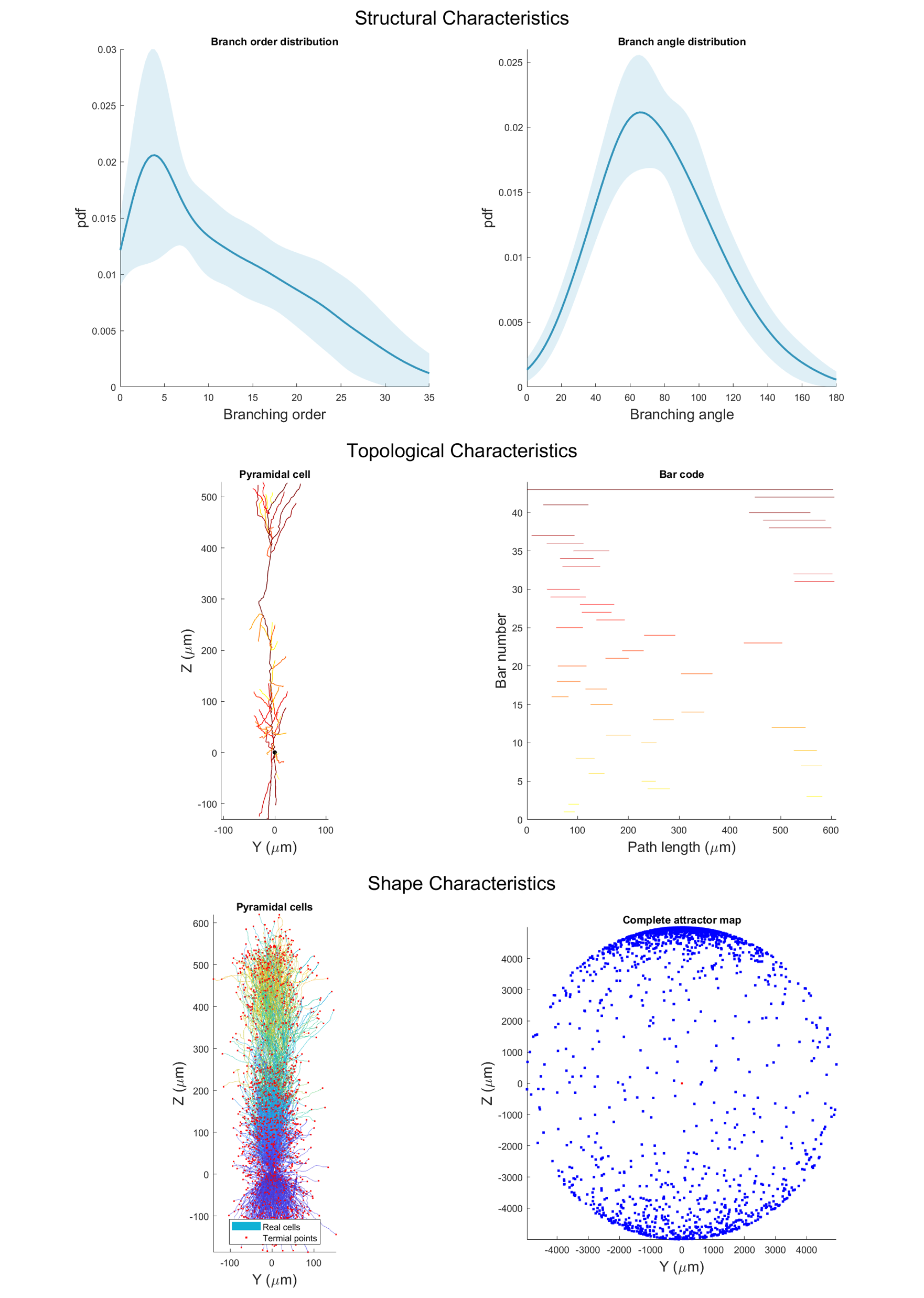
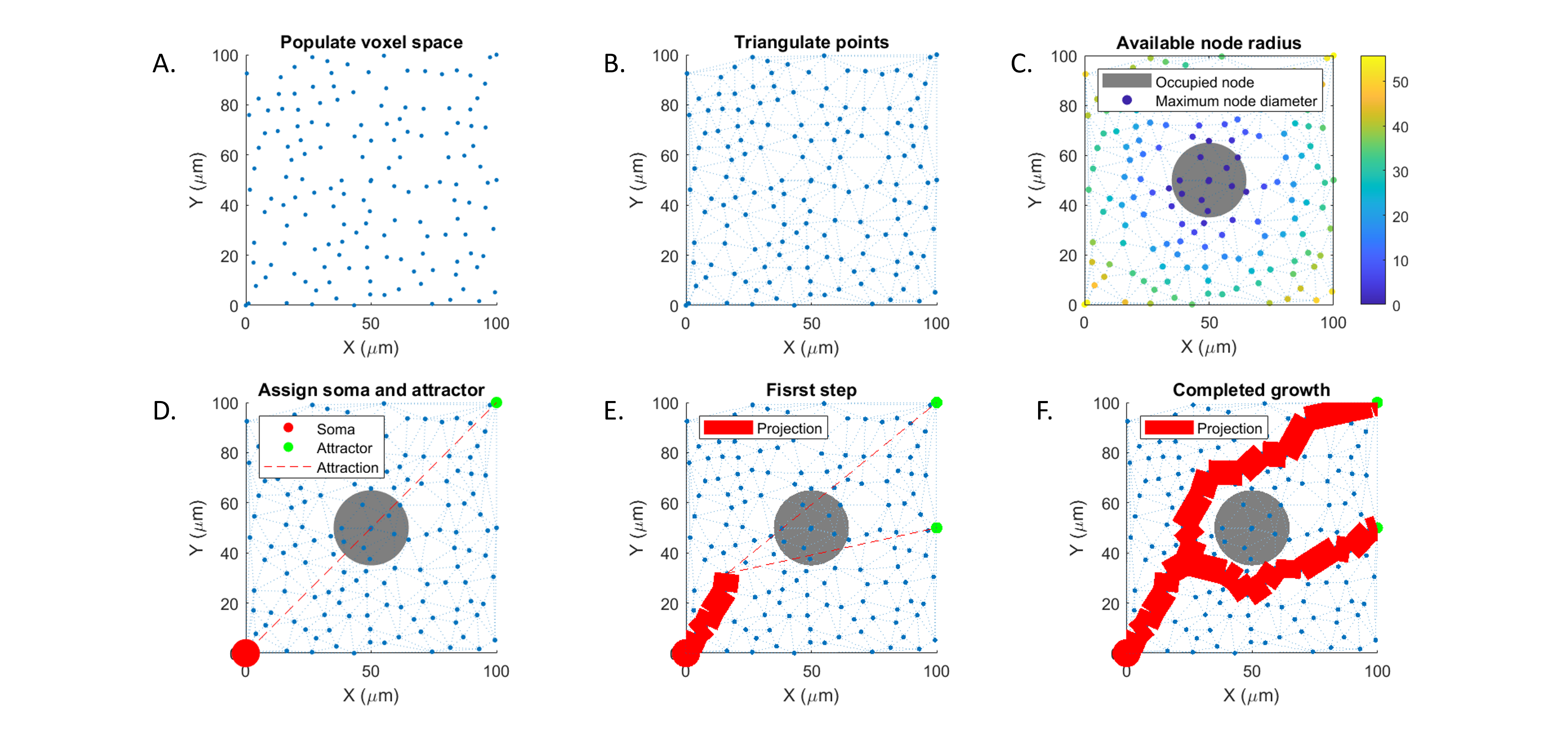
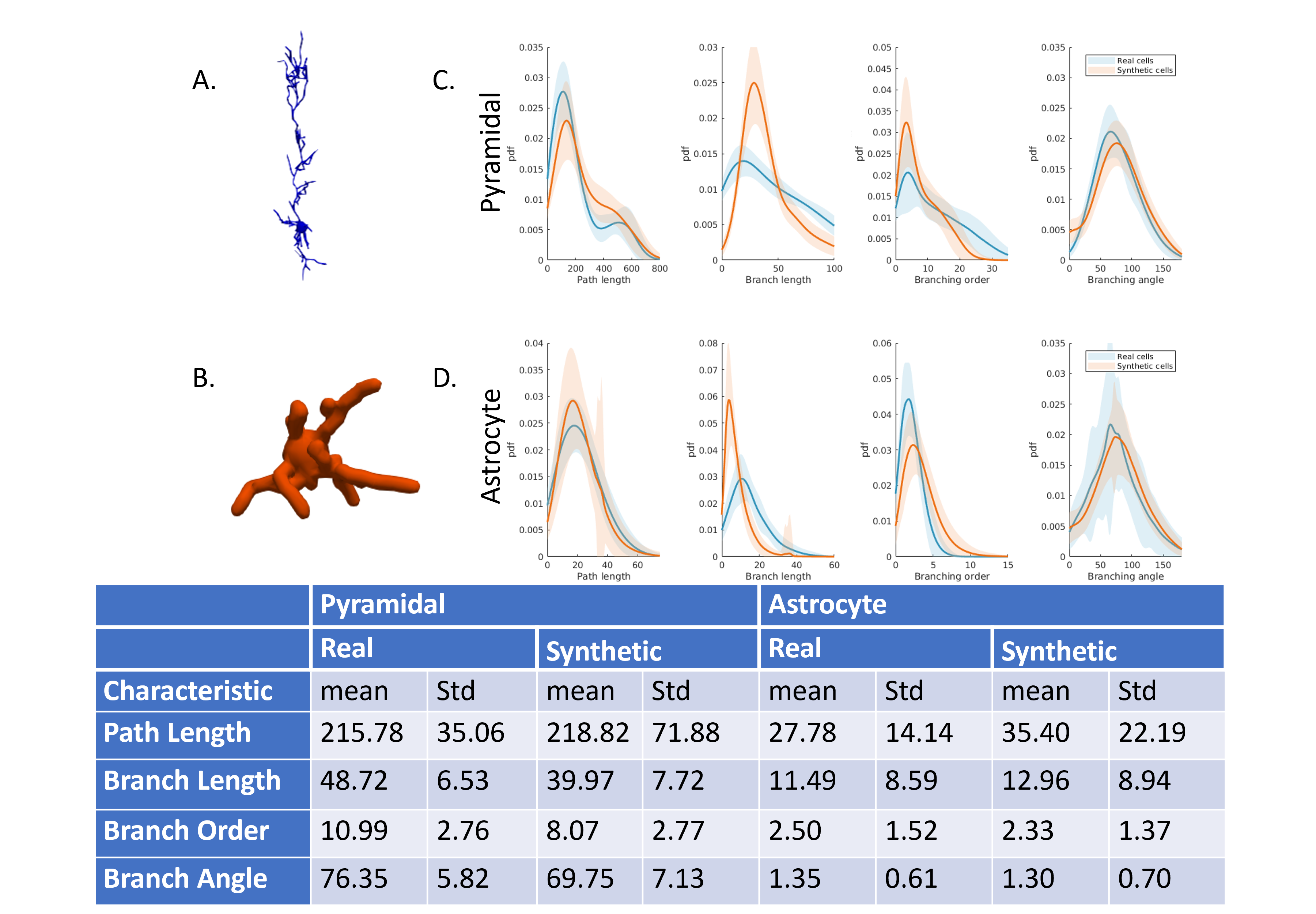
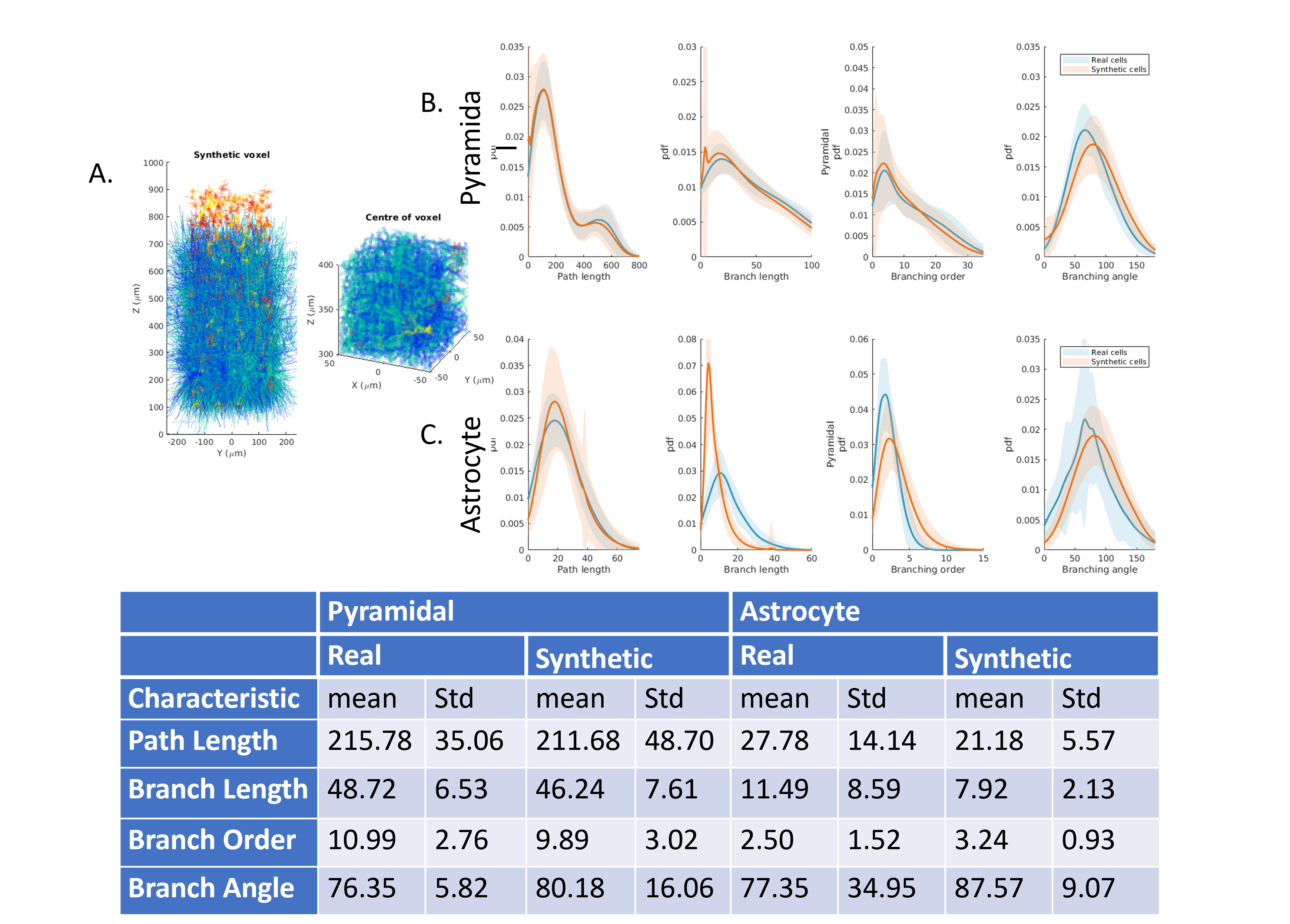
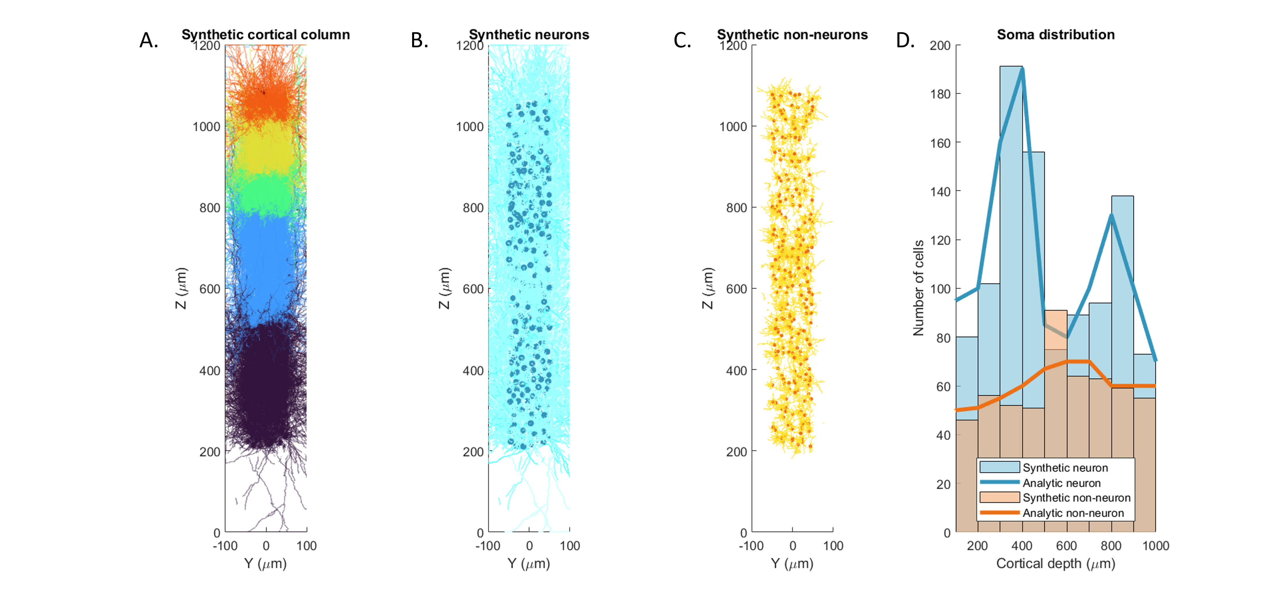
Fig 5. A column of synthesised mouse visual cortex. A. complete synthetic column, colours indicate the cell bodies layer location. B. Distribution of Neuronal cells (soma dark blue). C. Distribution of non-Neuronal cells (soma dark blue). D. Histogram of number of somas with respect depth for neurons and non-neurons overlaid with analytic al solutions given the volume and real distribution[12,13].