0589
Towards a Standard Model of Diffusion in White Matter with Phase and Relaxation – A Monte-Carlo Study1Center of Functionally Integrative Neuroscience, Department of Clinical Medicine, Aarhus University, Aarhus, Denmark, 2Division of Medical Physics, Department of Radiology, University Medical Center Freiburg, Freiburg, Germany, 3Champalimaud Research,Champalimaud Centre for the Unknown, Lisbon, Portugal, 4Department of Physics and Astronomy, Aarhus University, Aarhus, Denmark
Synopsis
Keywords: Microstructure, Microstructure
Motivation: Improving parameter estimation for the standard model of diffusion in white matter (SM) by modelling the subsequent decay of the spin-echo of the dMRI signal.
Goal(s): To numerically validate SMPR - an extension of SM incorporating orientation-dependent, susceptibility related relaxation rates and Larmor frequency shifts of the spin echo decay of the dMRI signal.
Approach: To perform Monte-Carlo (MC) simulations in orientationally dispersed, non-exchanging bundles of hollow magnetized cylinders, simulate a standard PGSE signal and its spin echo decay, and compare against the SMPR model.
Results: SMPR is in agreement with the MC simulations in both phase and signal magnitude.
Impact: Orientation-dependent susceptibility effects may improve parameter estimation of the Standard Model of diffusion in white matter and enable rotation-free mapping of susceptibility-related parameters.
Introduction
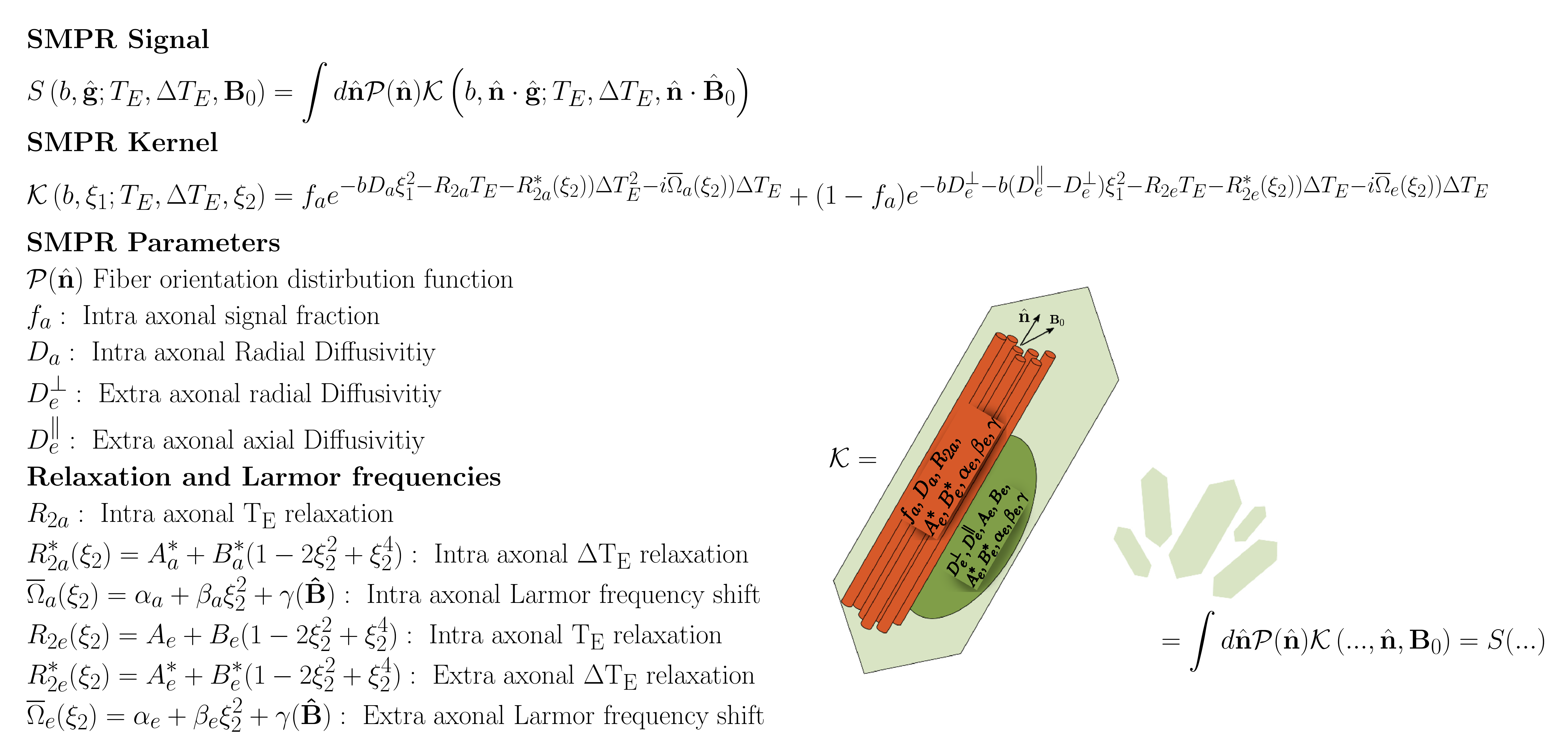
Estimating the parameters of the Standard Model (SM) of diffusion in white matter (WM) can be ill-posed1-4. We have previously proposed to incorporate orientation dependent susceptibility effects to SM to improve fitting by modelling the attenuation of a PGSE signal5 – the SMPR model. Simulations have so far demonstrated that SMPR may improve parameter estimation and aid in estimation of susceptibility parameters without having to rotate the sample. Here we extend the investigation by examining the accuracy of SMPR against Monte-Carlo simulations of a PGSE experiment with additional sampling of its spin echo decay (figure 1). This is done using a digital phantom consisting of non-communicating bundles of magnetized, hollow cylinders.Methods
TheoryWe consider the extended SM – SMPR – with diffusion weighting $$$(b,\hat{\mathbf{g}})$$$, echo time $$$T_E$$$ and additional sampling of the spin echo decay at times $$$\Delta{T_E}$$$. Figure 1 gives an overview of the signal $$$S(b,\hat{\mathbf{g}};T_E,\Delta{T_E},{\mathbf{B}}_0)$$$ and the added compartmental transverse relaxations rates6-9 $$$R_{2i},R_{2i}^*$$$ and Larmor frequency shifts10-14
$$$\overline\Omega_i$$$ for $$$i=e,a$$$ as defined in figure 1.
Monte-Carlo simulations
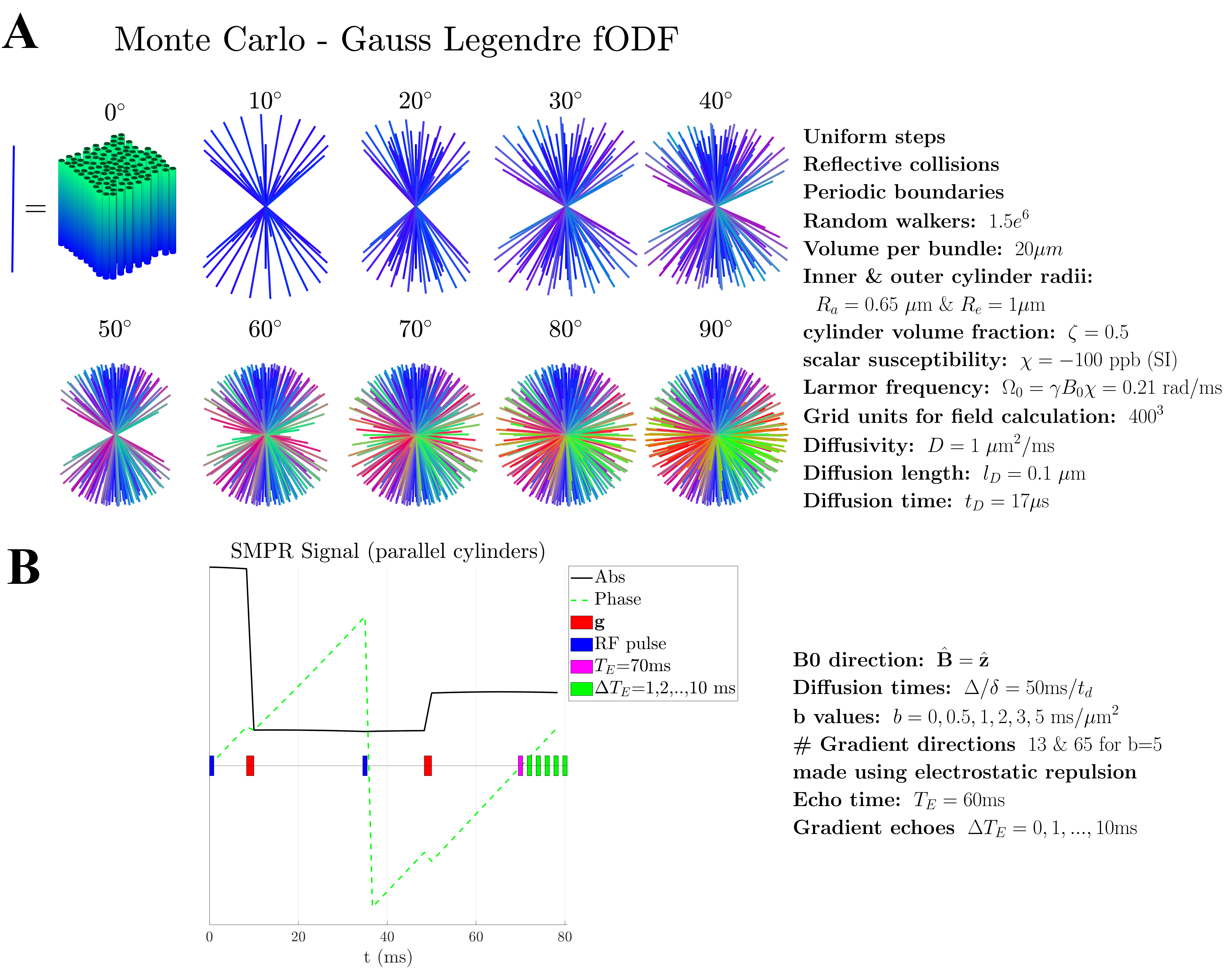
We performed MC simulations to validate SMPR (figure 2). The details are listed in figure 2A. We synthesized a digital phantom consisting of non-exchanging bundles of magnetized hollow cylinders resembling an ideal SM application. As a first step, we considered only cylinders with scalar susceptibility, and without spherical sources such that $$$\overline\Omega=\overline\Omega_e=\overline\Omega_a$$$. The bundle’s orientation distribution $$$\mathcal{P}(\hat{\mathbf{n}})$$$ (fODF) consists of 10 polar rings and 20 azimuthal points in each ring with fODF symmetry axis along $$$\hat{\mathbf{B}}_0)$$$. For this we calculated the signal kernel $$$\mathcal{K}(b,\hat{\mathbf{g}}\cdot\hat{\mathbf{n}};T_E,\Delta{T_E},\hat{\mathbf{B}}_0\cdot\hat{\mathbf{g}})$$$, as described in figure 2B including acquisition details. The SMPR signal $$$S(b,\hat{\mathbf{g}};T_E,\Delta{T_E},{\mathbf{B}}_0)$$$ for a given level of orientation dispersion was made by summing $$$\mathcal{K}(b,\hat{\mathbf{g}}\cdot\hat{\mathbf{n}};T_E,\Delta{T_E},\hat{\mathbf{B}}_0\cdot\hat{\mathbf{g}})$$$ from bundles up to a maximum polar angle $$$\theta_C$$$ (cf. figure 1A).
Results
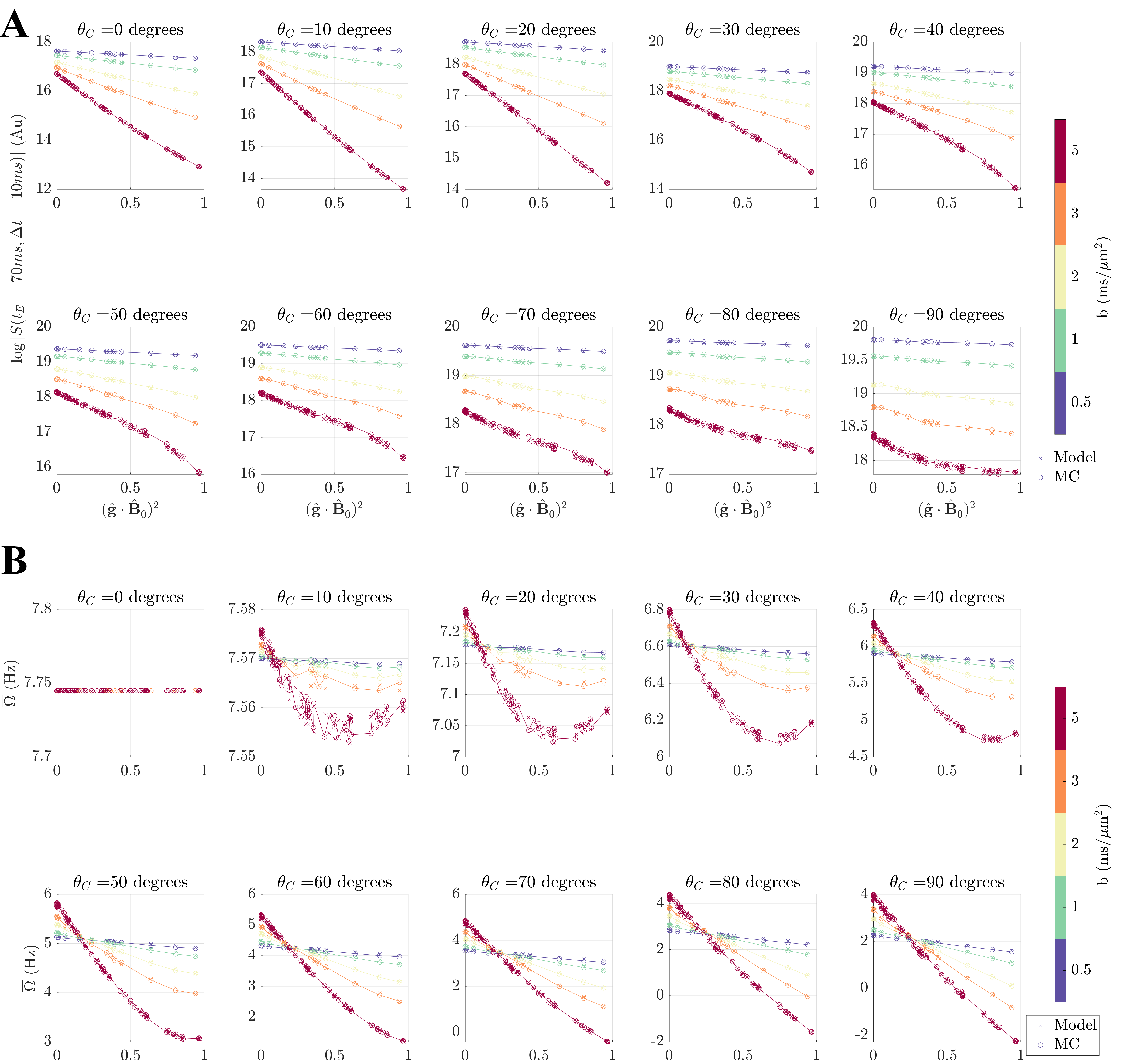
Figure 3A shows the signal magnitude for a given $$$T_E,\Delta{T_E}$$$ for varying levels of orientation dispersion, b-values and gradient directions, and figure 3B shows the overall Larmor frequency shift $$$\overline\Omega$$$. We find SMPR agrees with MC in all cases. The orientation dependence of $$$\overline\Omega$$$ depends on the amount of dispersion and angle between the fODF symmetry axis and $$$\hat{\mathbf{B}}_0$$$. $$$\overline\Omega$$$ varies little for low levels of dispersion since $$$\hat{\mathbf{B}}_0$$$ is parallel to the fODF symmetry axis, and because we only included a single weak bulk susceptibility, 0.05ppm.Discussion
Our simulations demonstrate that SMPR describes the spin echo decay of the PGSE signal for an ideal digital SM2 phantom well. Here we considered linear diffusion encoding, which unfortunately reduces the magnitude of the measured phase because the convolution $$$\mathcal{P}\otimes\mathcal{K}$$$ (cf. figure 1) is roughly taken over a plane perpendicular to the direction of the strong diffusion gradient. Planar encoding15,16 would restore the phase towards the range of values one would measure in a normal gradient echo as $$$\mathcal{P}\otimes\mathcal{K}$$$ is then centered around one direction instead of a plane. However, planar encoding comes at the expense of lower SNR17. The optimal compromise will be investigated in the future. We also plan to include susceptibility anisotropy of myelin and spherical inclusions. However, anisotropy of myelin will not affect the functional form of the relaxation rate and Larmor frequencies18,19, as it only adds an additional constant frequency shift in the intra-axonal space. Our phantom was fairly simple and constructed to be in agreement with the model assumptions, in order to test if the model did not leave out any important signal contributions, e.g., from 3rd and 4th order cumulants20. We therefore plan to test SMPR against MC simulations in more realistic phantoms including segmented WM from 3D electron microscopy21.Conclusion
We showed that our analytical SMPR model - incorporating orientation dependent susceptibility effects into the standard model of diffusion in white matter – agrees with Monte-Carlo simulations of PGSE in a phantom of dispersed cylinder bundles. We believe our results bring us one step closer to better SM parameter estimation and further enable susceptibility properties of WM to be measured without impractical sample rotations.Acknowledgements
This study is funded by the Independent Research Fund (grant 10.46540/3103-00144B)References
1. Novikov DS, Veraart J, Jelescu IO, Fieremans E. Rotationally-invariant mapping of scalar and orientational metrics of neuronal microstructure with diffusion MRI. Neuroimage. 2018;174:518-538. doi:10.1016/J.NEUROIMAGE.2018.03.006
2. Novikov DS, Fieremans E, Jespersen SN, Kiselev VG. Quantifying brain microstructure with diffusion MRI: Theory and parameter estimation. NMR Biomed. 2019;32(4):e3998. doi:10.1002/NBM.3998
3. Jelescu IO, Veraart J, Fieremans E, Novikov DS. Degeneracy in model parameter estimation for multi-compartmental diffusion in neuronal tissue. NMR Biomed. 2016;29(1):33-47. doi:10.1002/NBM.3450
4. Coelho S, Pozo JM, Jespersen SN, Jones DK, Frangi AF. Resolving degeneracy in diffusion MRI biophysical model parameter estimation using double diffusion encoding. Magn Reson Med. 2019;82(1):395-410. doi:10.1002/MRM.27714
5. Sandgaard AD, Kiselev VG, Shemesh N, Jespersen SN. Incorporating susceptibility effects into the standard model of diffusion in white matter. In: Joint Annual Meeting ISMRM-ESMRMB & ISMRT 32st Annual Meeting. ; 2023:0975.
6. Veronica P Dell’Acqua, Tax CMW, Malwina Molendowska, Jones DK, Kleban E, Greg D Parker. Measuring compartmental T2 and T2* orientation dependence in white matter. In: ISMRM Workshop on Diffusion MRI: From Research to Clinic. ; 2022.
7. Tax CMW, Kleban E, Chamberland M, Baraković M, Rudrapatna U, Jones DK. Measuring compartmental T2-orientational dependence in human brain white matter using a tiltable RF coil and diffusion-T2 correlation MRI. Neuroimage. 2021;236. doi:10.1016/J.NEUROIMAGE.2021.117967
8. Knight MJ, Dillon S, Jarutyte L, Kauppinen RA. Magnetic Resonance Relaxation Anisotropy: Physical Principles and Uses in Microstructure Imaging. Biophys J. 2017;112(7):1517-1528. doi:10.1016/j.bpj.2017.02.026
9. Kiselev VG, Novikov DS. Transverse NMR Relaxation in Biological Tissues. Vol 182.; 2018:149-168. doi:10.1016/j.neuroimage.2018.06.002
10. Sandgaard AD, Kiselev VG, Shemesh N, Jespersen SN. Incorporating the effect of white matter microstructure in the estimation of magnetic susceptibility in ex-vivo mouse brain. 2022;(August). doi:10.48550/arxiv.2208.02594
11. Sandgaard AD, Shemesh N, Kiselev VG, Jespersen SN. Larmor frequency shift from magnetized cylinders with arbitrary orientation distribution. NMR Biomed. 2023;36(3):e4859. doi:10.1002/nbm.4859
12. Kiselev VG. Larmor frequency in heterogeneous media. J Magn Reson. 2019;299:168-175. doi:10.1016/j.jmr.2018.12.008
13. He X, Yablonskiy DA. Biophysical mechanisms of phase contrast in gradient echo MRI. Proc Natl Acad Sci U S A. 2009;106(32):13558-13563. doi:10.1073/pnas.0904899106
14. Yablonskiy DA, Sukstanskii AL. Generalized Lorentzian Tensor Approach (GLTA) as a biophysical background for quantitative susceptibility mapping. Magn Reson Med. 2015;73(2):757-764. doi:10.1002/mrm.25538
15. Dhital B, Reisert M, Kellner E, Kiselev VG. Intra-axonal diffusivity in brain white matter. Neuroimage. 2019;189:543-550. doi:10.1016/J.NEUROIMAGE.2019.01.015
16. Westin CF, Szczepankiewicz F, Pasternak O, et al. Measurement tensors in diffusion MRI: Generalizing the concept of diffusion encoding. Lect Notes Comput Sci (including Subser Lect Notes Artif Intell Lect Notes Bioinformatics). 2014;8675 LNCS(PART 3):209-216. doi:10.1007/978-3-319-10443-0_27/COVER
17. Coelho S, Baete SH, Lemberskiy G, et al. Reproducibility of the Standard Model of diffusion in white matter on clinical MRI systems. Neuroimage. 2022;257:119290. doi:10.1016/J.NEUROIMAGE.2022.119290
18. Wharton S, Bowtell R. Fiber orientation-dependent white matter contrast in gradient echo MRI. Proc Natl Acad Sci U S A. 2012;109(45):18559-18564. doi:10.1073/pnas.1211075109
19. Sukstanskii AL, Yablonskiy DA. On the role of neuronal magnetic susceptibility and structure symmetry on gradient echo MR signal formation. Magn Reson Med. 2014;71(1):345-353. doi:10.1002/mrm.24629
20. Novikov DS, Reisert M, Kiselev VG. Effects of mesoscopic susceptibility and transverse relaxation on diffusion NMR. J Magn Reson. 2018;293:134-144. doi:10.1016/J.JMR.2018.06.007
21. Abdollahzadeh A, Belevich I, Jokitalo E, Sierra A, Tohka J. DeepACSON automated segmentation of white matter in 3D electron microscopy. Commun Biol 2021 41. 2021;4(1):1-14. doi:10.1038/s42003-021-01699-w
22. Jones DK, Horsfield MA, Simmons A. Optimal strategies for measuring diffusion in anisotropic systems by magnetic resonance imaging. Magn Reson Med. 1999;42(3):515-525. doi:10.1002/(SICI)1522-2594(199909)42:3<515::AID-MRM14>3.0.CO;2-Q
Figures