0585
A validated computational model of diffusion tensor imaging including cyclical strain and permeability1Imperial College London, London, United Kingdom, 2Cardiovascular Magnetic Resonance Unit, Royal Brompton Hospital, Guy’s and St Thomas’ NHS Foundation Trust, London, United Kingdom, 3National Heart and Lung Institute, Imperial College London, London, United Kingdom
Synopsis
Keywords: Diffusion Modeling, Modelling, strain, diffusion, permeability, random walk, finite volume
Motivation: To investigate the effects of strain on diffusion where existing studies are limited to isotropic unrestricted media.
Goal(s): The aim of this study is to develop and validate a novel methodology that extends the classic Monte Carlo random walk algorithm to incorporate the effects of strain within complex media.
Approach: Strain is included in the MCRW simulations displacing the geometry and the particles in each time step and is validated against established finite volume (FV) methods and an analytical solution for free diffusion.
Results: We demonstrate the ability to assess changes in the diffusion tensor due to cyclical strain and long diffusion times.
Impact: The updated MCRW model offers new capabilities for quantifying strain-induced biases in diffusion tensor CMR metrics enabling clinicians to more accurately interpret microstructural changes, particularly in patients with pathological alterations.
Introduction
Monte Carlo random walk (MCRW) techniques, alongside other finite element/volume methods (FEM/FV) have been used to simulate diffusion in complex biological tissue environments to elucidate the missing link between DTI (diffusion tensor) parameters and tissue microstructure1,2,3. While a few experimental studies have demonstrated the effect of strain on the measured diffusivity4,5 in simple media under conditions of isotropic unrestricted diffusion, strain effects within complex media remain poorly understood. This lack of understanding is particularly significant in DT cardiovascular magnetic resonance (DT-CMR) due to the continuous movement of the myocardium and the frequent use of stimulated echo acquisition mode (STEAM) sequences, where the cyclical strain of the heart could have a substantial effect on the measured DT-CMR parameters. In this study, we introduce and validate a novel methodology that extends the classic MCRW algorithm to incorporate the effects of strain within complex media characterized by the presence of semi-permeable membranes.Methods
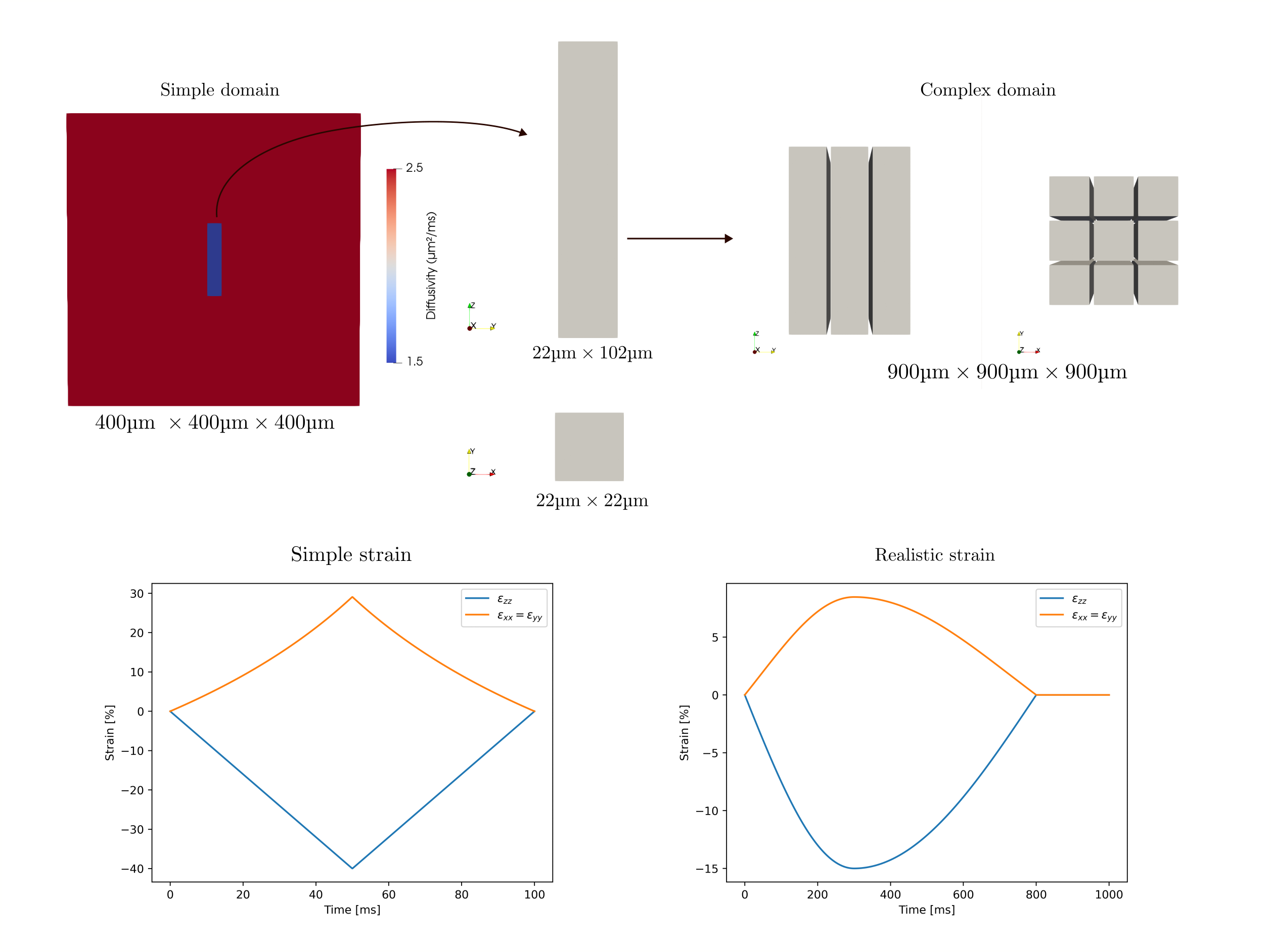
MCRW simulate the self-diffusion of water molecules by updating the particle positions through: $$$X(t + \Delta t)=X(t)+\sqrt{2D\Delta t}\vec{R}$$$, with $$$\vec{R}$$$ permitting unit movements with equal probability in any direction. In a strained medium, the deformation gradient tensor is represented as $$$\boldsymbol{F}_{iI}=\frac{\partial x_i}{\partial X_I}$$$ where $$$\boldsymbol{F}_{iI}$$$ denotes the components of the deformation gradient tensor, $$$X_I$$$ are the reference coordinates in the undeformed state, and $$$x_i$$$ are the corresponding deformed coordinates in the current configuration. For small deformations $$$\boldsymbol{F}(t)$$$ is well approximated by $$$F_{iI}(t)\approx\mathbf{I}+\boldsymbol{\epsilon}_{iI}(t)$$$, where $$$\mathbf{I}$$$ is the identity matrix and $$$\boldsymbol{\epsilon}_{iI}$$$ is the engineering strain tensor with components $$$\epsilon_{iI} = \frac{\delta_i}{L_I}$$$ where $$$\delta_i$$$ denotes the change in dimension along the axis $$$i$$$, for initial length $$$L_I$$$, along axis $$$I$$$. Conservation of volume implies $$$\det(\boldsymbol{F})=1$$$. Strain is included in the MCRW simulation displacing the geometry and the particle positions at each timestep where the displacement vector ( $$$\vec{\delta{\epsilon_i}}$$$) is given by $$$\vec{\delta{\epsilon_i}}=\left(\boldsymbol{\epsilon}(t + \Delta t)-\boldsymbol{\epsilon}(t)\right) \boldsymbol{F}^{-1}(t)\vec{X}(t)$$$.This study examines strain in two domains illustrated in figure 1: a single-cell with semi-permeable membranes for validation and a larger 3x3 cell array. Cardiomyocytes are modeled as 3D prisms, with ample extracellular space to prevent boundary effects. The single-cell domain has a higher membrane permeability of $$$0.5\text{µm}/\text{ms}$$$ and undergoes intense, brief strain (shown in figure 1, peak strain $$$-40\%$$$ in $$$z$$$ over $$$T=100\text{ms}$$$), while the multicellular domain uses a more realistic permeability of $$$0.025\text{µm}/\text{ms}$$$, with a more realistic strain ($$$T=1000\text{ms}$$$, peak strain in $$$z=-15\%$$$). Validation of the MCRW is performed via comparison with finite volume (FV) and an analytical solution (for free diffusion only). We model cyclical strain between short pulsed diffusion gradients, as would be present for STEAM DT-CMR in vivo.
Results
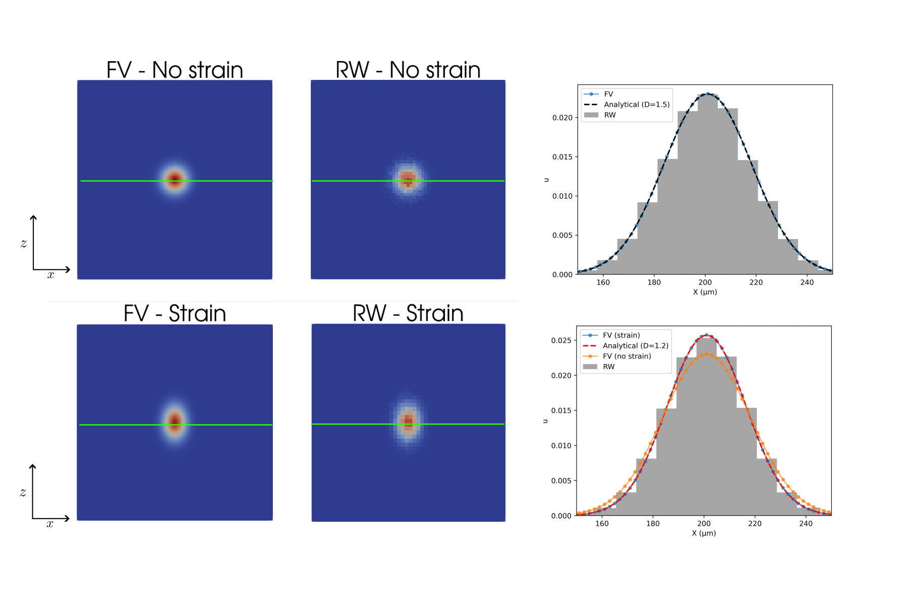
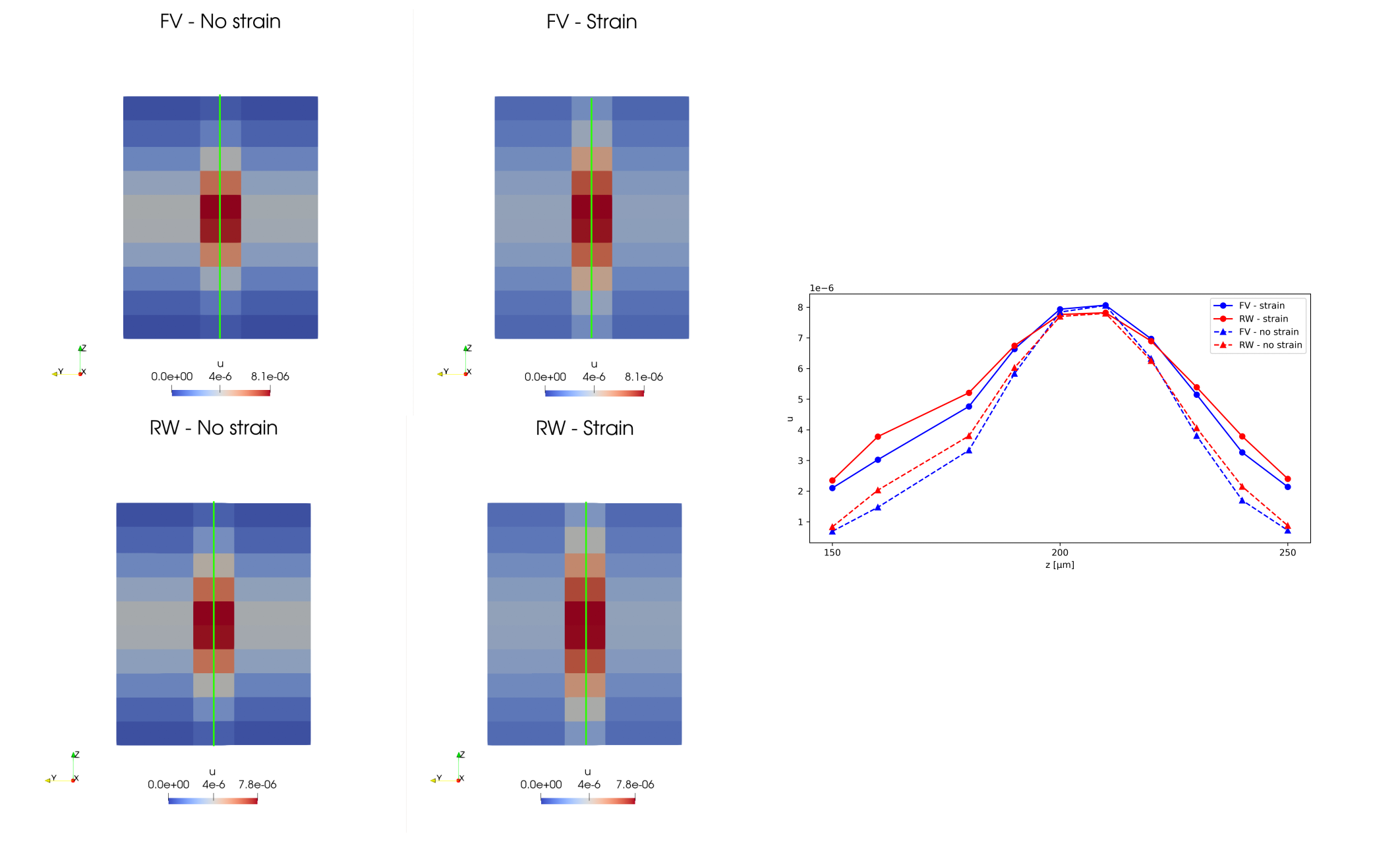
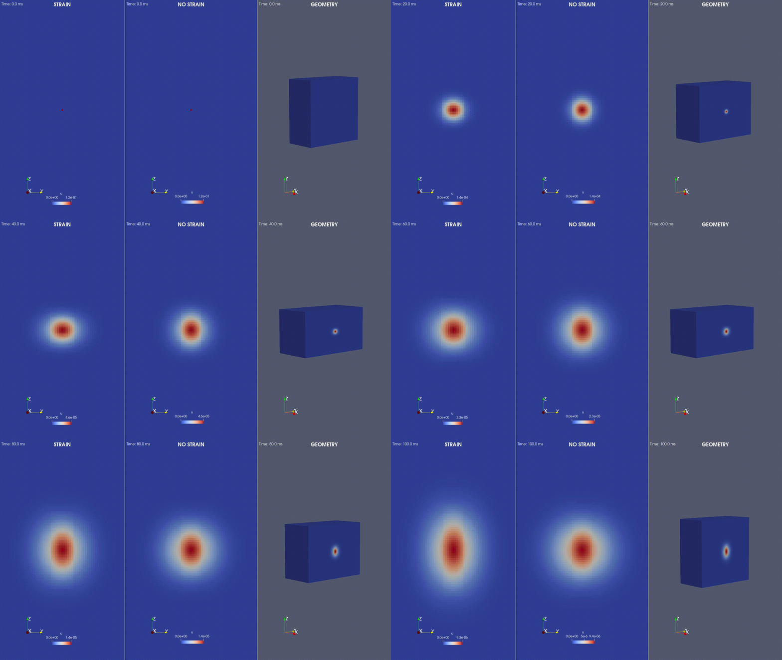
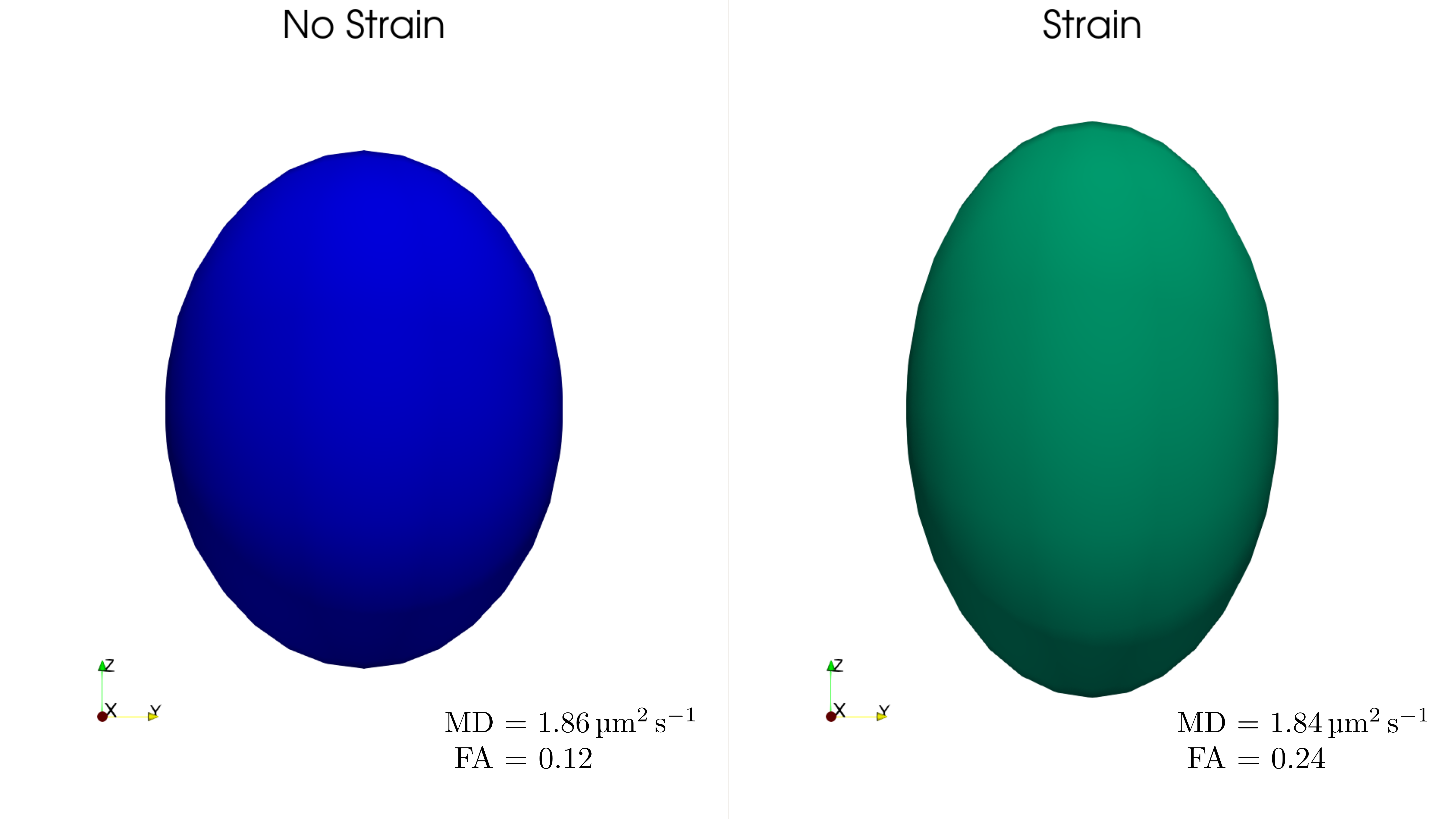
Figure 2 illustrates the impact of mechanical strain on the validation domain during free diffusion for $$$T=100\text{ms}$$$. Figure 3 and 4 elucidate the influence of strain on a complex domain characterised by semi-permeable membranes, where diffusion deviates from Gaussian behaviour. Figure 5 provides a visualization of the diffusion tensors and tensor parameters acquired within the large domain during a STEAM sequence.Discussion
The results obtained in free diffusion align with the model described by Reese et al.4,5 where $$$\boldsymbol{D}_{\text{strain}}=\frac{1}{T} \int_0^T{\boldsymbol{F}^{-1} \boldsymbol{D}_{\text{no strain}}\boldsymbol{F}^{-1} dt}$$$. For the validation case along the $$$x$$$ axis, $$$D_{\text{strain}}=0.8D_{\text{no strain}}=1.2\text{µm}^2/\text{ms}$$$. Stretching the domain during the diffusion time diminishes the measured diffusion while compressing it amplifies it, despite the fact that the geometry returns to its initial state at the end of the diffusion time. Permeability restricts particle mobility, more so along the $$$x$$$ and $$$y$$$ axis, where the narrower geometry of the cardiomyocyte results in greater restriction of the diffusing water molecule. Consequently, as depicted in figure 4, the strained simulations in the large domain reveal a substantial alteration due to strain along the cardiomyocyte long axis ($$$\lambda_1$$$) compared with the perpendicular directions $$$\lambda_2$$$, $$$\lambda_3$$$ increasing the overall fractional anisotropy of the tensor while maintaining a similar mean diffusivity.Conclusion
We have developed and validated a novel model that integrates bulk cyclical strain into the conventional MCRW validating it within a complex domain by comparison with a FV solution. The results for the larger domain reveal substantial alterations in the measured anisotropy of the diffusion tensor when incorporating strain and permeability over extended diffusion times. However, further work will need to investigate imaging in the contracted state (e.g. systole) and the effects of microstructural rearrangement (e.g. due to sheetlets) during the diffusion time6. This novel MCRW model will provide previously unobtainable quantification of any possible strain related systematic bias in DT-CMR measures obtained with STEAM, which will be important when analysing data from patient cohorts with pathological changes in myocardial strain and microstructure.Acknowledgements
This work was supported by the Engineering and Physical Sciences Research Council [grant number EP/X014010/1] and by British Heart Foundation (BHF) Grant/Award Number: RG/19/1/34160References
[1] - Ignasi Alemany, Jan N Rose, Pedro Ferriera, Dudley Pennell, Nielles-Vallespin Sonia, Andrew D. Scott, and Denis J. Doorly. Realistic numerical simulations of diffusion tensor CMR: The effects of perfusion and membrane permeability. 2023.
[2] - Jan N. Rose, Sonia Nielles-Vallespin, Pedro F. Ferreira, David N. Firmin,Andrew D. Scott, and Denis J. Doorly. Novel insights into in-vivo diffusion tensor cardiovascular magnetic resonance using computational modeling and histology-based virtual microstructure. Magnetic Resonance in Medicine,81(4), 2019. ISSN 15222594. doi: 10.1002/mrm.27561.
[3] - Mojtaba Lashgari, Nishant Ravikumar, Irvin Teh, Jing Rebecca Li, David L.Buckley, Jurgen E. Schneider, and Alejandro F. Frangi. Three-dimensional micro-structurally informed in silico myocardium—Towards virtual imaging trials in cardiac diffusion weighted MRI. Medical Image Analysis, 82, 112022. ISSN 13618423. doi: 10.1016/j.media.2022.102592.
[4] - Timothy G. Reese, Robert M. Weisskoff, R. Neil Smith, Bruce R. Rosen,Robert E. Dinsmore, and Van J. Wedeen. Imaging myocardial fiber architecture in vivo with magnetic resonance. Magnetic Resonance in Medicine, 34(6), 1995. ISSN 15222594. doi: 10.1002/mrm.1910340603.
[5] - Timothy G. Reese, J. Van Wedeen, and Robert M. Weisskoff. Measuring diffusion in the presence of material strain. Journal of Magnetic Resonance- Series B, 112(3), 1996. ISSN 10641866. doi: 10.1006/jmrb.1996.0139.6
[6] - Sonia Nielles-Vallespin, Zohya Khalique, Pedro F. Ferreira, Ranil de Silva,Andrew D. Scott, Philip Kilner, Laura Ann McGill, Archontis Giannakidis,Peter D. Gatehouse, Daniel Ennis, Eric Aliotta, Majid Al-Khalil, Peter Kell-man, Dumitru Mazilu, Robert S. Balaban, David N. Firmin, Andrew E.Arai, and Dudley J. Pennell. Assessment of Myocardial Microstructural Dynamics by In Vivo Diffusion Tensor Cardiac Magnetic Resonance. Journal of the American College of Cardiology, 69(6), 2017. ISSN 15583597. doi:10.1016/j.jacc.2016.11.051.6
Figures