0584
A modelling and experimental framework to investigate the sensitivity of steady-state diffusion MRI to microstructure1Wellcome Centre for Integrative Neuroimaging, FMRIB, Nuffield Department of Clinical Neurosciences, University of Oxford, Oxford, United Kingdom
Synopsis
Keywords: Microstructure, Diffusion/other diffusion imaging techniques, Diffusion acquisition
Motivation: Diffusion-weighted steady-state free precession (DW-SSFP) has demonstrated higher SNR-efficiency vs the diffusion-weighted spin-echo (DW-SE) in post-mortem tissue. However, its sensitivity to microstructural features has not been comprehensively investigated.
Goal(s): To develop an investigation framework to quantify DW-SSFP’s sensitivity to microstructure.
Approach: We combined Monte-Carlo simulations with phantom experiments incorporating diffusion hinderance/restriction and an ex-vivo mouse brain, comparing the estimated diffusion attenuation of DW-SSFP vs a DW-SE sequence with matched gradient waveforms and diffusion timings.
Results: DW-SSFP exhibited higher diffusion attenuation vs DW-SE in all tested substrates when gradient waveforms/timings are matched, suggesting its unique signal forming mechanisms may be highly sensitive to microstructure.
Impact: We present a framework combining Monte-Carlo simulations with experiments to characterise the sensitivity of diffusion-weighted steady-state free precession (DW-SSFP) to microstructure. DW-SSFP demonstrates greater signal attenuation versus a gradient waveform/timing-matched DW-SE across different substrates, demonstrating its potential for microstructural imaging.
Introduction
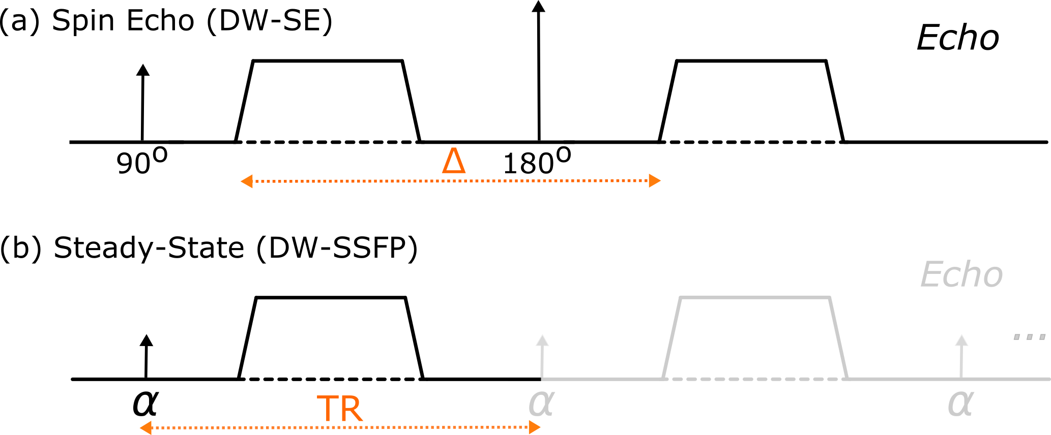
Diffusion-weighted steady-state free precession1 (DW-SSFP) (Figure 1) is an alternative diffusion imaging sequence that achieves high SNR-efficiency, strong diffusion weighting, and minimal image distortions. To date, DW-SSFP has primarily demonstrated its potential for post-mortem imaging2, achieving higher SNR-efficiency over the conventional diffusion-weighted spin-echo (DW-SE) sequence2,3. However, the sensitivity of DW-SSFP to microstructural features (e.g., restriction) has not been comprehensively investigated.The DW-SSFP sequence accumulates diffusion contrast over several TRs, with the measured signal combining different histories of magnetisation excitation and evolution. These signal-forming mechanisms lead to strong diffusion weighting that is expected to be highly sensitive to tissue microstructure. However, the complexity of these signal-forming mechanisms means the sequence is challenging to investigate analytically.
To address this, we are developing an investigation framework incorporating Monte-Carlo simulations4 to model DW-SSFP. Here, we use this framework to investigate the sensitivity of DW-SSFP to restriction, alongside experimental work in phantoms and an ex-vivo mouse brain. Our simulation findings reveal that the signal forming mechanisms of DW-SSFP achieve higher levels of signal attenuation for common restriction geometries vs DW-SE when considering matched gradient waveforms and diffusion timings. These findings are reflected experimentally, motivating future investigations to identify imaging regimes where the DW-SSFP sequence may achieve greater microstructural sensitivity vs an optimised DW-SE sequence.
Methods
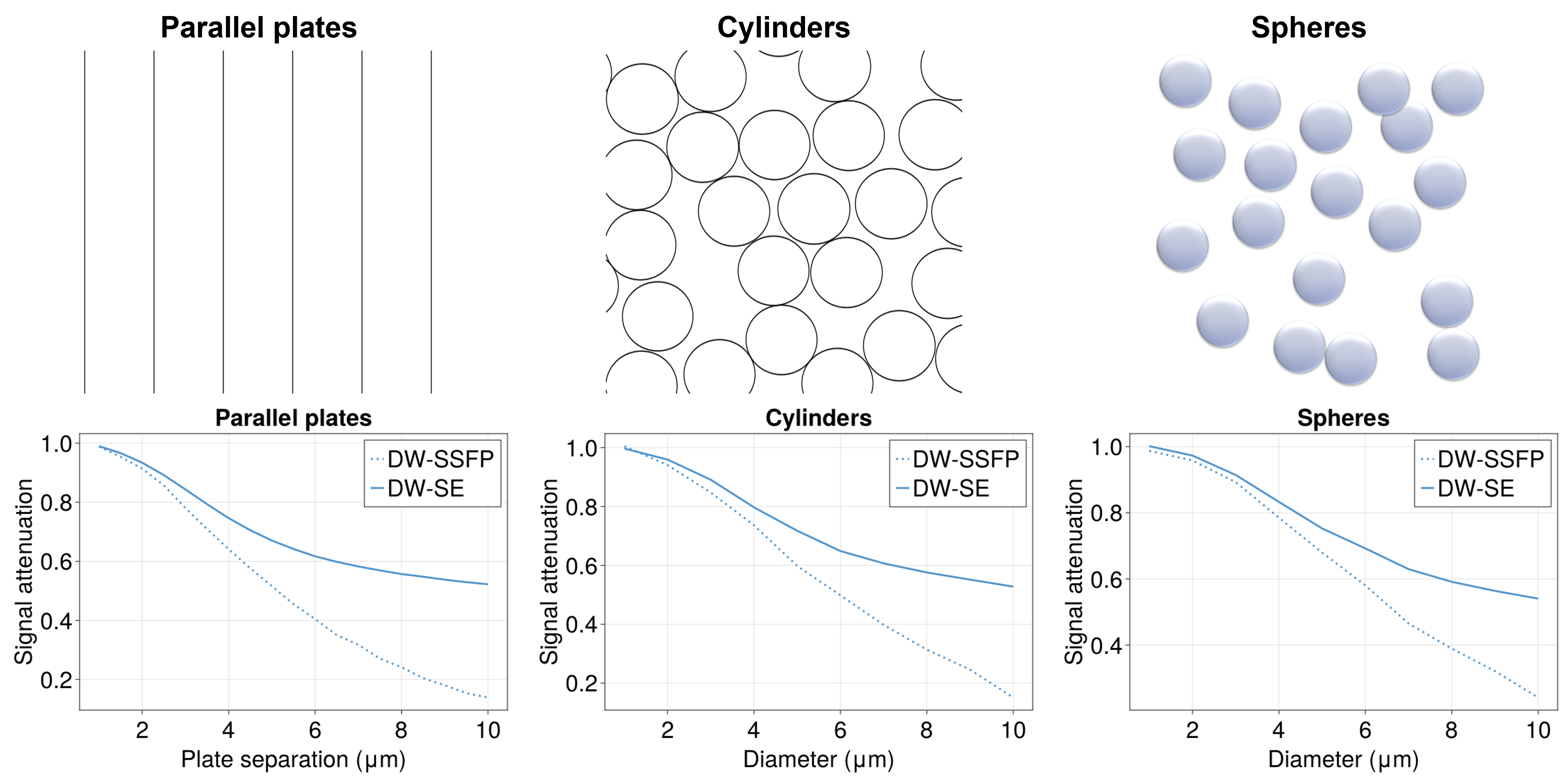
To investigate DW-SSFP’s sensitivity to restriction, Monte-Carlo simulations were performed using MCMRSimulator4 (v0.7). Three geometries were chosen for diffusion restriction: repeating equidistant parallel plates, randomly distributed identical cylinders and spheres (Figure 2). In all three cases, DW-SE and DW-SSFP signals from the restricted compartment were simulated for a single geometry size (1-10 µm). Sequences parameters and sample properties were matched to the ex-vivo experiments described below.For the experimental work, three samples were scanned using a 7T Bruker BioSpec preclinical scanner: A homogenous base liquid sample, a cell-mimicking microbead phantom consisting of spheres5 (<10 μm) which restrict and hinder diffusion, and an ex-vivo mouse brain. The samples were scanned using a DW-SSFP and DW-SE sequence with matched gradient waveforms and diffusion timings, where we consider the TR of the DW-SSFP sequence as a proxy for the DW-SE diffusion time Δ (Figure 1). Parameter optimisation was performed based on the DW-SSFP sequence, identifying the regime achieving optimal contrast-to-noise efficiency2,6 for estimated sample properties, assuming free diffusion. Analysis of experimental data was performed using FSL and in-house MATLAB scripts.
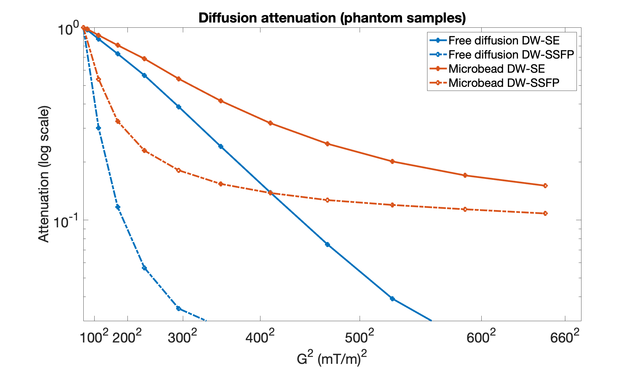
The liquid and microbead samples were scanned at 200 μm isotropic spatial resolution, with 11 measurements ranging from G = 58.74 to 646 mT/m, δ = 2 ms, and ΔDW-SE/TRDW-SSFP = 25 ms (fulfilling the 2π∙n DW-SSFP dephasing condition). For DW-SSFP, a = 28o and TE = 13.3 ms. For DW-SE, TE = 35 ms, TR = 5s. The gradient orientation was set to [0,0,1] for all scans.
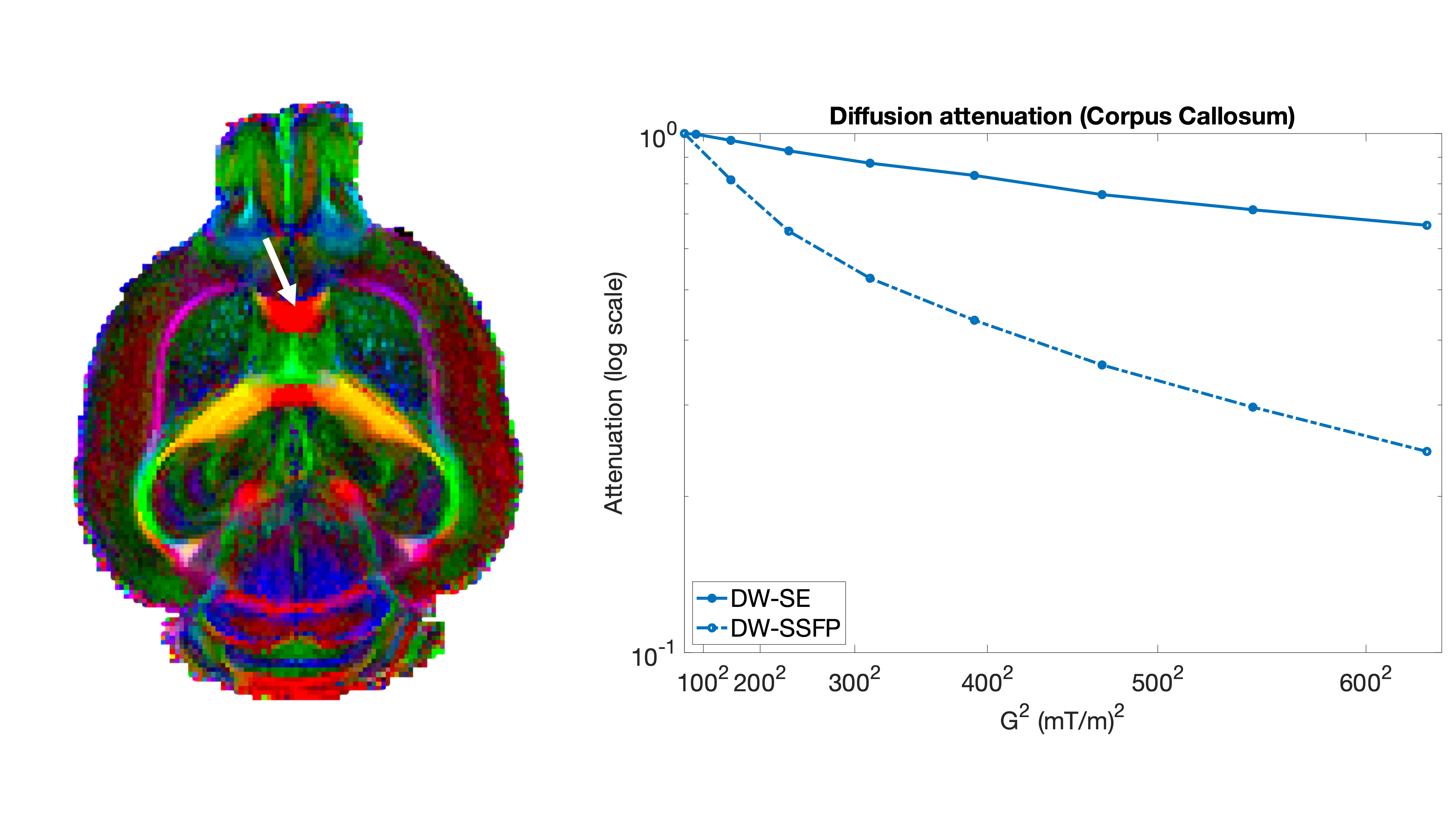
The ex-vivo mouse brain was scanned with 8 measurements ranging from G = 78.28 to 628.21 mT/m, δ = 3 ms, and ΔDW-SE/TRDW-SSFP = 20 ms. For DW-SSFP, a = 23o, TE = 11.2 ms and resolution = 100 μm isotropic. For DW-SE, TE = 35 ms, TR = 2.86s, in-plane resolution = 100x100 μm2 and slice thickness = 200 μm. The gradient orientation was set perpendicular to the corpus callosum along the ventral-dorsal axis. We additionally acquired multi-orientation DW-SSFP data (30 directions; G = 313.12 mT/m) to estimate a diffusion tensor.
Results
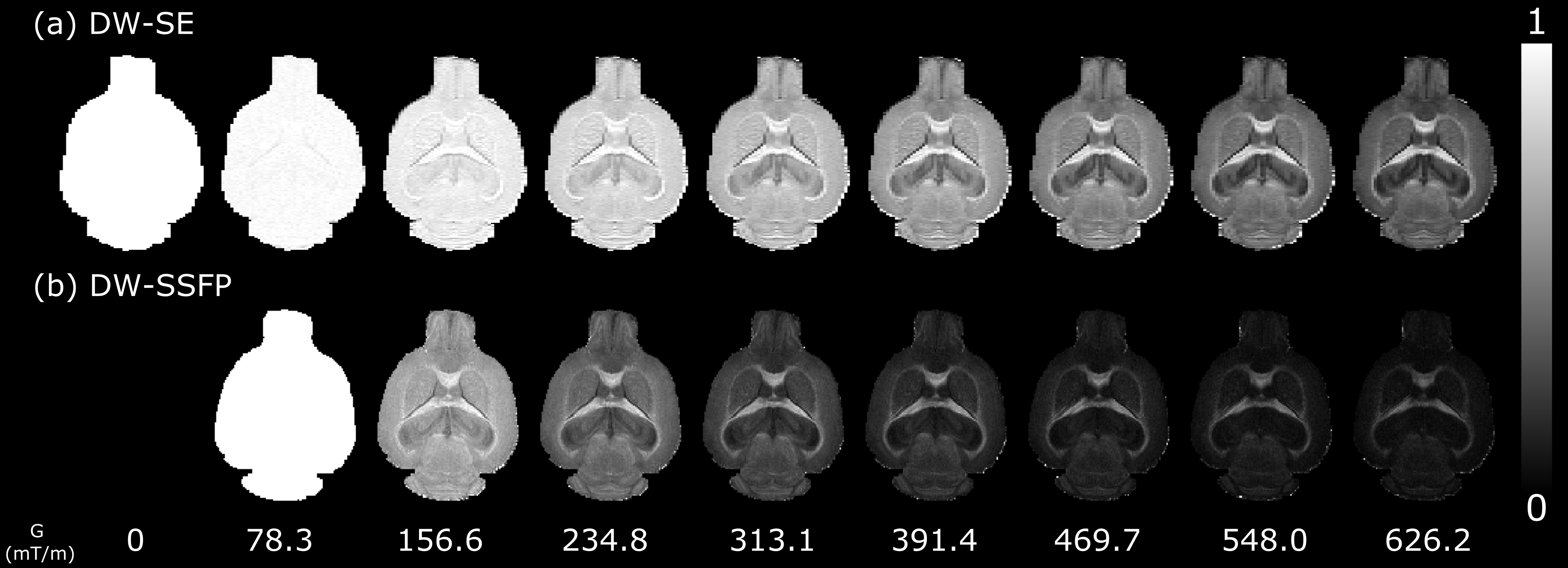
Figure 2 reveals that DW-SSFP achieves higher diffusion attenuation vs DW-SE for all three simulated restriction geometries. This finding agrees with experiment results in both phantoms (Figure 3) and the ex-vivo mouse corpus callosum (Figure 4). In the free diffusion sample, we estimated D = 2.03/1.99 μm/ms2 with DW-SSFP/DW-SE, demonstrating our DW-SSFP implementation can accurately estimate diffusivities. Figure 5 visualises the estimated signal attenuation over the ex-vivo mouse brain for the gradient/timings-matched DW-SE and DW-SSFP sequence as a function of gradient strength. The DW-SSFP sequence achieves considerably higher attenuation across the brain, with preserved signal limited to select white matter tracts at high G.Discussion
Our results show that DW-SSFP achieves higher diffusion attenuation vs gradient waveform/timings-matched DW-SE in a wide range of simulated and physical substrates. The attenuation difference suggests that DW-SSFP’s unique signal forming mechanism may confer additional sensitivity to microstructural features like restriction, with higher order signal-forming pathways providing complementary microstructural information.Our future work aims to leverage our findings from both our simulation and experimental framework, using Monte Carlo simulations to identify optimal parameter regimes to maximise microstructural sensitivity for both the DW-SSFP and DW-SE sequence, and using Monte-Carlo simulations to directly estimate the underlying microstructure from experimental diffusion MRI measurements.
Acknowledgements
ZZ is supported by University of Oxford and China Scholarship Council. MT is supported by core funding from the Wellcome Trust (203139/Z/16/Z and 203139/A/16/Z). KLM is supported by a Wellcome Trust Senior Research Fellowship (224573/Z/21/Z). MC is supported by a Wellcome Trust Collaborative Award (215573/Z/19/Z). BCT is supported by a Wellcome Trust Sir Henry Wellcome Fellowship (222829/Z/21/Z). The Wellcome Centre for Integrative Neuroimaging is supported by core funding from the Wellcome Trust (203139/Z/16/Z).
References
1. Merboldt, K.-D., Hxnicke, W., Gyngell, M. L., Frahm, J. & Bruhn, H. Rapid NMR imaging of molecular self-diffusion using a modified CE-FAST sequence. J. Magn. Reson. 1969 82, 115–121 (1989).
2. Miller, K. L., McNab, J. A., Jbabdi, S. & Douaud, G. Diffusion tractography of post-mortem human brains: Optimization and comparison of spin echo and steady-state free precession techniques. NeuroImage 59, 2284–2297 (2012).
3. McNab, J. A. & Miller, K. L. Sensitivity of diffusion weighted steady state free precession to anisotropic diffusion. Magn. Reson. Med. 60, 405–413 (2008).
4. Cottaar, M. MCMRSimulator.jl. (2022) doi:10.5281/zenodo.7318656.
5. McHugh, D. J. et al. A biomimetic tumor tissue phantom for validating diffusion‐weighted MRI measurements. Magn. Reson. Med. 80, 147–158 (2018).
6. Tendler, B. C. et al. Use of multi-flip angle measurements to account for transmit inhomogeneity and non-Gaussian diffusion in DW-SSFP. NeuroImage 220, 117113 (2020).
Figures