0580
Robust motion- and $$$\delta B_0$$$ -correction for high-resolution QSM at 7T1Biomedical Engineering Department, School of Biomedical Engineering and Imaging Sciences, King's College London, London, United Kingdom, 2Centre for the Developing Brain, School of Biomedical Engineering and Imaging Sciences, King's College London, London, United Kingdom, 3London Collaborative Ultra high field System (LoCUS), London, United Kingdom, 4Department for Forensic and Neurodevelopmental Sciences, Institute of Psychiatry, Psychology and Neuroscience, King's College London, London, United Kingdom, 5Biomedical Image Technologies, Universidad Politécnica de Madrid and CIBER-BNN, Madrid, Spain, 6Siemens Healthcare Limited, Frimley, United Kingdom, 7Guys and St Thomas’ NHS Foundation Trust, King's College London, London, United Kingdom, 8Department of Radiology, Stanford University, Palo Alto, CA, United States, 9Department of Electrical Engineering, Stanford University, Palo Alto, CA, United States
Synopsis
Keywords: Signal Modeling, Susceptibility
Motivation: Quantitative susceptibility mapping (QSM) provides valuable clinical information and is widely used, especially at ultra-high field (7T). Due to long echo times, QSM acquisitions are extra sensitive to the changes in B0 ($$$\delta\textbf{B}_0$$$), such as those secondary to motion.
Goal(s): To provide purely data-driven motion and $$$\delta\textbf{B}_0$$$ correction for QSM.
Approach: We use the self-navigated DISORDER k-space re-ordering, originally proposed for motion correction, to additionally estimate $$$\delta\textbf{B}_0$$$. Within-scan motion and $$$\delta\textbf{B}_0$$$ are then retrospectively corrected during image reconstruction.
Results: We show improved reconstruction in all 5 scanned volunteers when additionally correcting $$$\delta\textbf{B}_0$$$. This directly improves QSM.
Impact: The proposed method can result in improved image quality when scanning in presence of motion and , e.g. due to heavy breathing. Combining this approach with an optimized QSM protocol will provide motion- and -robust QSM.
Introduction
Quantitative susceptibility mapping (QSM) estimates local magnetic susceptibility ($$$\chi$$$) using the MRI-signal phase1, with clinical applications studying imaging biomarkers, brain development, and neurological diseases2-5. For high-resolution QSM, of special interest at 7T, long scan times make the sequence sensitive to motion6. Additionally, due to the long echo times (TE), QSM acquisitions are very sensitive to changes in the polarizing magnetic field ($$$\delta\textbf{B}_0$$$), which becomes more problematic at higher field strength (7T)7,8. We recently proposed a method to estimate both motion and $$$\delta\textbf{B}_0$$$ from navigators by appropriately designing the k-space trajectory and by using a robust optimizer9. In this work, we deploy this method to the self-navigated DISORDER trajectory10 to obtain within-scan $$$\delta\textbf{B}_0$$$ estimates in addition to the usually obtained motion estimates. Both motion and $$$\delta\textbf{B}_0$$$ are then retrospectively corrected during image reconstruction.Methods
AlignedSENSE:The alignedSENSE11 performs motion correction by dividing the k-space acquisition into temporal groups (shots) of readouts (each acquired per repetition time TR), where each shot $$$n$$$ has a different motion state with rigid motion parameters $$$\textbf{z}_n$$$, which can be jointly optimized together with the image:$$(\hat{\mathbf{x}},\hat{\mathbf{z}}_n)=argmin_{\textbf{x},\textbf{z}_n }\sum_{n}{||\textbf{A}_n\textbf{F}\textbf{ST}(\textbf{z}_n))\textbf{x}-\textbf{y}_n||^2_2}\ \ \ \ \ \ \ \ \ \ \ \ (1)$$where $$$\textbf{T,S,F,A}_n$$$ and $$$\textbf{y}_n$$$ respectively represent rigid motion, coil sensitivities, Fourier operator, sampling structure, and measured multi-coil k-space data. Using the DISORDER phase encoding scheme, which samples uniformly across k-space per shot, allows robust motion estimation and image reconstruction.
$$$\delta\textbf{B}_0$$$-informed alignedSENSE:
Assuming small or moderate motion levels, lower order $$$\delta\textbf{B}_0$$$ explains most of the B0 variations7,8. Therefore, we model $$$\delta\textbf{B}_0$$$ using 2nd-order solid harmonics with basis $$$\textbf{L}$$$ and coefficients $$$\textbf{c}$$$:$$$\delta\textbf{B}_0=\textbf{L}\textbf{c}$$$. For spoiled sequences, this model can be included in the alignedSENSE forward model12:$$\textbf{y}_n=\textbf{A}_n\textbf{F}\textbf{ST}(\textbf{z}_n)\textbf{P}( \textbf{c}_n)\textbf{x}\ \ \ \ \ \ \ \ \ \ \ \ (2)$$where $$$\textbf{P}(\textbf{c}_n)$$$ is the induced phase $$$e^{i2\pi\textbf{Lc}_nTE}$$$ for shot $$$n$$$. Image, motion, and $$$\textbf{c}_n$$$ are jointly estimated by performing an alternating optimization:$$(\hat{\mathbf{x}},\hat{\mathbf{z}}_n,\hat{\mathbf{c}}_n)=argmin_{\textbf{x},\textbf{z}_n,\textbf{c}_n}\sum_{n}{||\textbf{A}_n\textbf{F}\textbf{ST}(\textbf{z}_n)\textbf{P}( \textbf{c}_n)\textbf{x}-\textbf{y}_n||^2_2}\ \ \ \ \ \ \ \ \ \ \ \ (3)$$where $$$\textbf{z}_n$$$ and $$$\textbf{c}_n$$$ are estimated using the Levenberg-Marquardt algorithm9,11.
In-vivo data acquisition:
We used a single-echo GRE ($$$\Delta$$$=0.85x0.85x0.85mm3,TE/TR=20/31ms,FA=15°,FOV=224×224×157mm3,Head-Foot frequency encoding, acquisition time (TA)=11min) modified to allow for robust motion and $$$\delta\textbf{B}_0$$$ estimation: First, the DISORDER trajectory was used. Next, a high bandwidth (800Hz/voxel) was set to suppress differential distortion from varying $$$\delta\textbf{B}_0$$$. A moderate acceleration R=1.5x1.4 was used following previous recommendations10. 5 healthy volunteers (HV) were scanned at 7T (MAGNETOM Terra, Siemens Healthcare, Erlangen, Germany), where the HV was instructed to either be relaxed or to perform heavy breathing to induce temporal $$$\delta\textbf{B}_0$$$. Coil sensitivities were estimated from a reference scan (TA=18sec) using ESPIRiT13.
Image reconstruction:
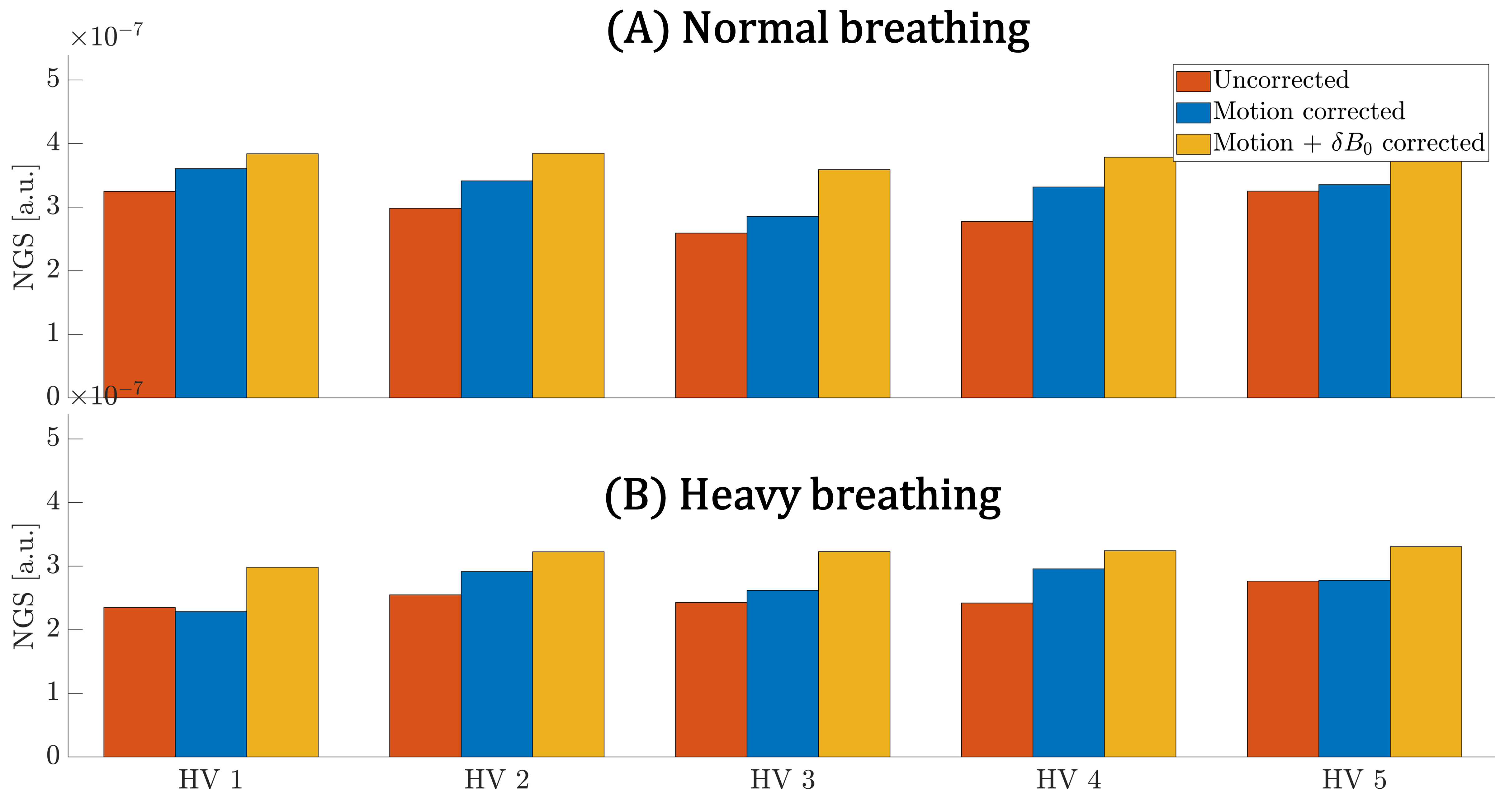
Acquired data was reconstructed without correction (SENSE14), with only motion correction (alignedSENSE), and with the proposed motion+$$$\delta\textbf{B}_0$$$ correction. Since no ground truth (GT) is available, image quality is quantified using the normalized gradient squared (NGS)15.
QSM processing:
Susceptibility maps were generated from the reconstructed image phase using the SEPIA toolbox16 with the following options: SEGUE phase unwrapping17, PDF background field removal18, and the threshold-based k-space division for $$$\chi$$$-computing19.
Results and discussion
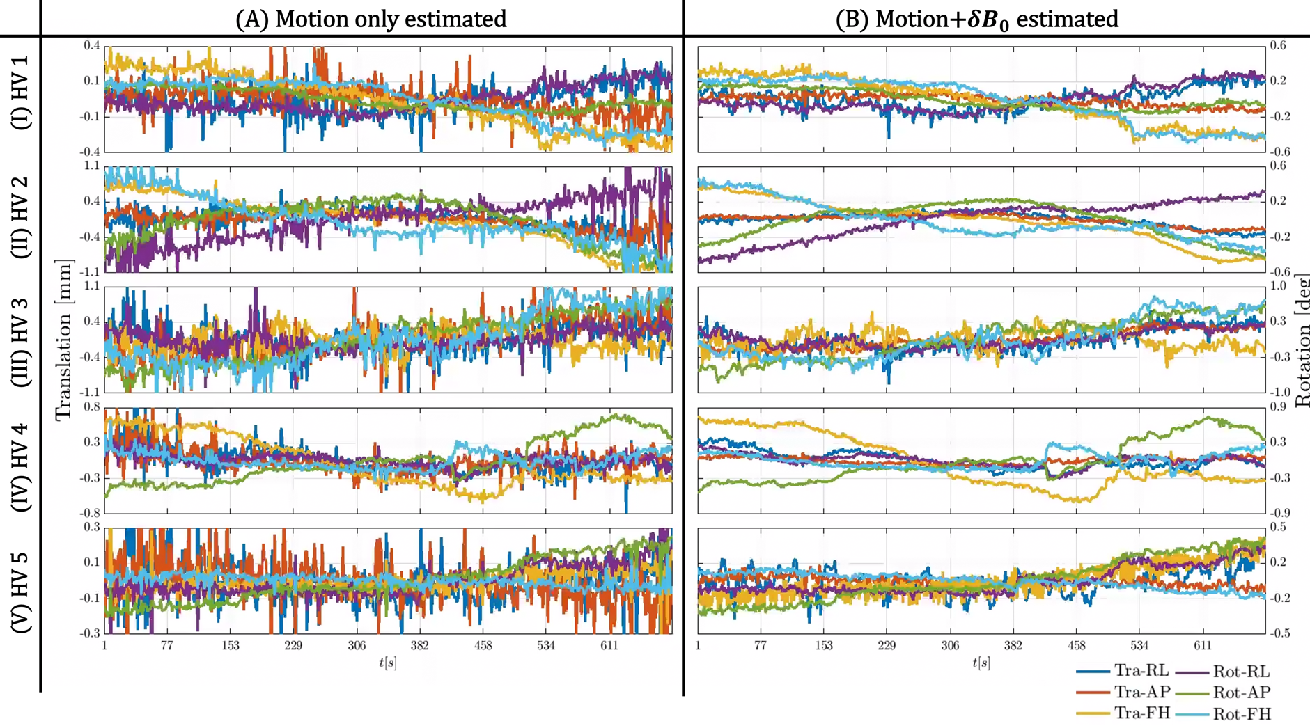
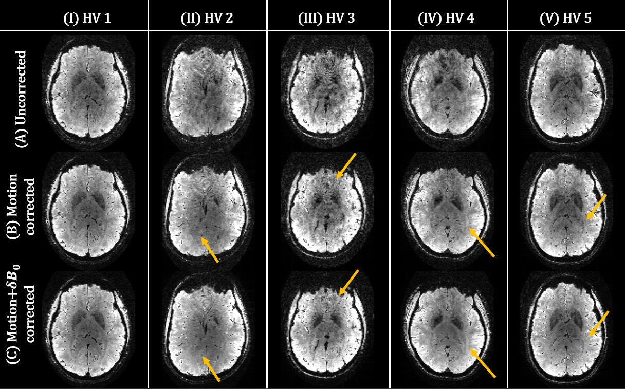
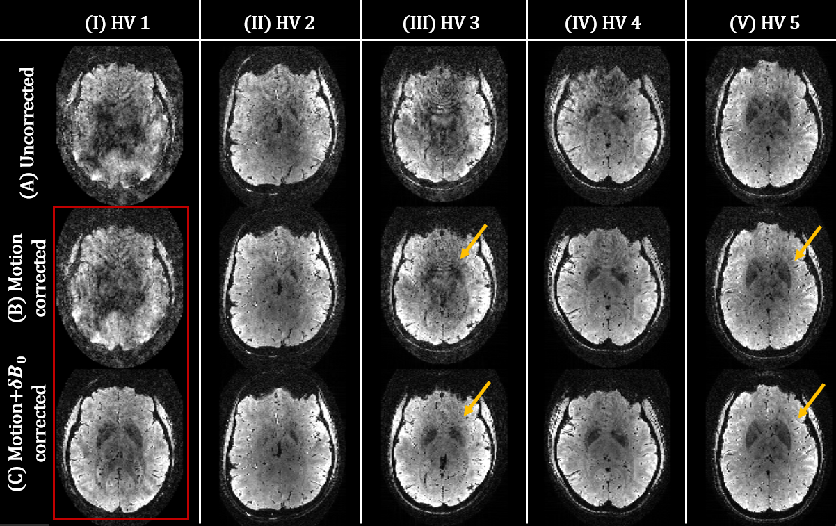
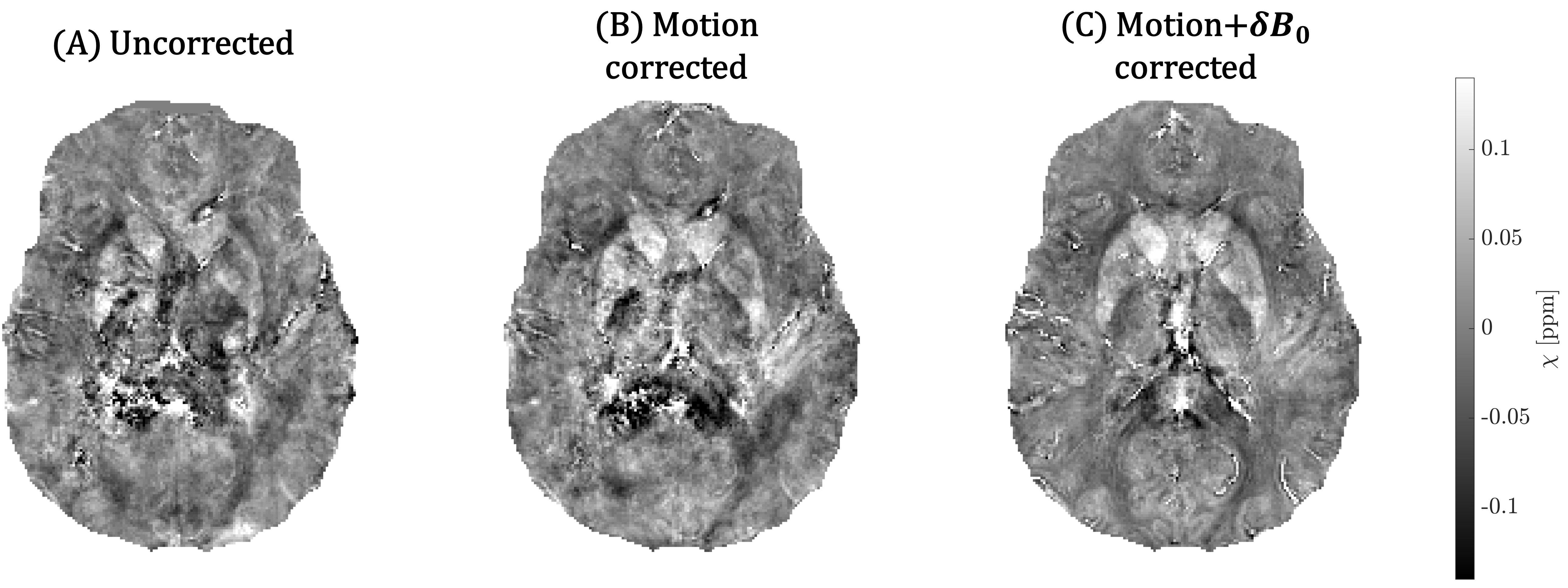
Figure 1 shows the motion traces for all 5 HVs when estimating motion only (A) and motion+$$$\delta\textbf{B}_0$$$ (B). The estimated motion traces became much more orderly when additionally modeling and estimating $$$\delta\textbf{B}_0$$$. The corresponding reconstructed images are shown in Figure 2 for all correction methods (rows) and HV (columns). Incremental improvements in image quality compared to the uncorrected case (A) are observed by estimating motion (B) and motion+$$$\delta\textbf{B}_0$$$ (C) (yellow arrows indicate areas of notable improved quality). Figure 3 shows the same figure for the “heavy breathing” experiment, where the same observations hold. A case of substantial improvement with the proposed method is indicated in red. A quantitative evaluation of the image quality is presented in Figure 4. In all cases, the proposed method results in higher quality scores. Finally, Figure 5 shows a pilot QSM reconstruction, performed on the “heavy breathing” acquisition in HV1. Consistent with the reconstructed image, substantial improvements are obtained by using the proposed reconstruction. Note that the acquisition was suboptimal for QSM processing and better QSM maps are expected with protocol parameters that balance the ability to correct motion/$$$\delta\textbf{B}_0$$$ as well as to perform QSM20.Conclusion
We have integrated a method to estimate motion and $$$\delta\textbf{B}_0$$$ into a retrospective motion+$$$\delta\textbf{B}_0$$$ corrected reconstruction, especially of interest in acquisitions with long TE. Robust performance was achieved using the self-navigated DISORDER trajectory. We show improved motion traces and image reconstruction in all in-vivo acquisitions, both qualitatively and quantitatively. A pilot QSM reconstruction shows the potential to use this method for QSM at 7T.Acknowledgements
This work was funded by the King’s College London & Imperial College London EPSRC Centre for Doctoral Training in Medical Imaging [EP/S022104/1], by core funding from the Wellcome/EPSRC Centre for Medical Engineering [WT203148/Z/16/Z], the Wellcome Trust Collaboration in Science grant [WT201526/Z/16/Z] and by the National Institute for Health Research (NIHR) Biomedical Research Centre based at Guy’s and St Thomas’ NHS Foundation Trust and King’s College London and/or the NIHR Clinical Research Facility. The views expressed are those of the author(s) and not necessarily those of the NHS, the NIHR or the Department of Health and Social Care.References
[1] Shmueli K, Zwart JA de, Gelderen P van, Li TQ, Dodd SJ, Duyn JH. Magnetic susceptibility mapping of brain tissue in vivo using MRI phase data. Magnetic Resonance in Medicine. 2009;62(6):1510-1522.
[2] Gillen, Kelly McCabe et al. “QSM is an imaging biomarker for chronic glial activation in multiple sclerosis lesions.” Annals of Clinical and Translational Neurology 8 (2021): 877 - 886.
[3] Uchida, Yuto et al. “Quantitative susceptibility mapping as an imaging biomarker for Alzheimer’s disease: The expectations and limitations.” Frontiers in Neuroscience 16 (2022).
[4] Lorio, Sara et al. “Quantitative MRI susceptibility mapping reveals cortical signatures of changes in iron, calcium and zinc in malformations of cortical development in children with drug-resistant epilepsy.” Neuroimage 238 (2020).
[5] Langkammer, Christian et al. “Quantitative Susceptibility Mapping in Parkinson's Disease.” PLoS ONE 11 (2016).
[6] Zaitsev M, Maclaren JR, Herbst M. “Motion artifacts in MRI: A complex problem with many partial solutions.” Journal of Magnetic Resonance Imaging (2015): 42(4):887-901.
[7] Wallace TE, Afacan O, Kober T, Warfield SK. “Rapid measurement and correction of spatiotemporal B0 field changes using FID navigators and a multi-channel reference image.” Magnetic Resonance in Medicine (2020): 83(2):575-589.
[8] Van de Moortele PF, Pfeuffer J, Glover GH, Ugurbil K, Hu X. Respiration-induced B0 fluctuations and their spatial distribution in the human brain at 7 Tesla. Magnetic Resonance in Medicine (2002): 47(5):888-95.
[9] Brackenier Y, Wang Nan, Liao Congyu, et al. Towards rapid and accurate navigators for motion and B0 estimation using QUEEN (QUantitatively-Enhanced parameter Estimation from Navigators). In: Proc Int Soc Mag Reson Med 23.
[10] Cordero-Grande L et al. Motion-corrected MRI with DISORDER: Distributed and incoherent sample orders for reconstruction deblurring using encoding redundancy. Magn Reson Med. 2020;84(2):713-726.
[11] Cordero-Grande, Lucilio, Rui Pedro A. G. Teixeira, Emer J. Hughes, Jana Hutter, Anthony N. Price and Joseph V. Hajnal. “Sensitivity Encoding for Aligned Multishot Magnetic Resonance Reconstruction.” IEEE Transactions on Computational Imaging 2 (2016): 266-280.
[12] Brackenier, Yannick et al. “Data‐driven motion‐corrected brain MRI incorporating pose‐dependent B0 fields.” Magnetic Resonance in Medicine 88 (2022): 817 - 831.
[13] Uecker, Martin et al. “ESPIRiT—an eigenvalue approach to autocalibrating parallel MRI: Where SENSE meets GRAPPA.” Magnetic Resonance in Medicine 71 (2014).
[14] Pruessmann, Klaas Paul et al. “SENSE: Sensitivity encoding for fast MRI.” Magnetic Resonance in Medicine 42 (1999).
[15] McGee, Kiaran P. et al. “Image metric‐based correction (Autocorrection) of motion effects: Analysis of image metrics.” Journal of Magnetic Resonance Imaging 11 (2000).
[16] Chan, Kwok-Shing and José P. Marques. “SEPIA—Susceptibility mapping pipeline tool for phase images.” NeuroImage 227 (2020).
[17] Karsa, Anita and Karin Shmueli. “SEGUE: A Speedy rEgion-Growing Algorithm for Unwrapping Estimated Phase.” IEEE Transactions on Medical Imaging 38 (2019): 1347-1357.
[18] Liu, Tian et al. “A novel background field removal method for MRI using projection onto dipole fields (PDF).” NMR in Biomedicine 24 (2011).
[19] Wharton, Sam J. et al. “Susceptibility mapping in the human brain using threshold‐based k‐space division.” Magnetic Resonance in Medicine 63 (2010).
[20] Rua, Catarina et al. “Multi-centre, multi-vendor reproducibility of 7T QSM and R2* in the human brain: Results from the UK7T study.” Neuroimage 223 (2020)
Figures