0578
2D T1, T2, T2* and PDFF mapping in the kidney with rosette MRF using Hermitian low-rank and dictionary-patch based regularization1Radiology, University of Michigan, Ann Arbor, MI, United States, 2Biomedical Engineering, University of Michigan, Ann Arbor, MI, United States
Synopsis
Keywords: MR Fingerprinting, Kidney, MRF; low-rank;
Motivation: Subjective qualitative T2-weighted, T1-weighted (with and without contrast) and fat suppressed images are currently used to characterize renal masses. Characterization could be improved and standardized by using objective, generalizable, quantiative criteria.
Goal(s): In this work, 2D single-breathhold, high-resolution T1/T2/T2*/PDFF mapping rosette MRF is deployed for kidney tissue characterization.
Approach: A novel MRF reconstruction is introduced to enable reduced MRF data collection time, incorporating separate low-rank models along the TE and TR domains, Hermitian symmetry via virtual coils, and a dictionary-patch based regularization.
Results: In vivo results in healthy subjects demonstrate 2D 1x1x5 mm3 T1/T2/T2*/PDFF MRF kidney mapping in a single breath-hold.
Impact: Simultaneous mapping of T1/T2/T2*/PDFF in the kidney in a single high-resolution breath-hold scan via the proposed MRF approach is feasible. This technique could bolster traditional pre-/post- contrast renal mass protocols with objective characterization methods.
INTRODUCTION:
MR Fingerprinting (MRF)1 employs highly undersampled acquisitions to sample transient state signals and provide high-quality tissue property maps. Advanced MRF reconstructions can minimize residual aliasing and noise amplification. Global low-rank (LR) models2,3,4 and regularization approaches based on compressed sensing9, dictionary-based4,11, locally low-rank10 and patch-based methods12,13 have been demonstrated for this purpose, improving map quality. Hermitian symmetry5 has been proposed for steady-state imaging via PFPP6, LORAKS7 and Virtual Coil Concept (VCC).8 Here, we propose to enable high resolution T1/T2/T2*/Proton Density Fat (PDFF) kidney mapping within a breath-hold by incorporating Hermitian symmetry via VCC, and combining a global low-rank model along the TR domain, with a pixel-wise low-rank model along the TE domain. Furthermore, we develop a novel regularizer that combines patch-based locally low-rank with prior knowledge of the MR signal model, named LRH-DP. These improvements enable 2D T1/T2/T2*/PDFF mapping in the kidneys with a resolution of 1x1x5 mm3 and improved apparent precision relative to conventional mapping methods.METHODS:
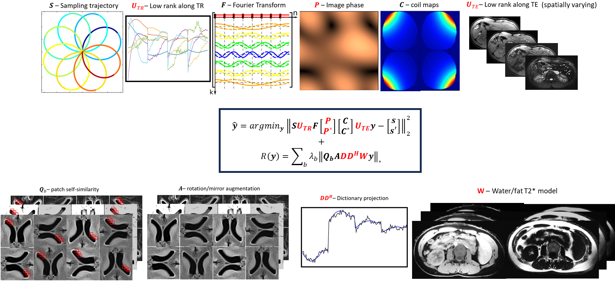
The proposed approach combines low-rank Hermitian symmetry (LRH) in the forward model with a dictionary-patch (DP) based regularizer. The proposed LRH-DP reconstruction (Fig.1) is solved by the following optimization problem:$$\widehat{y}=argmin_{y}\left\|SU_{TR}F\binom{P}{P^{*}}\binom{C}{C^{*}}U_{TE}y\binom{s}{s'}\right\|_{2}^{2}+\sum_b\lambda_b\left\|Q_bADD^HWy\right\|_{*} $$
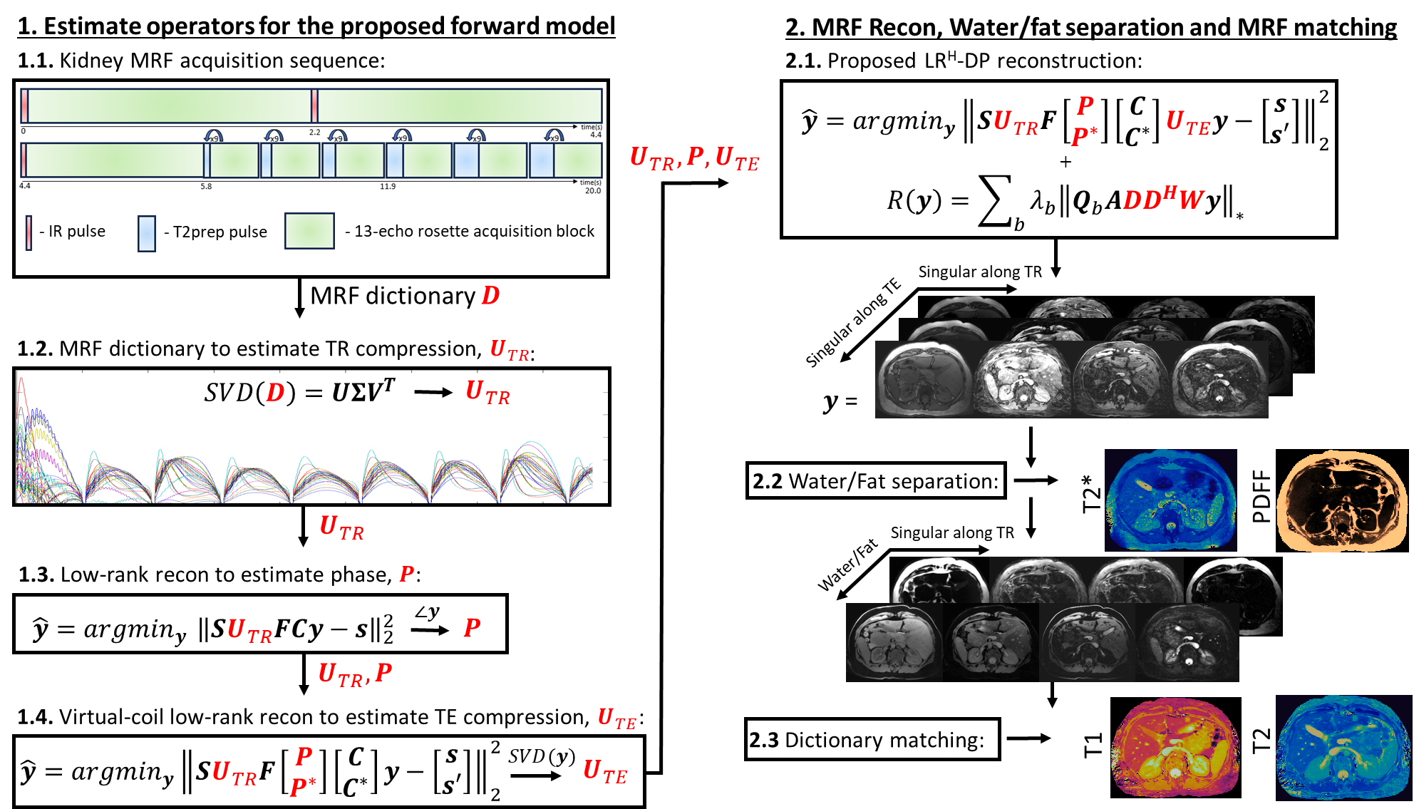
where S is the sampling trajectory, UTR is a global low-rank basis along the TR domain, F is the Fourier transform, P is the image phase of the MRF series, C are coil sensitivities, UTE is a pixel-wise low-rank basis along the TE domain, y are the so-called singular images, s are the acquired data, $$$s'(k)=s^*(-k)$$$ (where k denotes k-space coordinate), $$$\lambda_b$$$ is the regularization strength for a given pixel b, Qb identifies patches similar to the patch around b and sorts them in a Casorati matrix, A creates replicas of y mirrored and rotated about the center, D is the MRF dictionary capturing the dynamics along the TR, and W synthesizes images according to the water/fat/T2* model14 that captures dynamics along the TE. The operation DDH synthesizes an MRF series with the signal evolution along the TR (i.e. changes in T1/T2 primarily due to inversion and T2prep pulse), according to the best dictionary match. The operation within W performs water/fat separation and T2* mapping following a standard signal model and synthesizes an MRF series with the signal evolution along the TE (i.e. changes along water/fat precession and T2* due to the multi-echo Rosette readout) based on the estimated water, fat, B0, and T2* values. The operators in the proposed forward model are estimated via preliminary processes: UTR comes from the dictionary, P comes from a low-rank reconstruction and UTE comes from a virtual-coil low-rank reconstruction (Fig.2). Following this initial estimation, LRH-DP is performed, the reconstructed images undergo water/fat separation and T2* mapping via a graph-cut algorithm,14 and the water signal is matched to the dictionary for T1/T2 mapping.
EXPERIMENTS:
The proposed approach was evaluated in five healthy subjects (age 38.4±14.6 years, F:M 3:2) at 1.5T (Magnetom Sola, Siemens Healthineers, Erlangen, Germany). Imaging settings include: field of view (FOV)=300 mm2; 5 mm slice thickness; resolution=1.0×1.0 mm2; TE/ΔTE/TR=0.84/1.3/21.4 ms; flip angle=25º; FISP readout; 13-echo rosette trajectory; 20 second breath-hold. MRF data were reconstructed with both the originally proposed low-rank reconstruction4 and the proposed LRH-DP. Conventional mapping was performed with MOLLI (T1), T2prep-bSSFP (T2) and 12-echo GRE (T2*/PDFF). Mean and standard deviation (precision surrogate) was measured with manual ROIs in the kidney cortex and medulla.RESULTS:
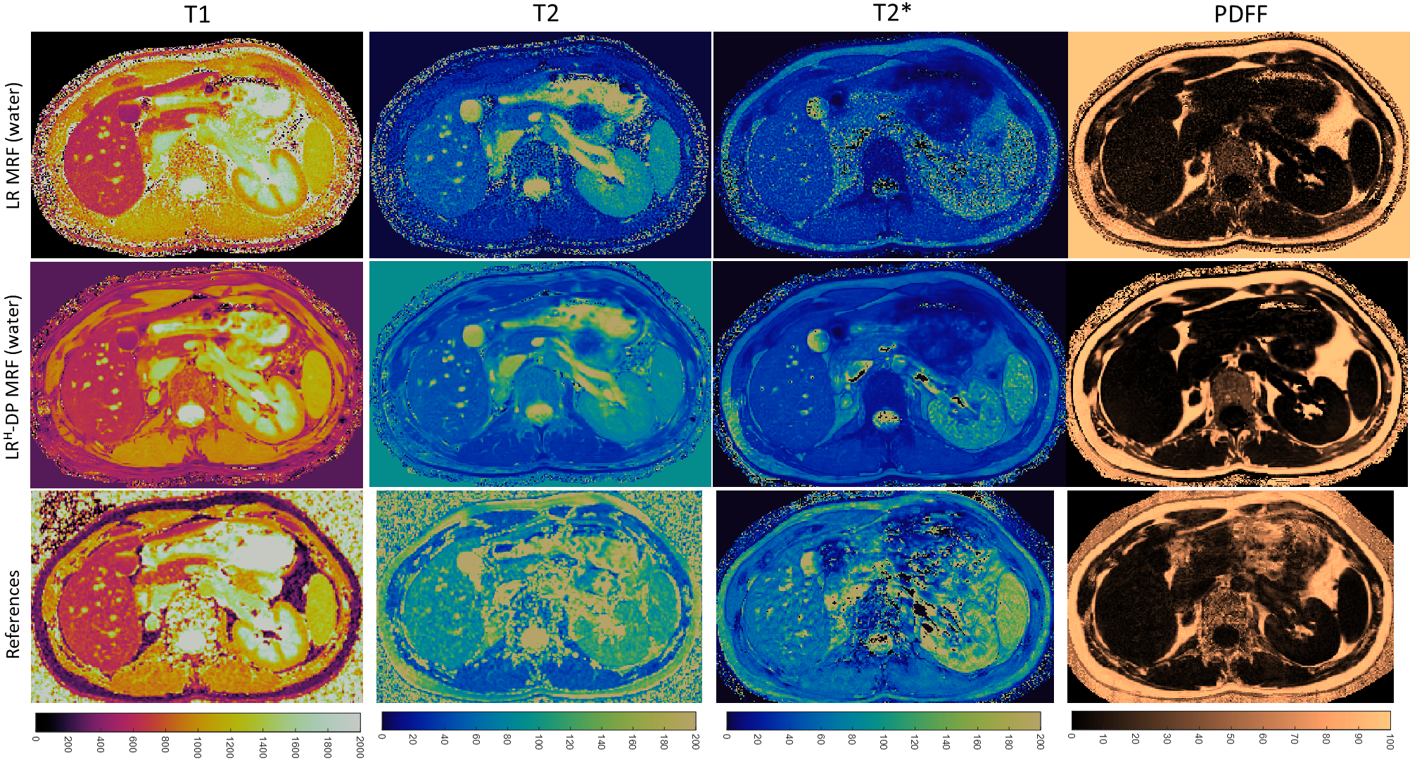
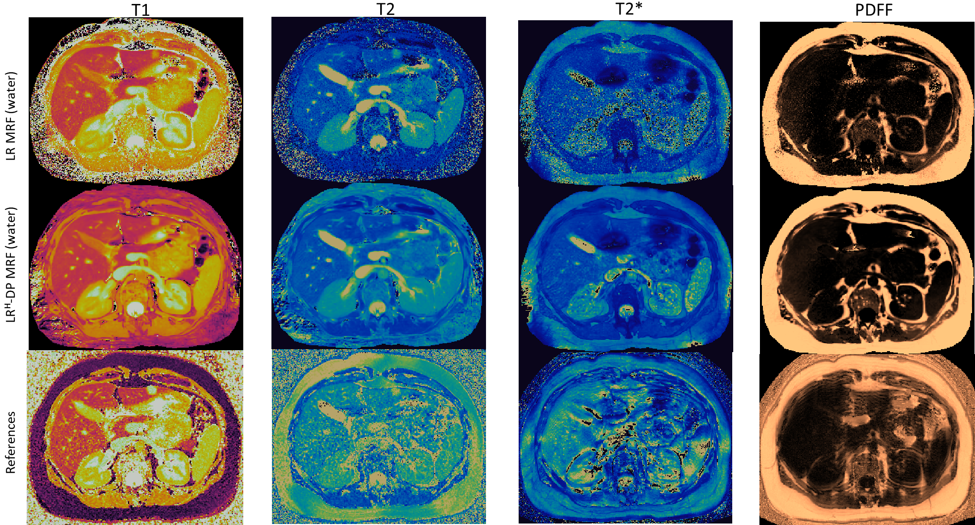
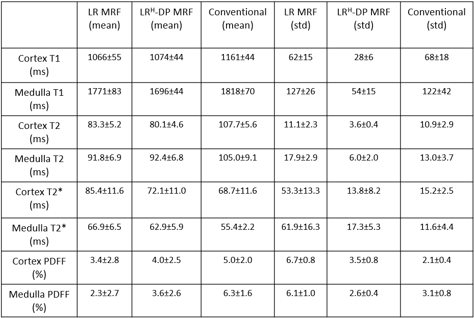
The original low-rank MRF suffered from residual aliasing and noise amplification in the maps, in addition to loss of details around water/fat boundaries. The proposed LRH-DP shows improved map quality with better detailed structures. Conventional maps exhibited noise amplification due to the relatively high resolution (Figs. 3 and 4). T1 and T2 values measured with MRF agree with literature values15,16 and are lower than the conventional measurements acquired here, possibly due to magnetization transfer17 effects. Differences in T2* were also observed; however, both conventional and MRF sequences are less sensitive to high (>30ms) T2* values. Good agreement was observed for PDFF. The proposed LRH-DP had the lowest standard deviation for all measurements (Fig. 5).CONCLUSION:
A novel MRF reconstruction (LRH-DP) combining low-rank models, Hermitian symmetry, and dictionary-patch based regularization was deployed to enable 2D T1/T2/T2*/PDFF kidney mapping in a single breath-hold with a resolution of 1x1x5 mm3. LRH-DP demonstrated superior performance to conventional methods and may facilitate renal mass characterization via the analysis of these four co-registered tissue properties.Acknowledgements
This work was supported in part by the NIH (R01 HL153034, R01HL163991, R01HL163030) and Siemens Healthineers.References
1. Ma D, Gulani V, Seiberlich N, Liu K, Sunshine JL, Duerk JL, Griswold MA. Magnetic resonance fingerprinting. Nature. 2013 Mar;495(7440):187-92.McGivney DF, Pierre E, Ma D, et al. SVD compression for magnetic resonance fingerprinting in the time domain. IEEE Trans. Med. Imaging 2014;33:2311–2322 doi: 10.1109/TMI.2014.2337321.
2. McGivney DF, Pierre E, Ma D, et al. SVD compression for magnetic resonance fingerprinting in the time domain. IEEE Trans. Med. Imaging 2014;33:2311–2322 doi: 10.1109/TMI.2014.2337321.
3. Zhao B, Setsompop K, Adalsteinsson E, et al. Improved magnetic resonance fingerprinting reconstruction with low-rank and subspace modeling. Magn. Reson. Med. 2018;79:933–942 doi: 10.1002/mrm.26701.
4. Assländer J, Cloos MA, Knoll F, Sodickson DK, Hennig J, Lattanzi R. Low-rank alternating direction method of multipliers reconstruction for MR fingerprinting. Magn. Reson. Med. 2018;79:83–96 doi: 10.1002/mrm.26639
5. Noll DC, Nishimura DG, Macovski A. Homodyne detection in magnetic resonance imaging. IEEE transactions on medical imaging. 1991 Jun;10(2):154-63.
6. Bydder M, Robson MD. Partial Fourier partially parallel imaging. Magnetic Resonance in Medicine: An Official Journal of the International Society for Magnetic Resonance in Medicine. 2005 Jun;53(6):1393-401.
7. Haldar JP. Low-rank modeling of local k-space neighborhoods (LORAKS) for constrained MRI. IEEE transactions on medical imaging. 2013 Dec 5;33(3):668-81.
8. Blaimer M, Gutberlet M, Kellman P, Breuer FA, Köstler H, Griswold MA. Virtual coil concept for improved parallel MRI employing conjugate symmetric signals. Magnetic Resonance in Medicine: An Official Journal of the International Society for Magnetic Resonance in Medicine. 2009 Jan;61(1):93-102.
9. Davies M, Puy G, Vandergheynst P, Wiaux Y. A compressed sensing framework for magnetic resonance fingerprinting. Siam journal on imaging sciences. 2014;7(4):2623-56.
10. Lima da Cruz G, Bustin A, Jaubert O, Schneider T, Botnar RM, Prieto C. Sparsity and locally low-rank regularization for MR fingerprinting. Magnetic resonance in medicine. 2019 Jun;81(6):3530-43.
11. Cline CC, Chen X, Mailhe B, Wang Q, Pfeuffer J, Nittka M, Griswold MA, Speier P, Nadar MS. AIR-MRF: accelerated iterative reconstruction for magnetic resonance fingerprinting. Magnetic resonance imaging. 2017 Sep 1;41:29-40.
12. Akçakaya M, Basha TA, Goddu B, Goepfert LA, Kissinger KV, Tarokh V, Manning WJ, Nezafat R. Low‐dimensional‐structure self‐learning and thresholding: regularization beyond compressed sensing for MRI reconstruction. Magnetic Resonance in Medicine. 2011 Sep;66(3):756-67.
13. Bustin A, Lima da Cruz G, Jaubert O, Lopez K, Botnar RM, Prieto C. High‐dimensionality undersampled patch‐based reconstruction (HD‐PROST) for accelerated multi‐contrast MRI. Magnetic resonance in medicine. 2019 Jun;81(6):3705-19.
14. Hernando D, Kellman P, Haldar JP, Liang ZP. Robust water/fat separation in the presence of large field inhomogeneities using a graph cut algorithm. Magnetic Resonance in Medicine: An Official Journal of the International Society for Magnetic Resonance in Medicine. 2010 Jan;63(1):79-90.
15. Cox EF, Buchanan CE, Bradley CR, Prestwich B, Mahmoud H, Taal M, Selby NM, Francis ST. Multiparametric renal magnetic resonance imaging: validation, interventions, and alterations in chronic kidney disease. Frontiers in physiology. 2017 Sep 14;8:696.
16. De Bazelaire CM, Duhamel GD, Rofsky NM, Alsop DC. MR imaging relaxation times of abdominal and pelvic tissues measured in vivo at 3.0 T: preliminary results. Radiology. 2004 Mar;230(3):652-9.
17. Hilbert T, Xia D, Block KT, Yu Z, Lattanzi R, Sodickson DK, Kober T, Cloos MA. Magnetization transfer in magnetic resonance fingerprinting. Magnetic resonance in medicine. 2020 Jul;84(1):128-41.
Figures