0575
Accelerated Preclinical UHF Abdominal T1 Mapping using Novel Rosette Ultrashort Echo Time (PETALUTE)1School of Health Sciences, College of Health and Human Sciences, Purdue University, West Lafayette, IN, United States, 2College of Engineering, Purdue University, West Lafayette, IN, United States, 3High Field MR Centre, Department of Biomedical Imaging and Image-guided Therapy, Medical University of Vienna, Vienna, Austria, 4Department of Physiology, Development, and Neuroscience, University of Cambridge, Cambridge, United Kingdom, 5Department of Clinical Neurosciences, Wolfson Brain Imaging Centre, Cambridge, United Kingdom, 6Radiology and Biomedical Imaging, University of California San Francisco, San Francisco, CA, United States, 7Weldon School of Biomedical Engineering, Purdue University, West Lafayette, IN, United States, 8Department of Radiology, Medical Physics, Medical Center-University of Freiburg, Faculty of Medicine, University of Freiburg, Freiburg, Germany, 9Nuffield Department of Clinical Neurosciences, University of Oxford, Oxford, United Kingdom, 10Physical Sciences Platform, Sunnybrook Research Institute, Toronto, ON, Canada, 11Department of Medical Biophysics, University of Toronto, Toronto, ON, Canada
Synopsis
Keywords: Preclinical Image Analysis, Preclinical, Abdomen, T1 Mapping
Motivation: Well established techniques for fast 3D T1 mapping with cartesian/radial trajectories are prone to respiratory artifacts.Previously established non-cartesian sequences have mitigated the influence of motion artifacts, though still suffer from long measurement times.
Goal(s): Implementation of a novel 3D dual-echo rosette k-space trajectory for preclinical UTE MRI(PETALUTE) for abdominal imaging of both anatomical and quantitative T1 measurements and retrospective 4-fold acceleration.
Approach: PETALUTE(resolution 0.24x0.24x0.24mm3,accelerated scan-time 2:15min) acquisition for T1 mapping via variable flip angle method and evaluation of T1 values and acceleration effects.
Results: High-resolution non-gated abdominal imaging with the ability to clearly distinguish anatomy and T1 values,that did not deprecate when accelerated.
Impact: Well established methods for T1 mapping using cartesian/radial trajectories suffer from motion artifacts due to long acquisition duration.PETALUTE,a novel 3D dual-echo rosette k-space trajectory for preclinical UTE-MRI,is able to generate high-resolution non-gated abdominal anatomical images and T1 mapping in ~2min.
Introduction
VTR, VFA and inversion recovery with cartesian and radial trajectories [1]–[3] are established techniques for fast 3D T1 mapping. Long acquisition times with Cartesian trajectoriesare prone to artifacts due to respiratory motion. Non-cartesian encoding variants[4] have been developed to overcome this drawback, though still suffer from long measurement times. Recent advances in UTE MRI[5]–[7] have implemented novel rosette trajectory measurements for 3D ultrashort echo time (UTE) MR(SI). Compared to radial acquisition schemes, the novel rosette k-space trajectory samples more inner and outer k-space data with less coherence. This improves both SNR and the point spread function, and allows for further acceleration via undersampling or using compressed sensing during reconstruction. The aim of our study was thus, to implement a novel 3D dual-echo rosette k-space trajectory for abdominal T1 mapping using preclinical UTE MRI and to test the performance of a non-accelerated and retrospectively 4-fold accelerated PETALUTE sequence.Materials & Methods
Four mice with 4T1 mammary and flank tumors (dual tumor model) were scanned in a 7T horizontal-bore small animal MRI system (BioSpec 70/30; Bruker Instruments; gradient insert: maximum gradient: 660 mT/m; maximum slew rate: 4570 T/m/s) and a volume Tx/Rx 1H RF coil (40mm). The imaging protocol was as follows: (1) vendor-provided RAREvrt (resolution: 0.234x0.234x1mm3; FOV: 30x30mm; slice thickness: 1mm (20 slices); variable TR: 933.9ms,1787.7ms, 2985.8ms, 5011.1ms, 15000.0ms, TE 20.288ms, scan time: 6.8min), (2 & 3) PETALUTE acquisition with: TE of 16 µs, resolution of 0.24x0.24x0.24mm3 with a matrix of 256×256×256 for a field of view of 60x60x60 mm3, which was achieved with 36,864 petals consisting of 206 points per petal (103 points per echo) acquired in 1.6 ms. We investigated the T1 relaxation time as measured using the variable flip angle method, flip angle: 4° (2) and 20° (3). The total acquisition time for dual-echo PETALUTE (2 & 3 combined) was 9 minutes with a TR of 7 ms. The data was further undersampled 4-times (18,192 petals, 2.15 minutes) retrospectively to evaluate precision of T1 relaxation with acceleration. Both non-accelerated and accelerated the dual-echo PETALUTE reconstruction was performed in MATLAB (MathWorks, USA) and BART (Berkeley Advanced Reconstruction Toolbox) by regular regridding applying a density-compensated nonuniform fast Fourier transformation. T1 maps were created using the respective first echoes of the 4° Ernst-angle and 20° measurements using FSL FLIRT and FSLmath[8].T1 = 2TR ((S1/α1 - S2/α2) / (S2/α2 - S1/α1))
To ensure good visual comparability, the 4° measurements and T1 maps were co-registered to the T1 FLASH images using FSLeyes. ROIs in kidney, thigh muscles and tumors were segmented and evaluated using 3DSlicer in order to determine the correctness of T1 mapping via PETALUTE and the influence of acceleration.
Results
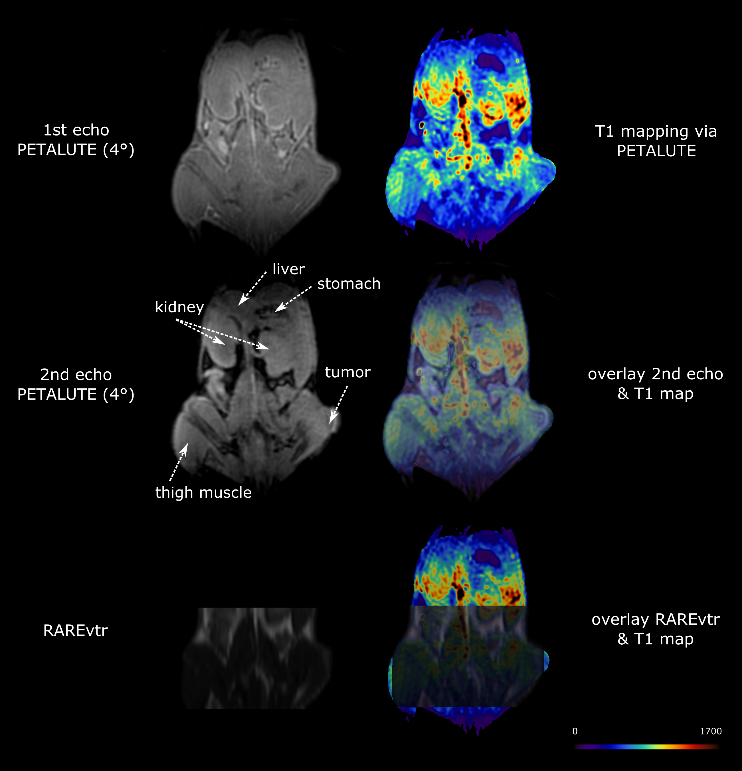
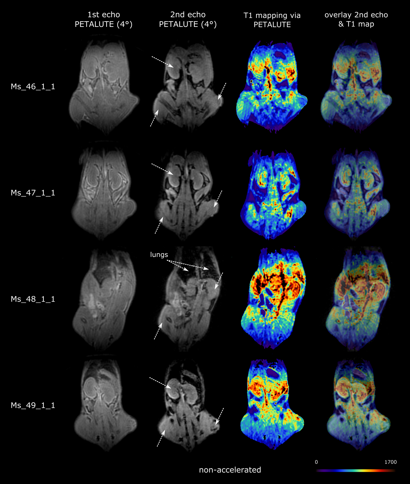
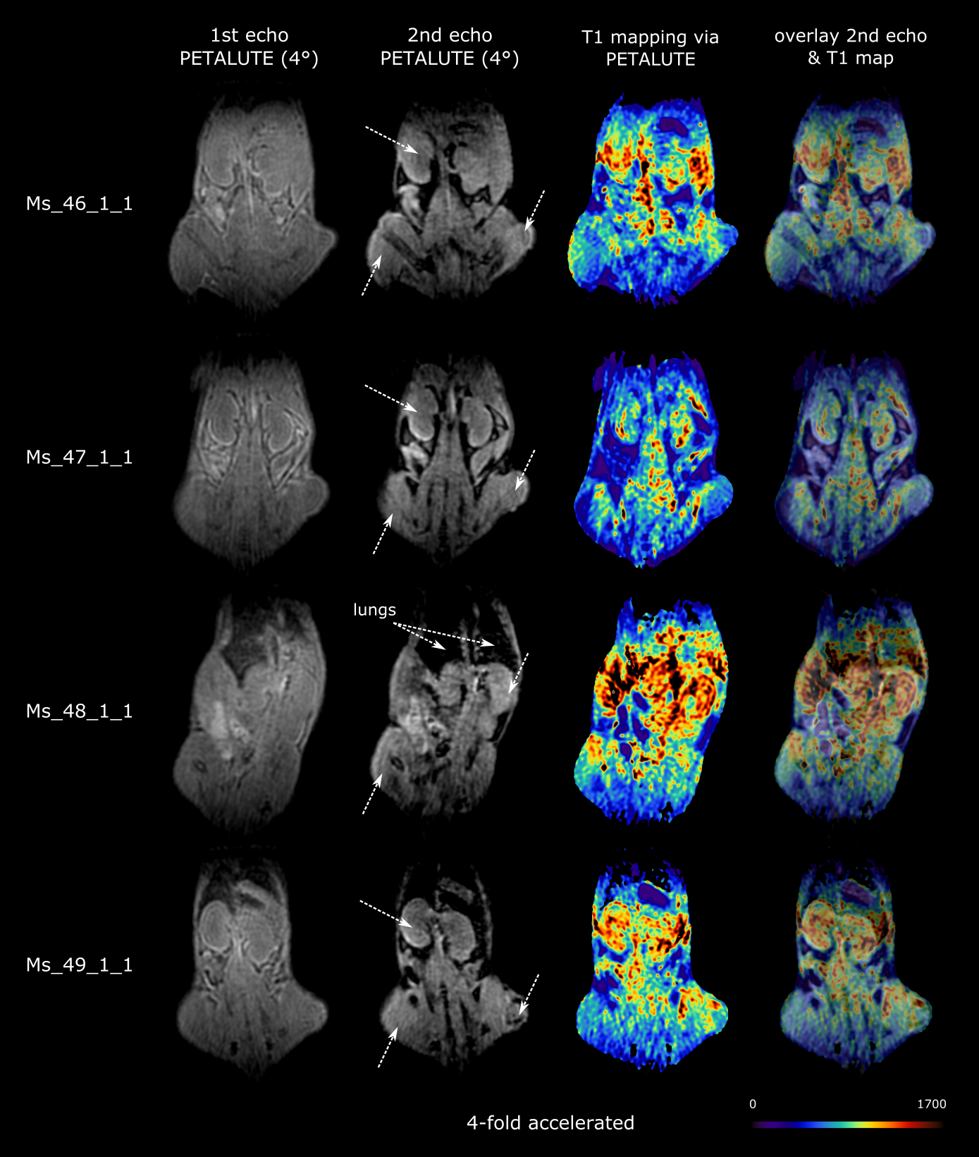
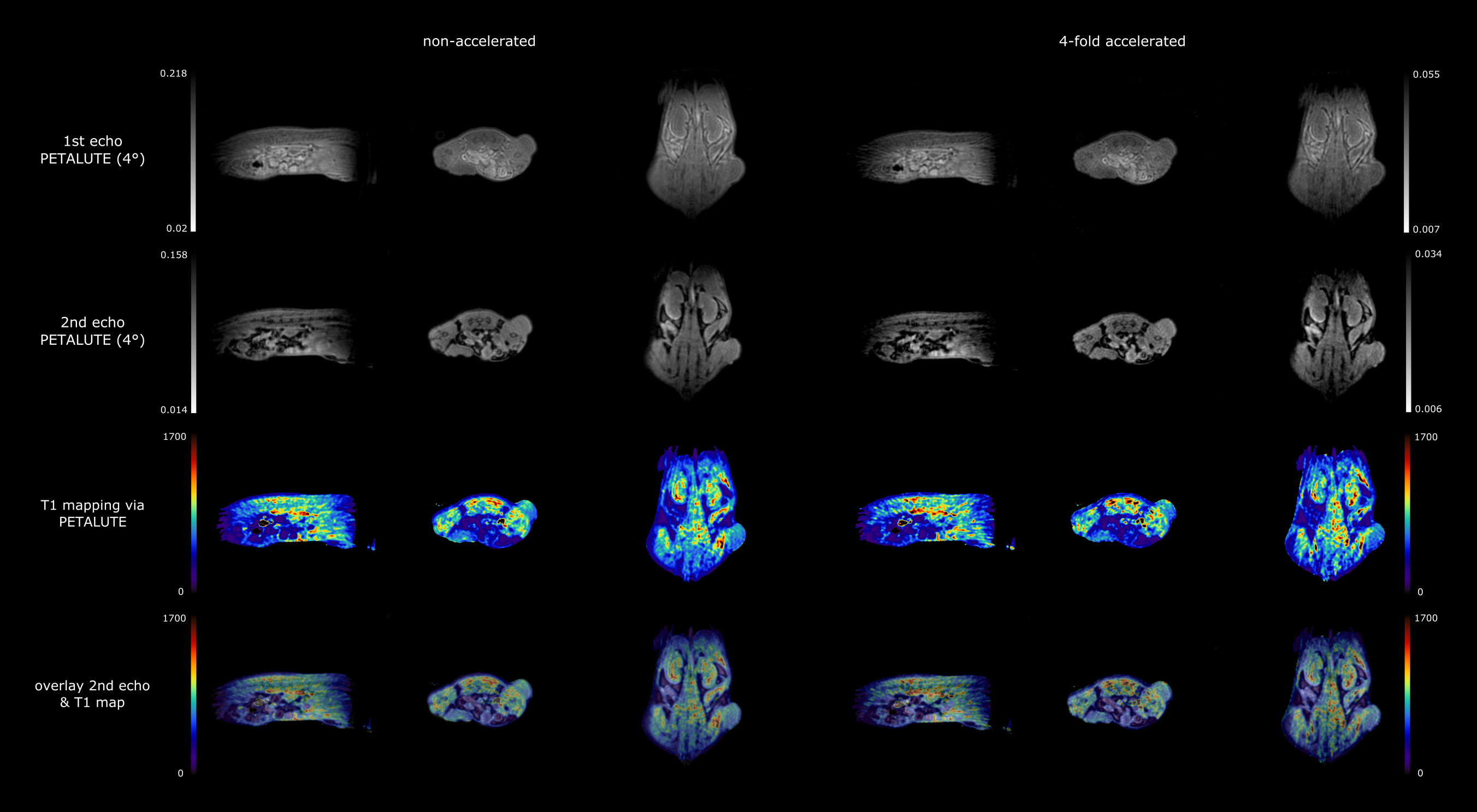
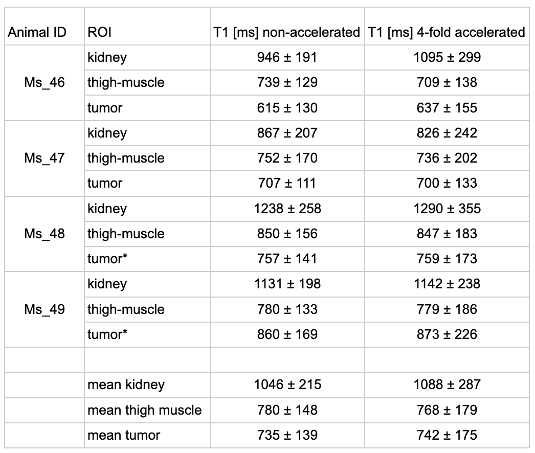
Reconstructed PETALUTE images as well as the resulting T1 map allow to clearly distinguish anatomical features of the mice (Figure 1). ROIs within the kidney, thigh muscle and tumors allow the determination of T1 in non-accelerated (Figure 2) and retrospectively 4-fold accelerated (Figure 3) PETALUTE images. The mean T1 values (Table 1) are as follows: 1046±215ms and 1088±287ms for kidney, 780±148ms and 768±179ms for thigh muscle, 735±139ms and 742±175ms for tumors (non-accelerated and accelerated, respectively). Furthermore, non-accelerated and 4-fold accelerated T1 values were not significantly different (paired two-tailed t-Test, p=0.399), thus not having been degraded (Figure 4).Discussion & Conclusion
We were able to develop and acquire high-resolution non-gated anatomical abdominal images via accelerated PETALUTE (a resolution of 0.24x0.24x0.24mm3 and a total duration of 2:15min for the 4-fold acceleration) that were able to depict organs such as the kidney and lung. This further allowed for T1 mapping and it was shown that no significant differences between the non-accelerated acquisition and 4-fold acceleration could be found, thus indicating that undersampling does not deprecate the estimated T1 maps. Although the estimated T1 values are lower than those previously reported with conventional acquisition strategies [9]–[13], it was previously shown that ultra short T2* together B1 inhomogeneity leads to substantially shorter T1 values for VFA UTE acquisitions[14]. As resolving the T2* -dependency has to – the best of our knowledge – not yet been investigated due to its complexity, future work will try to partially mitigate these influences by implementing a T2* correction before T1 calculation. Overall, we demonstrated the potential utilization of PETALUTE for abdominal imaging of both anatomical and quantitative measurements.Acknowledgements
Wellcome Trust, Canada Research Chairs Program
References
[1] P. Garteiser et al., “Experimental Protocols for MRI Mapping of Renal T1,” in Preclinical MRI of the Kidney: Methods and Protocols [Internet], Humana Press, 2021. doi: 10.1007/978-1-0716-0978-1_22.
[2] P. Garteiser et al., “Analysis Protocols for MRI Mapping of Renal T1,” in Preclinical MRI of the Kidney: Methods and Protocols, A. Pohlmann and T. Niendorf, Eds., in Methods in Molecular Biology. , New York, NY: Springer US, 2021, pp. 577–590. doi: 10.1007/978-1-0716-0978-1_35.
[3] S. J. Hectors et al., “MRI Mapping of Renal T1: Basic Concept,” in Preclinical MRI of the Kidney: Methods and Protocols, A. Pohlmann and T. Niendorf, Eds., in Methods in Molecular Biology. , New York, NY: Springer US, 2021, pp. 157–169. doi: 10.1007/978-1-0716-0978-1_9.
[4] T. L. Faller, A. J. Trotier, S. Miraux, and E. J. Ribot, “Radial MP2RAGE sequence for rapid 3D T1 mapping of mouse abdomen: application to hepatic metastases,” Eur. Radiol., vol. 29, no. 11, pp. 5844–5851, Nov. 2019, doi: 10.1007/s00330-019-06081-3.
[5] X. Shen, “Development and Applications of 3D Ultra-short Echo Time MRI with Rosette k-Space Pattern,” Purdue University Graduate School, 2022. [Online]. Available: https://doi.org/10.25394/PGS.20323671.v1
[6] X. Shen, M. Chiew, and U. Emir, “Development of 3D Rosette K-Space Trajectory in Ultra-Short Echo Time MRI Applications,” presented at the MEDICAL PHYSICS, Jun. 2021.
[7] X. Shen et al., “Ultra-short T2 components imaging of the whole brain using 3D dual-echo UTE MRI with rosette k-space pattern,” Magn. Reson. Med., vol. 89, no. 2, pp. 508–521, 2023, doi: 10.1002/mrm.29451
[8] G. Helms, H. Dathe, and P. Dechent, “Quantitative FLASH MRI at 3T using a rational approximation of the Ernst equation,” Magn. Reson. Med., vol. 59, no. 3, pp. 667–672, 2008, doi: 10.1002/mrm.21542.
[9] H. Lu et al., “A pH-responsive T1-T2 dual-modal MRI contrast agent for cancer imaging,” Nat. Commun., vol. 13, no. 1, Art. no. 1, Dec. 2022, doi: 10.1038/s41467-022-35655-x.
[10] T. L. Faller, A. J. Trotier, A. F. Rousseau, J.-M. Franconi, S. Miraux, and E. J. Ribot, “2D multislice MP2RAGE sequence for fast T1 mapping at 7 T: Application to mouse imaging and MR thermometry,” Magn. Reson. Med., vol. 84, no. 3, pp. 1430–1440, 2020, doi: 10.1002/mrm.28220.
[11] N. D. Bryant et al., “Multi-parametric MRI characterization of inflammation in murine skeletal muscle,” NMR Biomed., vol. 27, no. 6, pp. 716–725, Jun. 2014, doi: 10.1002/nbm.3113.
[12] S. Erdogan, Z. O. Medarova, A. Roby, A. Moore, and V. P. Torchilin, “Enhanced tumor MR imaging with gadolinium-loaded polychelating polymer-containing tumor-targeted liposomes,” J. Magn. Reson. Imaging, vol. 27, no. 3, pp. 574–580, 2008, doi: 10.1002/jmri.21202.
[13] S. Pickup, M. Romanello, M. Gupta, H. K. Song, and R. Zhou, “Dynamic Contrast-Enhanced MRI in the Abdomen of Mice with High Temporal and Spatial Resolution Using Stack-of-Stars Sampling and KWIC Reconstruction,” Tomography, vol. 8, no. 5, pp. 2113–2128, Aug. 2022, doi: 10.3390/tomography8050178
[14] D. F. Alamidi et al., “Variable flip angle 3D ultrashort echo time (UTE) T1 mapping of mouse lung: A repeatability assessment,” J. Magn. Reson. Imaging, vol. 48, no. 3, pp. 846–852, 2018, doi: 10.1002/jmri.25999.
Figures