0570
A MULTIPLEX-Lite method for fast 3D multi-parametric imaging with MR Angiography1UIH America, Inc., Houston, TX, United States, 2United Imaging Research Institute of Innovative Medical Equipment, Shenzhen, China, 3United Imaging Healthcare, Shanghai, China
Synopsis
Keywords: Pulse Sequence Design, Multi-Contrast
Motivation: Existing 3D multi-parametric imaging methods suffers from long scan times, and decent MRA contrasts are yet to be provided.
Goal(s): To develop a 3-minute 3D high resolution multi-parametric imaging method that offeres whole brain imaging with TOF quality MRA results.
Approach: A simplified version of the MULTIPLEX method, namely MULTIPLEX-Lite, is proposed. Compared to the original MULTIPLEX method, addtional MRA results are offered, while removing the T1 and PD mappings to further improve scan time.
Results: The proposed MULTIPLEX-Lite method offered whole brain high resolution (<1mm3 voxel volume) MRA, PDW, T1W, aT1W, SWI, R2* maps and QSM in one 3-minute scan.
Impact: The proposed MULTIPLEX-Lite method is among the most feasible and practical solutions for routine clinically friendly 3D high-resolution multi-parametric imaging practices.
Introduction
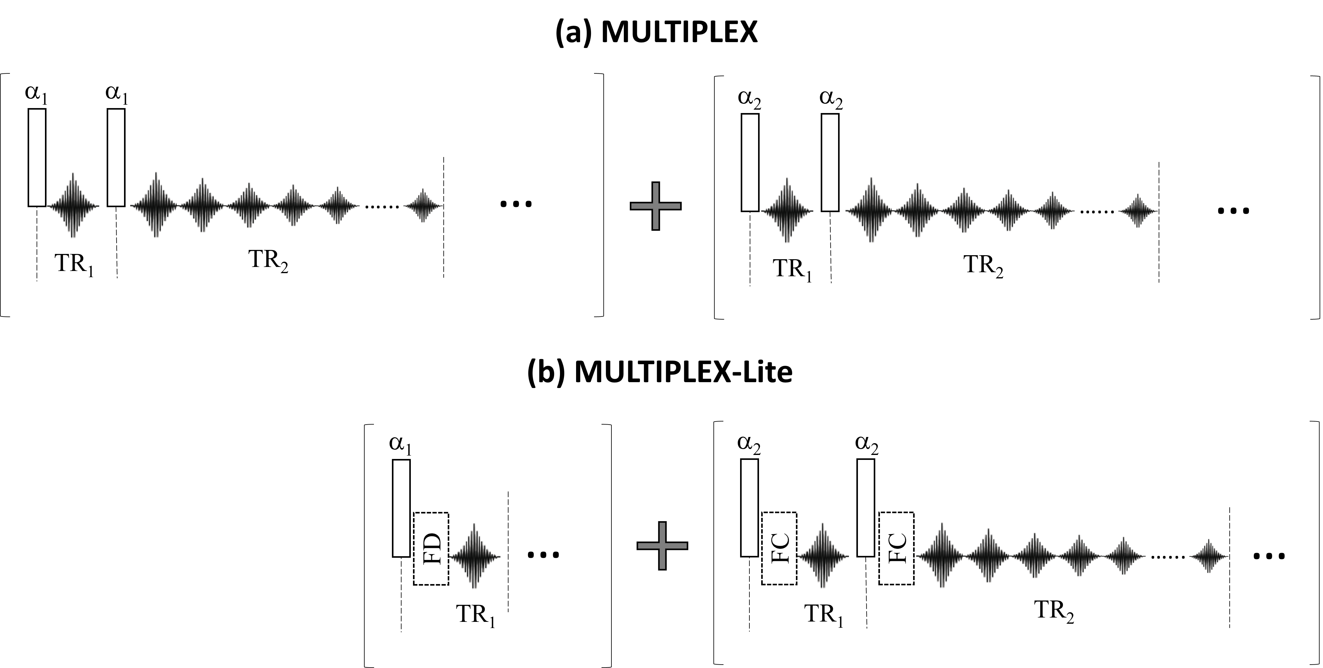
Recently, a MULTIPLEX method was proposed for 3D high-resolution multi-parametric MR imaging1. The basic design of the original method (Fig.1a) comprised of two separate dual-TR acquisition blocks, with different flip angles (FA) in each block to generate different T1 (and PD) weightings, as well as multi-echo acquisition for susceptibility imaging. When using high resolution (e.g. voxel volume<1mm3), scan time are usually on the longer side (5~8 minutes) for daily clinical use. Also, decent MR angiography (MRA) results are not readily available.In this work, we proposed a simplified version of the original MULTIPLEX method, namely MULTIPLEX-Lite, to achieve improved MRA results within the MULTIPLEX framework. Also, since T1 and PD mapping are generally not a must in routine scans, taking out the function of these mappings while keeping most of the other MULTIPLEX results, the scan time can be significantly reduced to a more clinically dedicated and friendly level.
Methods
On the basis of the original MULTIPLEX design (Fig.1a), the proposed MULTIPLEX-Lite strategy (Fig.1b), the first dual-TR block was replaced by a single-TR GRE acquisition with FD module and a FA of α1, while employing FC modules in the second dual-TR block. The flip angle α1 and α2 in MULTIPLEX-Lite were set as such (4°/16°) to generate PD- and T1-weighted signals, respectively.For image reconstruction, MULTIPLEX results were calculated as previously described (1). For MULTIPLEX-Lite, the α1 images served as both PDW and dark blood images; the α2 images were averaged for the T1W images; both the α1 and α2 images were used to generate the augmented-T1W (aT1W) images2 using the multi-dimensional integration method3; the multi-echo images in TR2 were used for generating SWI, R2* mapping and QSM images. Finally, MRA images were generated via non-linear weighted subtraction between the FC and FD images4.
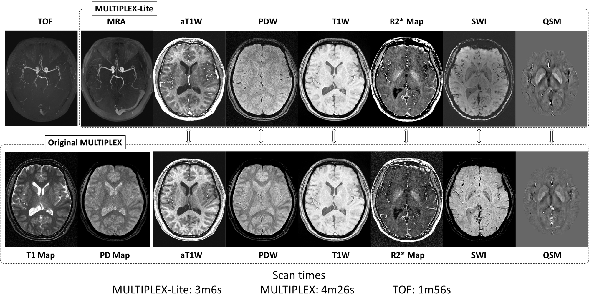
Five healthy volunteers (3 males, 32~45y/o) were scanned on a 3T system (uMR 790, United Imaging Healthcare, Shanghai, China) using a 32-channel head coil. The key parameters for both MULTIPLEX and MULTIPLEX-Lite scans were: echo number=5, TE=4.06~21.34ms, TR1/TR2=8.2/28.1ms, α1/α2=4°/16°, voxel size=0.6x0.6x1.5mm3. A deep learning-based acceleration method5 was employed to provide four-fold acceleration effects, reducing the scan times of MULTIPLEX-Lite and MULTIPLEX to 3m6s and 4m26s, respectively. For MRA reference, product TOF images were also obtained using the same spatial resolution settings, with scan time of 1m56s.
Results
The results of a representative subject are shown in Fig.2.Both MULTIPLEX-Lite and MULTIPLEX generated aT1W, PDW, T1W, R2* map, SWI and QSM images. All vessels appeared dark in the MULTIPLEX-Lite PDW images, and thus the corresponding aT1W (calculated as T1W/PDW) images showed brighter vessels. Some MULTIPLEX-Lite images showed relatively reduced SNR, while others had similar SNR and image contrasts. MULTIPLEX-Lite additionally generated MRA images with similar quality to routine TOF images, while trading away the T1 and PD maps.
The scan time of MULTIPLEX-Lite was ~70% of MULTIPLEX’s, or ~48% of the total duration of MULTIPLEX plus TOF scans.
Discussion & Conclusion
A MULTIPLEX-Lite method was proposed to achieve 3D high resolution multi-parametric imaging with clinically friendly scan time. Compared to the original MULTIPLEX method, the proposed method offers additional high-quality MRA results, minus the T1/PD mapping results, while achieving ~30% reduction in scan times. By integrating a separate single-TR acquisition block to obtain dark blood images while collecting bright blood images in the dual-TR block, it has been demonstrated in literature4,6 and this work to provide TOF-like bright blood MRA, making it a valuable extra contrast. However, since inflowing venous blood was not saturated in current implementation, veins and sinuses were also visible but with reduced contrasts by the non-linear subtraction algorithm4.As MULTIPLEX-Lite collects only one echo as the source of PDW and dark blood contrasts, the resultant PDW and aT1W images thus have reduced SNR. Similarly, as only one set of multi-echoes was collected, the resultant R2* map also had reduced SNR. However, the T1W images remained unchanged, as well as SWI and QSM due to the use of phase information. Nevertheless, the MULTIPLEX-Lite design reduced the total acquisition time by at least 30%. If considering the additionally available MRA results, the efficiency of the MULTIPLEX-Lite can be as high as 50%.
With state-of-the-art acceleration schemes such as deep learning networks5, we have demonstrated it is possible to achieve 3D whole brain multi-parametric imaging, including MRA, PDW, T1W, aT1W, SWI, R2* maps and QSM, in one single scan of mere ~3 minutes, making the proposed method highly clinically practical and useful.
Acknowledgements
No acknowledgement found.References
1. Ye Y, Lyu J, Hu Y, Zhang Z, Xu J, Zhang W. MULTI-parametric MR imaging with fLEXible design (MULTIPLEX). Magn Reson Med 2022;87(2):658-673.
2. Ye Y, Lyu J, Hu Y, et al. Augmented T(1) -weighted steady state magnetic resonance imaging. NMR Biomed 2022;35(8):e4729.
3. Ye Y, Lyu J, Sun W, et al. A multi-dimensional integration (MDI) strategy for MR T2 * mapping. NMR Biomed 2021;34(7):e4529.
4. Ye Y, Hu J, Wu D, Haacke EM. Noncontrast-enhanced magnetic resonance angiography and venography imaging with enhanced angiography. J Magn Reson Imaging 2013;38(6):1539-1548.
5. Chen EZ, Zhang C, Chen X, Liu Y, Chen T, Sun S. Computationally Efficient 3D MRI Reconstruction with Adaptive MLP. In: Greenspan H, Madabhushi A, Mousavi P, et al., editors. Medical Image Computing and Computer Assisted Intervention – MICCAI 2023. Cham: Springer Nature Switzerland; 2023. p. 195-205.
6. Kimura T, Ikedo M, Takemoto S. Hybrid of opposite-contrast MR angiography (HOP-MRA) combining time-of-flight and flow-sensitive black-blood contrasts. Magn Reson Med 2009;62(2):450-458.
Figures