0562
Nonlinear storage modulus quantification with MR-elastography: from in vitro to in vivo assessment1Biomaps, CEA, CNRS, Orsay, France, 2Laboratory of Biomarker Imaging, INSERM, Paris, France
Synopsis
Keywords: Elastography, Elastography
Motivation: Estimating the nonlinear coefficient in MR elastography could provide a relevant mechanical parameter for tumor characterization.
Goal(s): The goal of this study was to develop a method for assessing the nonlinear coefficient in MR elastography.
Approach: We developed a specific MR elastography setup and a post-processing pipeline to quantify nonlinear coefficients in phantoms and mice with a tumor implanted subcutaneously.
Results: In phantoms, we observed that nonlinear coefficient was able to provide a higher contrast than storage modulus to distinguish different structure. In addition, in mice, nonlinear coefficient was correlated to the entropy which is a marker of collagen distribution irregularity.
Impact: The estimation of the nonlinear coefficient provides a new biomarker to assess tissue mechanical parameters. The relation between this parameter and tissue structure could be relevant in tumor investigation, as tissue microarchitecture is an important marker of tumor severity.
Introduction
Assessing simple mechanical parameters, such as shear, storage and loss moduli with ultrasound (US) or magnetic resonance (MR) elastography, has been shown to be useful for diagnosing various fibro-inflammatory diseases and cancer1, 2. However, evaluating nonlinear elasticity may improve tumor characterization3, 4. In US-elastography, the acoustoelasticity approach enables to determine the nonlinear coefficient of the shear modulus (A)5. A first approach shows the feasibility of the method in clinical MR-elastography6. The purpose of this study is to develop a preclinical method for assessing the nonlinear coefficient with MR-elastography and explore its use in cancer investigation.Methods
Theory: According to the acoustoelasticity theory, the experiment involves measuring the storage modulus of a medium subjected to a uniaxial stress from a shear wave propagating in a specific direction. Under these conditions, Gennisson et al.5 developed the equations to estimate the nonlinear coefficient in three configurations of shear wave displacement (Fig.1).$$G_{12}^{'} = G_{0}^{'}-\sigma_{2}(1+\frac{A}{12G^{'}}) (1)$$
$$G_{21}^{'} = G_{0}^{'} -\sigma_{2}(\frac{A}{12G^{'}}) (2)$$
$$ G_{13}^{'} = G_{0}^{'} +\sigma_{2}(1+\frac{A}{6G^{'}}) (3)$$
Where G’ is the storage modulus with the first subscript being the direction of polarization and the second subscript the direction of propagation, G0' is the basal stress-free storage modulus, $$$\sigma_{2}$$$ is the uniaxial stress in the direction 2 (Fig.1), and A is the nonlinear storage modulus.
Phantoms and mice: We performed MRI in two homogeneous phantoms containing 1% and 1.5% agar concentrations (AGAR_1 and AGAR_2) and in a heterogeneous phantom consisting of bovine liver sample embedded in gel (1.5% agar, 2% gelatin). We also performed MRI in seven mice with patient-derived cholangiocarcinomas implanted in the right hypochondrium.
MRI: We developed a specific compression setup to uniaxially compress a phantom or small animal inside the MRI tunnel (Fig.1). MRI of the phantoms and mice was performed in a 7T MRI scanner (Pharmascan, Bruker, Ettlingen, Germany). MR-elastography was obtained using 300 Hz mechanical vibrations synchronized with a modified spin-echo sequence. Nine slices were acquired with a volumetric resolution of 0.4 mm3, TE 23 ms, TR 1023 ms, four dynamic scans per vibration period, and acquisition time of 6 min for each of the three spatial directions. T2-weighted MRI (TE 15 ms, TR 1300 ms) was performed with the same spatial resolution as MR-elastography. The morphological T2-weighted images and the functional MR-elastography images were acquired without compression and were repeated after compression steps of 0.5 mm.
Reconstruction: To obtain storage modulus maps according to equation (1), a spatio-temporal filter was applied along direction 2. Phantom deformation ($$$\epsilon$$$) maps were calculated by performing 3D affine registration on the T2 images. Stress ($$$\sigma$$$) was calculated with the Hooke law, as $$$\sigma = 3G^{'}.\epsilon$$$. The slope between the storage modulus for each propagation axis and the applied stress was estimated and the nonlinear coefficient A was computed using (1). The same reconstruction process was used for 21 and 13 subscripts.
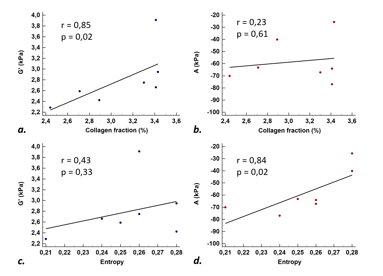
MR-elastography storage modulus and nonlinear coefficient in mice were correlated to collagen fraction and entropy (a marker of collagen distribution irregularity), obtained on picrosirius red stained histological tumor slices7.
Results
The dependence of the storage moduli to uniaxial stress in agar phantoms are shown in Fig.2. Similar nonlinear coefficients were obtained, independently of the chosen configuration (Fig.2.c).Fig.3 shows T2-weighted images, storage modulus and nonlinear coefficient maps of the bovine liver phantom. At zero stress, the gel storage modulus G0’ was 1.5 higher than the liver storage modulus (1.5±0.9 kPa versus 1.0±0.4 kPa), whereas, the average nonlinear coefficient across three configurations revealed a fourfold difference between the gel and bovine liver (AGel = -6.6 kPa versus ALiver = -25.7 kPa).
Parametric maps of xenografted cholangiocarcinoma G0’ and A are shown in Fig 4. In contrast to A, G’ was significantly correlated to collagen fraction(G’: Spearman r = 0.85, p = 0.02; A: r = 0.23, p = 0.61). However, in contrast to G’, a significant correlation was observed between A and entropy (G’: r = 0.43, p = 0.33; A: r = 0.84, p = 0.02) (Fig.5).
Discussion
At zero stress, the phantoms are isotropic. When a stress is applied, the storage modulus evolves differently according to the propagation direction and the phantoms become anisotropic. Moreover, the changes of storage modulus under compression reveal a high gel-liver contrast on the nonlinear coefficient maps. The in vivo tumor results suggest that assessing nonlinear coefficient may help characterizing tissue structure.Conclusion
The results of our study show the feasibility of measuring in vitro and in vivo the nonlinear storage modulus with MR-elastography and suggest that the method may be useful for characterizing tumor tissue structure.Acknowledgements
This work was partly funded by France Life Imaging, project Quantum (grant ANR-11-INBS-0006).References
1. Deffieux, T., Gennisson, J. L., Bousquet, L., Corouge, M., Cosconea, S., Amroun, D., et al. (2015). Investigating liver stiffness and viscosity for fibrosis, steatosis and activity staging using shear wave elastography. Journal of hepatology, 62(2), 317-324.
2. Pagé, G., Garteiser, P., & Van Beers, B. E. (2022). Magnetic resonance elastography of malignant tumors. Frontiers in Physics, 10, 910036.
3. Kim, M. Y., Choi, N., Yang, J. H., Yoo, Y. B., & Park, K. S. (2015). False positive or negative results of shear-wave elastography in differentiating benign from malignant breast masses: analysis of clinical and ultrasonographic characteristics. Acta Radiologica, 56(10), 1155-1162.
4. Bernal, M., Chamming’s, F., Couade, M., Bercoff, J., Tanter, M., & Gennisson, J. L. (2015). In vivo quantification of the nonlinear shear modulus in breast lesions: Feasibility study. IEEE transactions on ultrasonics, ferroelectrics, and frequency control, 63(1), 101-109.
5. Gennisson, J. L., Rénier, M., Catheline, S., Barrière, C., Bercoff, J., Tanter, M., & Fink, M. (2007). Acoustoelasticity in soft solids: Assessment of the nonlinear shear modulus with the acoustic radiation force. The Journal of the Acoustical Society of America, 122(6), 3211-3219.
6. Pagé, G., Bied, M., Garteiser, P., Van Beers, B., Etaix, N., Fraschini, C., Bel-Brunon, A. & Gennisson, J. L. (2023). Comparison of ultrasound elastography, magnetic resonance elastography and finite element model to quantify nonlinear shear modulus. Physics in Medicine & Biology, 68(20), 205003.
7. Li, J., Zormpas-Petridis, K., Boult, J. K., Reeves, E. L., Heindl, A., Vinci, M.,et al. (2019). Investigating the contribution of collagen to the tumor biomechanical phenotype with noninvasive magnetic resonance elastography. Cancer Research, 79(22), 5874-5883.
Figures
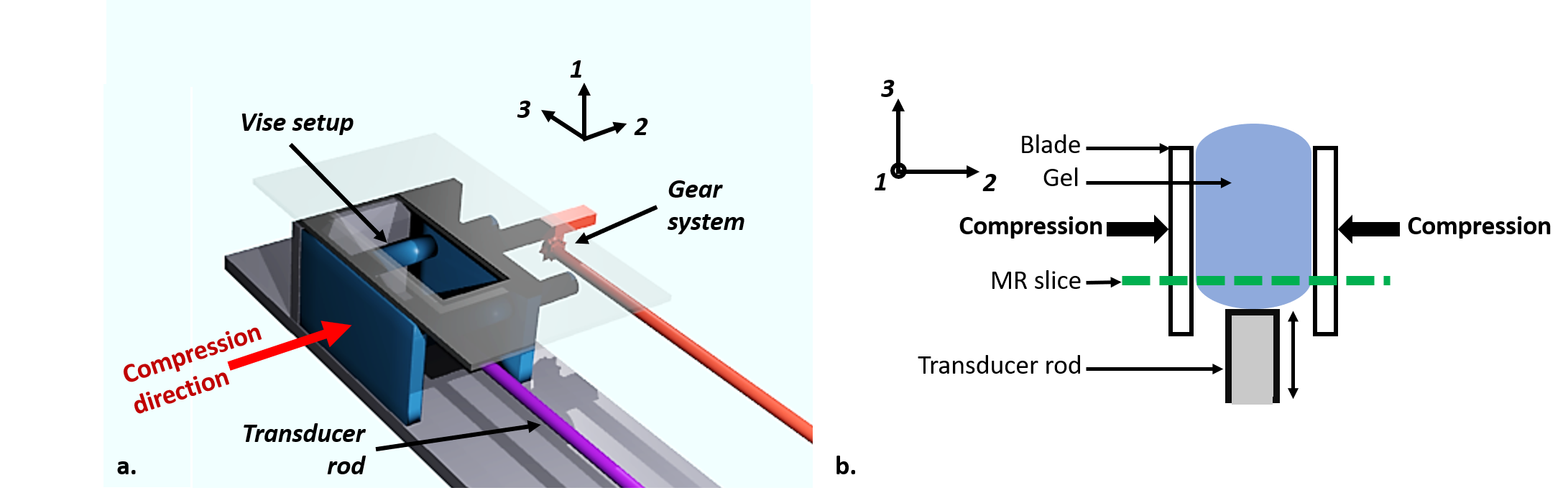
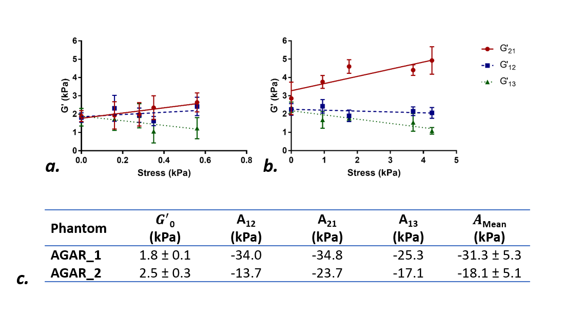
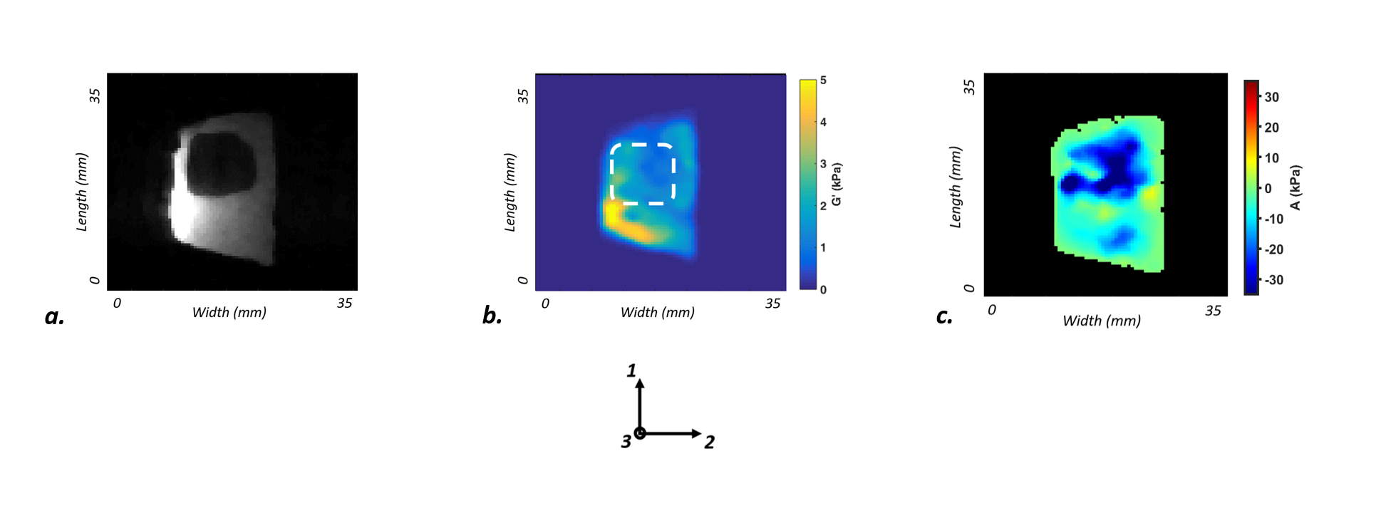
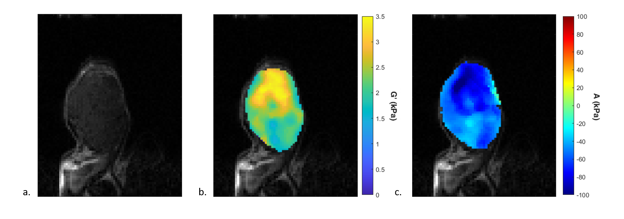
Fig.4: A. T2-weighted image of cholangiocarcinoma implanted subcutaneously in mouse. B. Storage modulus map of the tumor. C. Nonlinear coefficient map of the tumor.