0559
Biomechanical tumor hallmarks for the clinical diagnosis of glioma by high-resolution multifrequency MR elastography1Department of Radiology, Charité - Universitätsmedizin Berlin, Berlin, Germany, 2Faculty of Physics and Earth Sciences, Peter Debye Institute, Leipzig University, Leipzig, Germany, 3Department of Neuropathology, Charité - Universitätsmedizin Berlin, Berlin, Germany, 4Institute of Neuroradiology, Charité - Universitätsmedizin Berlin, Berlin, Germany, 5Department of Neurosurgery, University Hospital Frankfurt, Frankfurt am Main, Germany
Synopsis
Keywords: Elastography, Elastography
Motivation: Previous studies using MR Elastography (MRE) have suggested that gliomas exhibit reduced stiffness and viscosity. However, the interplay between micromechanical tumor changes that determine the macroscopic mechanical properties measured by MRE remains unclear.
Goal(s): This study aims to investigate the relationship between viscoelastic parameters measured MRE in-vivo and histopathologically quantified parameters in glioma.
Approach: High-resolution multifrequency MRE with quantified histopathology was prospectively performed in 23 patients with glioma.
Results: Stiffness and viscosity in gliomas are associated with increased cell elongation, micro-vessel density, and apoptotic rate suggesting unjamming, neovascularization and cell proliferation as biomechanically sensitive tumor hallmarks for clinical diagnosis.
Impact: In this study, we demonstrated that viscoelastic parameters, quantified by MR Elastography, provide insights into cell mobility, cellularity, mitotic and apoptotic rates, as well as vascularization of gliomas in-vivo. This technique holds promise for future clinical diagnosis of neurotumors.
Introduction
Gliomas, the most common type of primary brain tumors, are characterized by a dismal survival rate.1 MRI is the standard diagnostic tool for therapy planning and follow-up monitoring of patients with brain tumors.2 Despite the availability of these diverse imaging sequences for tumor characterization, differentiation remains a challenge. Therefore, histopathology and genetic analysis are typically required to establish a diagnosis and understand tumor subtypes and mutation states.3MR Elastography (MRE) non-invasively measures the mechanical properties of soft tissue in-vivo.4 Previous studies have indicated that high-grade gliomas demonstrate a decrease in stiffness and loss angle values.5-7 Additionally, it has been demonstrated that considerable heterogeneity can be observed both within and among brain tumors.5 However, the correlation between tumor biomechanical parameters assessed by MRE and the histopathological manifestation of the tumor remains poorly understood.
In this study, we combine multifrequency MRE with quantified histopathology in glioma to understand the micromechanical changes that determine the coarse-grained mechanical properties.
Methods
A total of 23 glioma patients (3 grade II, 7 grade III, 13 grade IV) were included. MRI scans consisting of contrast-enhanced MRI, DWI and multifrequency MRE were performed pre-surgically. Multifrequency MRE was performed on a 3-Tesla MRI scanner (Magnetom Lumina, Siemens Healthineers) using a single-shot, spin-echo EPI sequence with externally induced vibrations of 20, 25, 30 and 40Hz. Eight equidistant phase steps over a full vibration cycle were acquired with 3D flow-compensated motion-encoding-gradients. 36 images slices with a field-of-view of 201×201mm2 and a resolution of 1.6×1.6×2mm3 were acquired. After 2D motion correction, kMDEV inversion algorithm was used.8 Maps of shear wave speed (SWS), penetration rate (PR) and loss angle were generated. To investigate the correlation between mechanical parameters and histopathology, the manually drawn volume-of-interests (VOIs) were segmented into three subregions based on stiffness (SWS) values determined by k-means clustering. Post-surgical histological tumor stains including hematoxylin and eosin (HE) stains for cell aspect ratio (AR) and density, CD31 for vascularization, p53 for apoptosis, Ki-67 for mitosis, and alcian blue (AB)/PAS for glycosaminoglycan analysis were used and cells, cellular nuclei and AB/PAS were segmented and quantified.Results
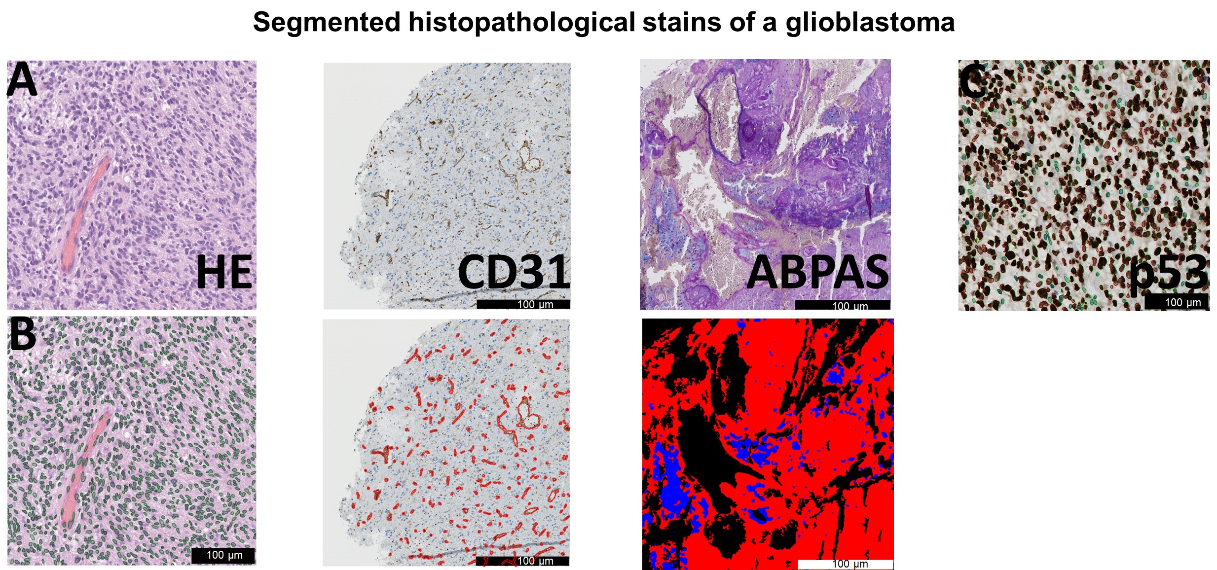
Figures 1,2, and 3 show representative slices of patients with glioma grade II, III and IV, respectively. The soft, medium and stiff subregions are delineated making the mechanical heterogeneity of the tissue apparent. Figure 4 shows an example of automatic feature segmentation in HE, CD31, p53, as well as ABPAS stains of a glioblastoma.For regions of low, medium and high stiffness mean SWS was 0.87±0.23m/s, 1.14±0.8m/s, and 1.36±0.08m/s, mean PR was 0.56±0.23m/s, 0.77±0.19m/s, and 0.83±0.16m/s, and mean loss angle was 0.57±0.08rad, 0.53±0.09rad, and 0.57±0.09rad, respectively.
In regions of medium and high stiffness, AB was correlated with PR and the loss angle (p<0.05). In all regions, PR was correlated with p53 (p<0.05). In regions of medium to high tumor stiffness, p53 was correlated with loss angle (p<0.05) while in regions of low and medium stiffness, SWS was correlated with AR of tumor cells (p<0.05). In soft tumor regions, SWS was correlated with CD31 (p<0.05). Scatter plots of the correlation analysis are shown in Figure 5.
Discussion and Conclusion
Previous studies have proposed that the mechanical properties of tumors are linked to their composition.5,9-12 Gliomas, due to their heterogeneity, can be classified into soft, medium, and stiff subregions, each exhibiting distinct histopathological characteristics. In regions with medium and high stiffness, an increase in glycosaminoglycans was associated with an increase in PR and a decrease in the loss angle, suggesting that glycosaminoglycans shape the solid-elastic properties of the tumors. Overall, an increase in apoptosis was found to correlate with an increase in PR. In regions with medium to high tumor stiffness, an increase in apoptosis was also linked to a decrease in the loss angle, indicating that viscosity-related parameters could serve as potential biomarkers for the malignancy of brain tumors, in agreement with previous results.5,6,11 In soft tumor regions, an increase in micro-vessel density was associated with reduced stiffness, which contradicts previous findings in intracranially implanted tumors in mice.10,11 Otherwise, in patients with high-grade gliomas, which are characterized by increased neoangiogenesis, softer properties than in low-grade gliomas were reported supporting our observation.7 Furthermore, in regions with soft and medium stiffness, an increase in cell shape aspect ratio, and consequently cell elongation, was associated with decreased stiffness, suggesting shape-induced cancer cell unjamming and infiltrative growth of softer high-grade gliomas.13The identified mechanical hallmarks might be related to cancer cell mobility, neoangionesis and mitotic activity – tumor characteristics which are clinically highly relevant and potentially available by non-invasive multifrequency MRE for improved tumor characterization and therapy monitoring.
Acknowledgements
Funding from the German Research Foundation (Sa901/17-2, RTG2260 BIOQIC, CRC1340 Matrix in Vision, CRC1540, FOR5628) is gratefully acknowledged.References
summary. Neuro-Oncology, 2021. 23(8): p. 1231-1251.
2. Wen, P.Y., et al., Updated Response Assessment Criteria for High-Grade Gliomas: Response Assessment in Neuro-Oncology Working Group. Journal of Clinical Oncology, 2010. 28(11): p. 1963-1972.
3. Weller, M., et al., European Association for Neuro-Oncology (EANO) guideline on the diagnosis and treatment of adult astrocytic and oligodendroglial gliomas. The Lancet Oncology, 2017. 18(6): p. e315-e329.
4. Muthupillai, R., et al., Magnetic resonance elastography by direct visualization of propagating acoustic strain waves. Science, 1995. 269(5232): p. 1854-7.
5. Streitberger, K.-J., et al., How tissue fluidity influences brain tumor progression. Proceedings of the National Academy of Sciences, 2020. 117(1): p. 128-134.
6. Schregel, K., et al., Magnetic Resonance Elastography reveals effects of anti-angiogenic glioblastoma treatment on tumor stiffness and captures progression in an orthotopic mouse model. Cancer Imaging, 2020. 20(1): p. 35.
7. Pepin, K.M., et al., MR Elastography Analysis of Glioma Stiffness and <em>IDH1</em>-Mutation Status. American Journal of Neuroradiology, 2018. 39(1): p. 31.
8. Herthum, H., et al., Cerebral tomoelastography based on multifrequency MR elastography in two and three dimensions. Frontiers in Bioengineering and Biotechnology, 2022. 10.
9. Sauer, F., et al., Changes in Tissue Fluidity Predict Tumor Aggressiveness In Vivo. Advanced Science, 2023. 10(26): p. 2303523.
10. Jamin, Y., et al., Exploring the biomechanical properties of brain malignancies and their pathologic determinants in vivo with magnetic resonance elastography. Cancer Res, 2015. 75(7): p. 1216-1224.
11. Schregel, K., et al., Characterization of glioblastoma in an orthotopic mouse model with magnetic resonance elastography. NMR Biomed, 2018. 31(10): p. e3840.
12. Svensson, S.F., et al., MR elastography identifies regions of extracellular matrix reorganization associated with shorter survival in glioblastoma patients. Neuro-Oncology Advances, 2023. 5(1).
13. Grosser, S., et al., Cell and Nucleus Shape as an Indicator of Tissue Fluidity in Carcinoma. Physical Review X, 2021. 11(1): p. 011033.
Figures
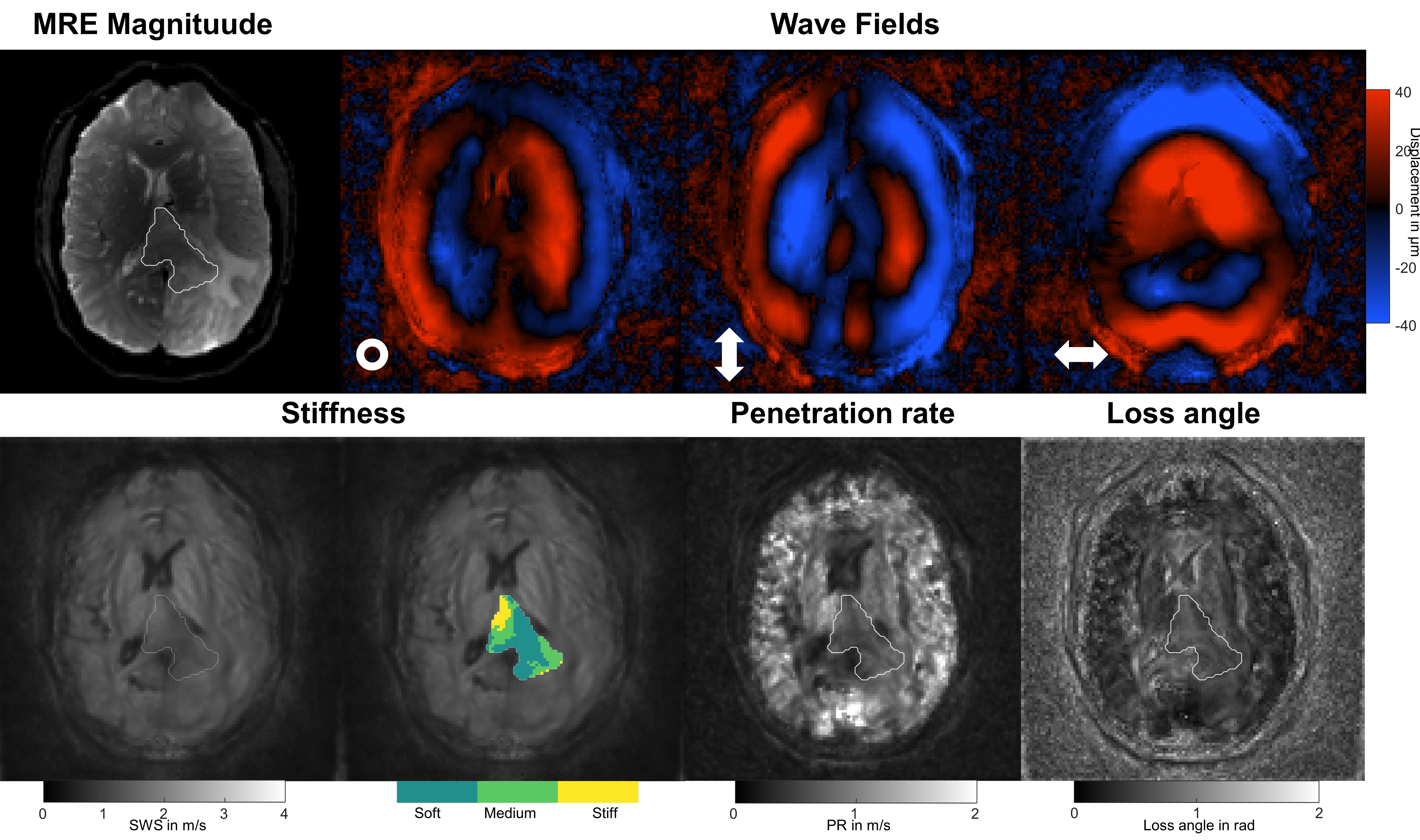
The MRE magnitude, wave fields of a 30 Hz vibration frequency for the three motion encoding components, and the mechanical maps of a patient with a grade II oligodendroglioma (indicated by the VOI) are presented. The tumor exhibits heterogeneous mechanical properties and has been segmented into three regions (indicated by the colored VOI) based on variations in stiffness values.The symbols ʘ, ↔, ↕ illustrate head-feet, left-right, and ventral-dorsal deflections, respectively. SWS, shear wave speed in m/s; PR, penetration rate in m/s; VOI, volume-of-interest.
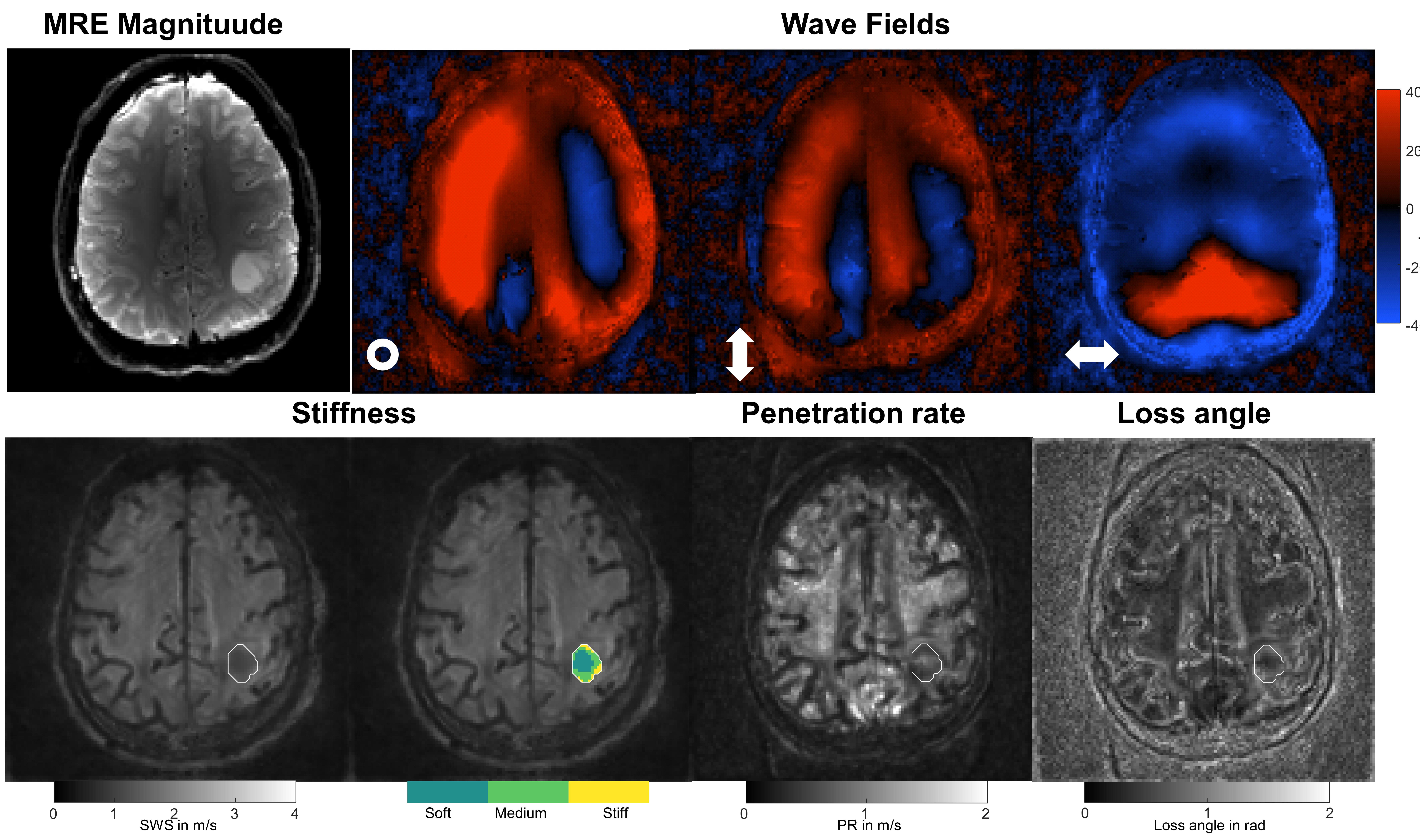
MRE magnitude, wave fields of a 30 Hz vibration frequency for the three motion encoding components, and the mechanical maps of a patient with a grade III oligodendroglioma (indicated by the white VOI) are presented. The tumor exhibits reduced SWS, PR and loss angle values compared to the surrounding tissue. The symbols ʘ, ↔, ↕ illustrate head-feet, left-right, and ventral-dorsal deflections, respectively. SWS, shear wave speed in m/s; PR, penetration rate in m/s; VOI, volume-of-interest.
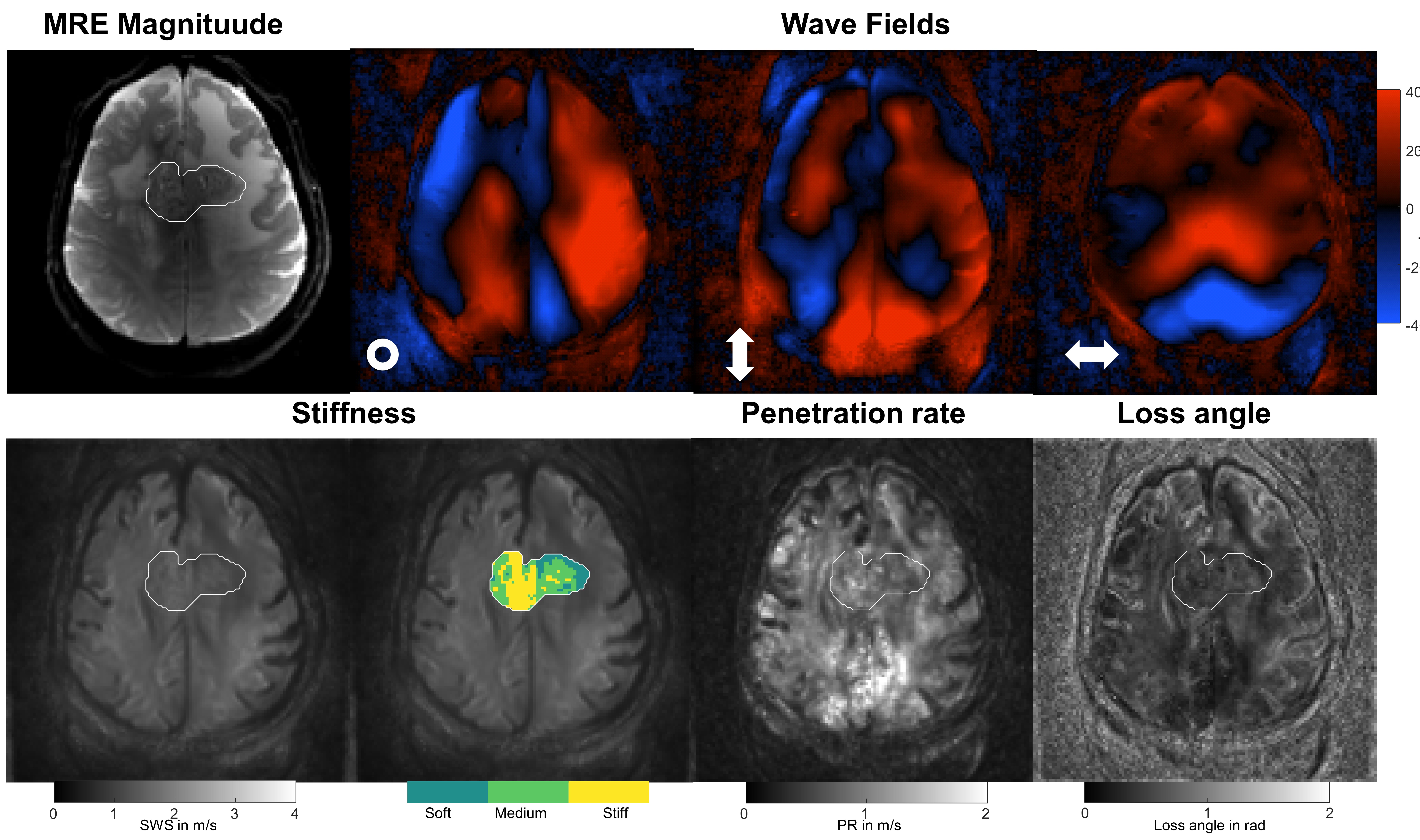
MRE magnitude, MRE wave fields pf a 30 Hz vibration frequency for the three motion encoding components, and the mechanical maps of a patient with glioblastoma (indicated by the white VOI) are presented. The tumor exhibits heterogeneous mechanical properties and appears to have areas with both increased and decreased stiffness compared to the surrounding tissue. The symbols ʘ, ↔, ↕ illustrate head-feet, left-right, and ventral-dorsal deflections, respectively. SWS, shear wave speed in m/s; PR, penetration rate in m/s; VOI, volume-of-interest.
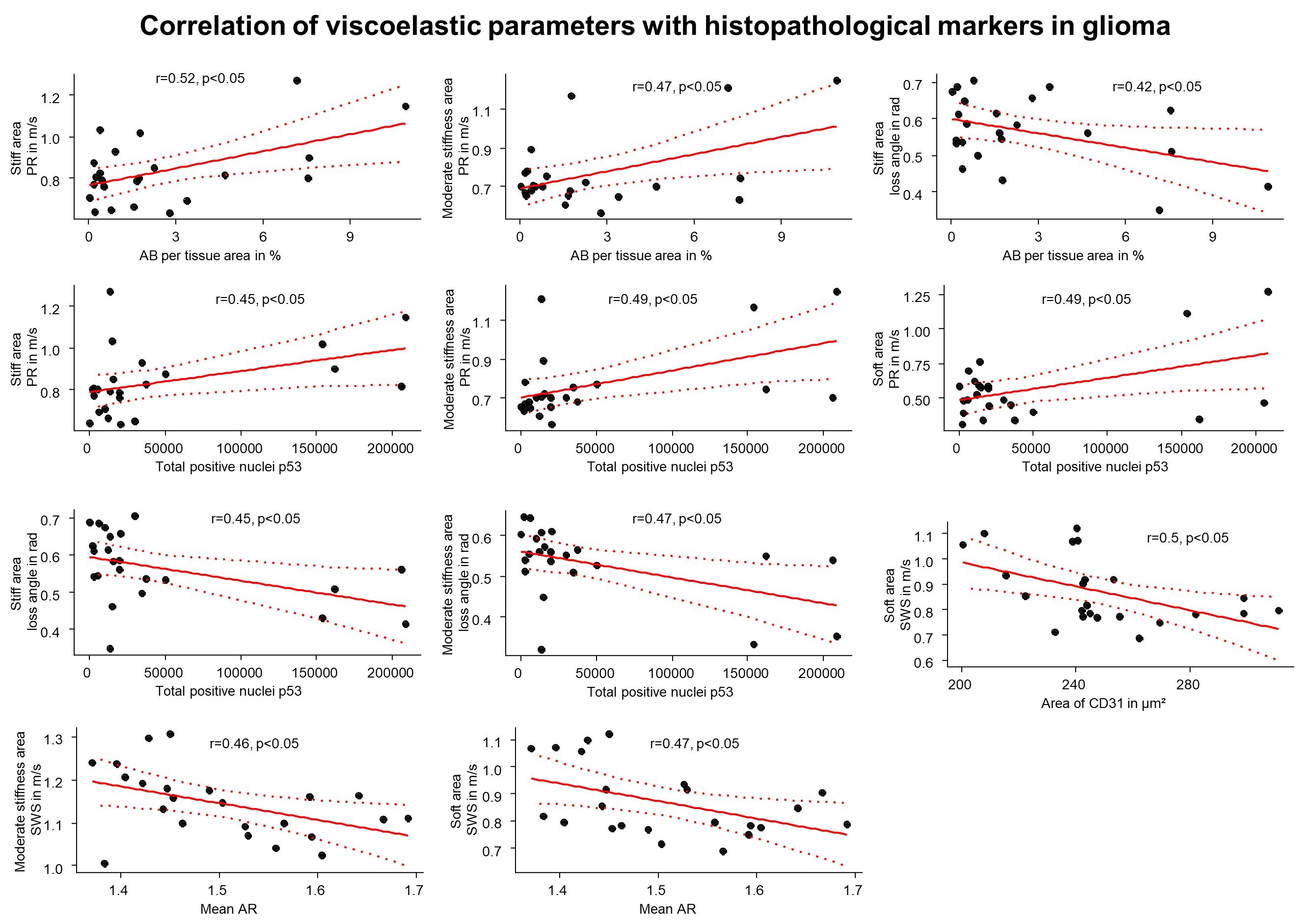
Correlation analyses of quantified histopathological stains and MRE. r- and p-values of the linear regression model with n = 23 given in each panel. 95% confidence intervals are indicated by the red dotted lines. SWS, shear wave speed in m/s; PR, penetration rate in m/s; AB, alcian blue; AR, aspect ratio.