0558
Data-Driven Analysis of Brain Tissue Mechanics using Magnetic Resonance Elastography and Non-Negative Matrix Factorization1Department of Biomedical Engineering, University of Delaware, Newark, DE, United States, 2Department of Mechanical Engineering, University of Delaware, Newark, DE, United States
Synopsis
Keywords: Elastography, Brain
Motivation: There exists a need for a comprehensive method to analyze regional brain tissue mechanics that accounts for variability across subject populations.
Goal(s): Here we aimed to implement a multivariate data-driven technique to capture brain mechanical properties across a wide population while preserving small-scale differences between subjects.
Approach: Non-negative matrix factorization was used to reduce mechanical properties derived from magnetic resonance elastography (MRE) into a low-dimensional form to generate unconfined regions of the brain that demonstrate high covariance across all subjects.
Results: This technique was able to capture recognizable anatomical regions in the brain without structural input to determine weightings on the population average.
Impact: This low-dimensional representation of brain tissue mechanics acquired from non-negative matrix factorization and MRE will help define baseline properties that accurately represent a wide range of subject populations while minimizing variability across imaging studies and contributing to improved statistical models.
Introduction
Magnetic resonance elastography (MRE) is a noninvasive imaging technique used to measure precise mechanical properties of the brain that can inform researchers about microstructural brain tissue health in aging, injury, and disease1. Current MRE applications are based on average property maps acquired from a wide range of subjects that fail to capture individual differences. More recent studies use subject-specific properties to model brain injury mechanics2,3, but could benefit from a compact description of normal mechanical properties and their variability to create more generalized models. Here we seek to describe population variability in brain mechanical properties in a tractable, low-dimensional form4. Non-negative matrix factorization (NMF) can be used to decompose this data into spatial factors and calculate loadings to determine contributions to average mechanical properties, across subjects, from different regions of the brain. NMF has been shown to capture patterns of neuroanatomical variation, and this study will expand that approach to analyze differences in mechanical properties5,6. Individual factors correspond to regions that co-vary between subjects, and we examine which of these factors is most affected by age, which has been shown to correlate with a reduction in brain stiffness1.Methods
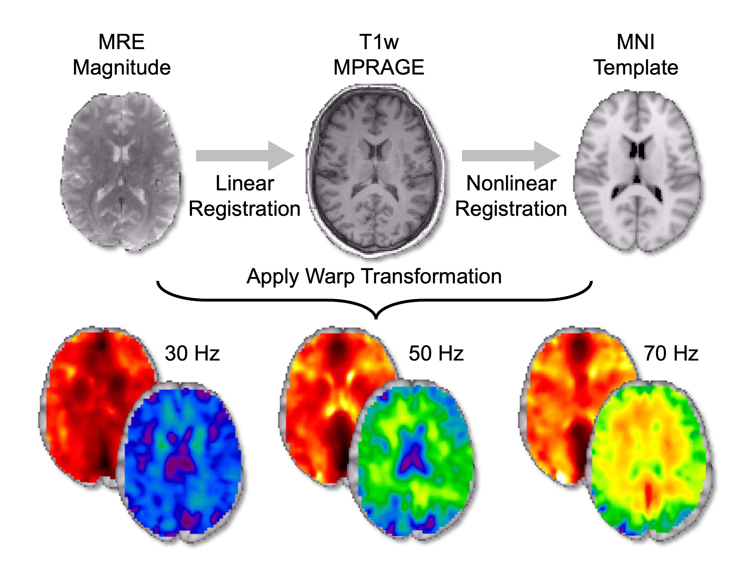
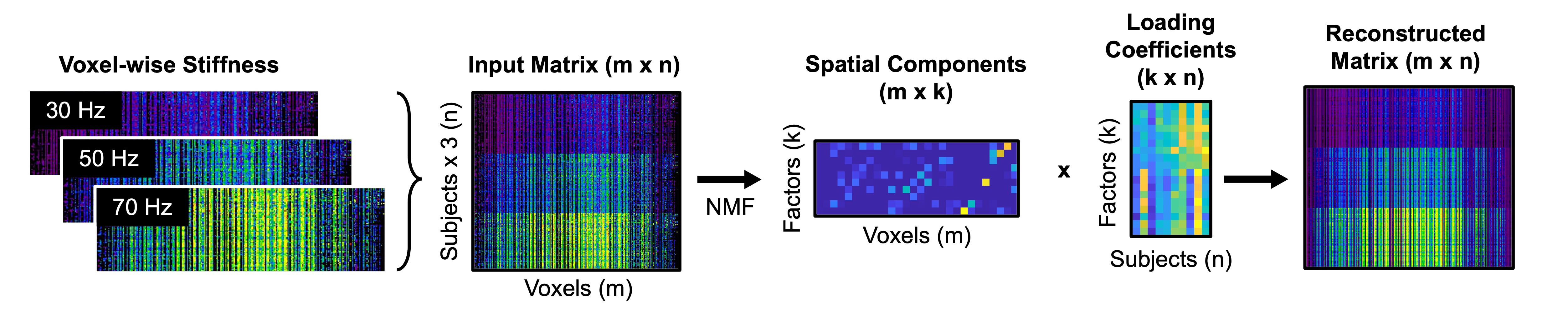
MRE data was collected using a Siemens 3T Prisma scanner with a 64-channel head/neck coil. Vibrations were generated using a Resoundant pneumatic actuation system delivered through a soft pillow driver at 30, 50, and 70 Hz. A 3D multiband, multishot spiral sequence allowed for data collection with an OSCILLATE7 encoding scheme to accelerate acquisition time to under 5 minutes per frequency. This study used data collected from 81 healthy subjects ranging in age from 14-80 years old. A nonlinear inversion algorithm8 was used to estimate material properties of the brain tissue. All data is publicly available at nitrc.org/projects/bbir9.All subject data was registered to standard space using the MNI-152 atlas (Figure 1). Maps of shear stiffness for all subjects at each actuation frequency were combined into a single matrix, $$$X$$$, which was decomposed using an orthogonal projective NMF10 to estimate both spatial factors, $$$W$$$, and factor loadings, $$$H$$$ (Figure 2). We performed NMF using 8 factors and reconstructed the data matrix as $$$\hat{X }=WH$$$. Normalized reconstruction error was determined as $$$\parallel X-\hat{X}\parallel\div{\parallel X\parallel}$$$.
To test the generalizability of the factorization, resulting spatial factors were used to analyze a separate, novel MRE dataset, $$$X_{novel}$$$. Data from a group of healthy older adults (N=61; 60-90 years old)11 was registered and organized in a similar fashion, then projected onto the original factors to estimate a new set of loadings, $$$H_{novel}=W^{T}X_{novel}$$$.
Results and Discussion
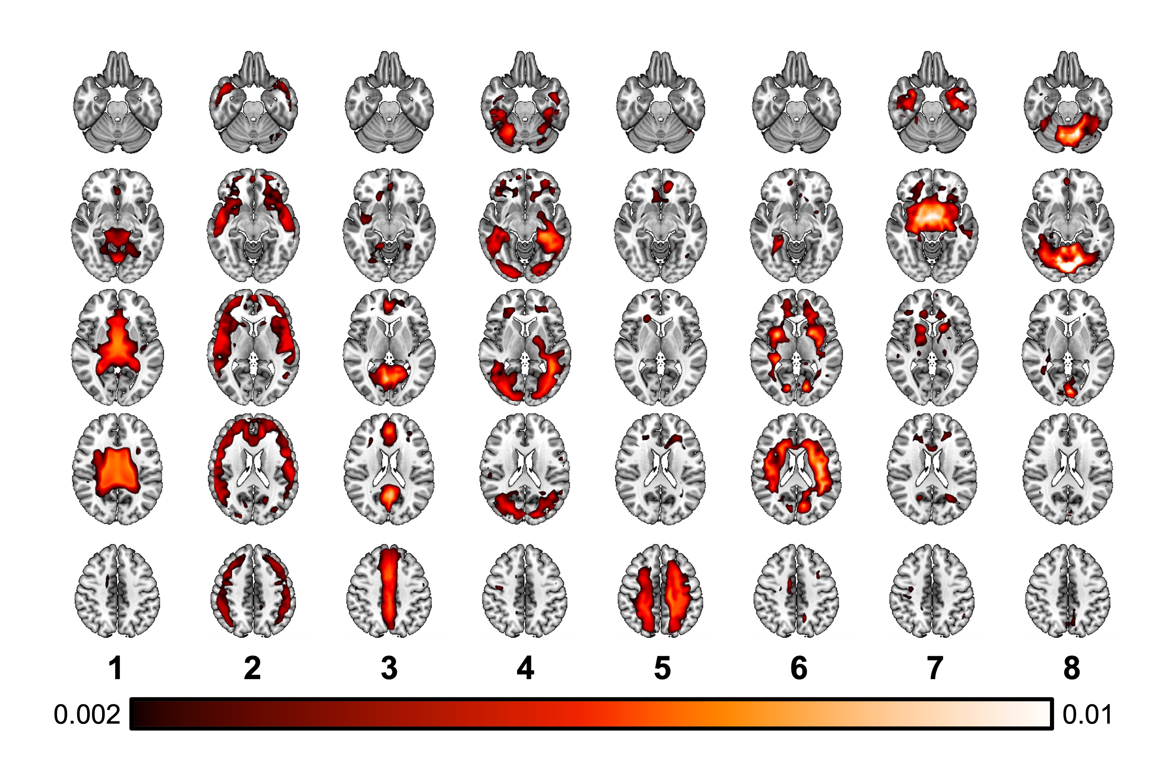
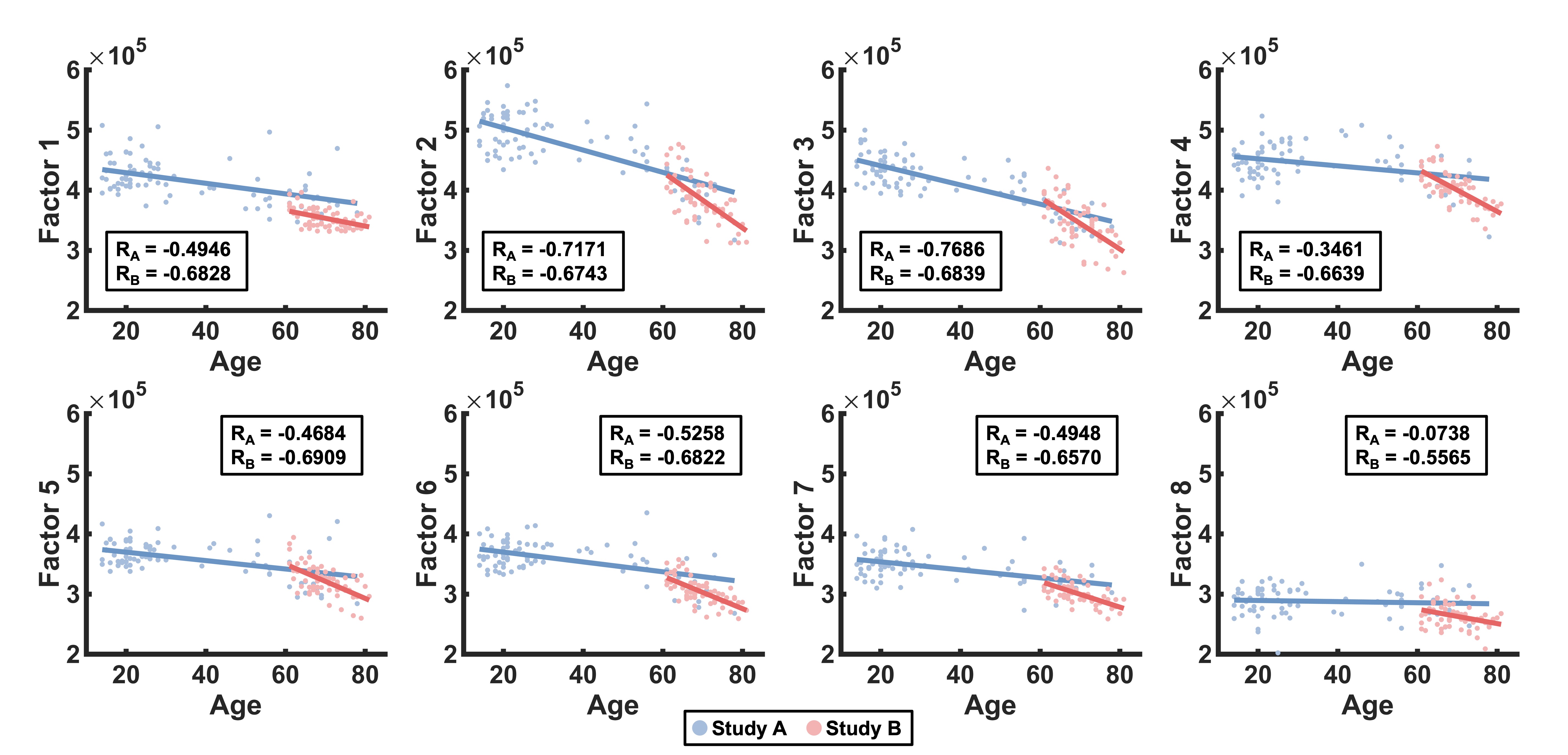
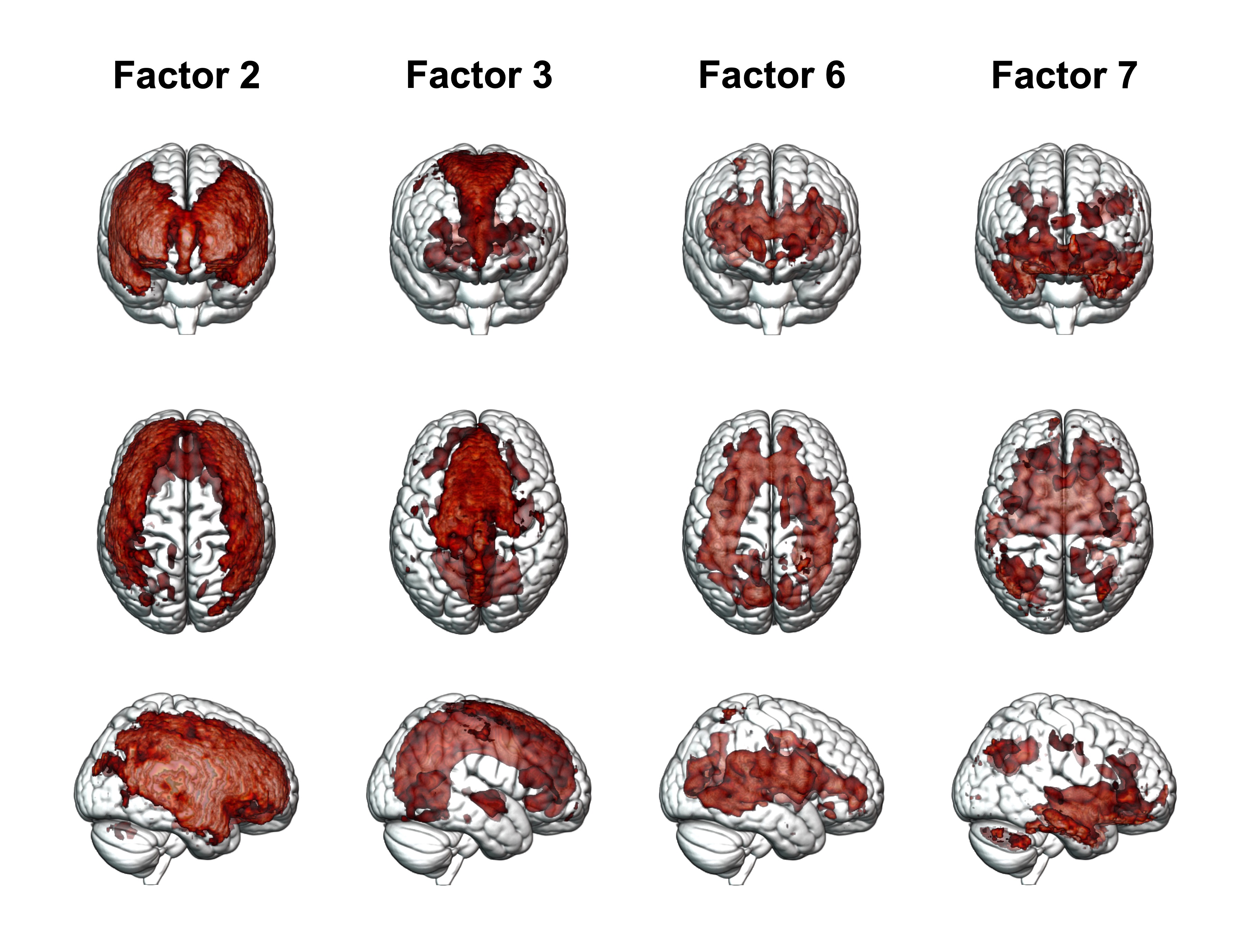
The NMF produced 8 factors that appeared to capture identifiable anatomical regions in the brain which co-vary in shear stiffness across the population. Figure 3 shows resulting spatial components that can be combined to exhibit full-brain coverage with minimal overlap. The cortex of the brain was captured by Factor 2 (frontal/parietal) and Factor 4 (temporal/occipital). The remaining factors tend to correspond with common brain regions such as the ventricles (1), midline (3), white matter tracts (5), subcortical structures (6), brainstem (7) and the cerebellum (8). The projection of these factors onto another subject population was assessed by conducting the same 8-factor NMF on the novel dataset. This demonstrated that the original spatial components were able to successfully reconstruct the novel input matrix within 5% of its baseline reconstruction error. Figure 4 shows how subject loadings are associated with age for both populations. While all regions decreased with age, as expected, some exhibited a stronger correlation. Stiffness values associated with the frontal cortex and periventricular white matter are highly correlated with age (Figure 5), whereas the ventricles and cerebellum remain more constant over time, which was observed in both the original and novel datasets. This is consistent with previous MRE research that did not detect age-related differences in the ventricles and cerebellum1. This technique provides an innovative approach for studying regional variation in the structural significance of brain tissue, and its relation to aging, without being confined to predetermined regions of interest that are often used in this type of analysis.Conclusions
Matrix factorization enables the decomposition of image space to determine contributions from specific brain regions, allowing for standardization across protocols, resulting in decreased variability. Reducing data in this way and applying it to various MRE studies will allow researchers to define baseline mechanical properties of the brain for a given subject population and identify potentially valuable regions for analyzing aging or disease. This analysis can also provide new information for creating computational models of brain biomechanics to understand traumatic brain injury and inform surgical planning.Acknowledgements
This project was supported by NIH grants U01-NS112120 and R01-AG058853, and ONR grant N00014-22-1-2198.References
- Hiscox, LV, Schwarb, H, McGarry, MDJ, & Johnson, CL. Aging brain mechanics: Progress and promise of magnetic resonance elastography. NeuroImage 2021; 232: 117889.
- Upadhyay, K, Alshareef, A, Knutsen AK, Johnson, CL, Carass, A, Bayly, PV, Pham, DL, Prince, JL, & Ramesh, KT. Development and validation of subject-specific 3D human head models based on a nonlinear visco-hyperelastic constitutive framework. J. R. Soc. Interface 2022; 19(195): 20220561.
- Alshareef, AA, Knutsen, AK, Johnson, CL, Carass, A, Upadhyay, K, Bayly, PV, Pham, DL, Prince, JL, & Ramesh, KT. Integrating material properties from magnetic resonance elastography into subject-specific computational models for the human brain. Brain Multiphysics 2021; 2: 100038.
- Lam, F, Li, Y, & Peng, X. Constrained Magnetic Resonance Spectroscopic Imaging by Learning Nonlinear Low-Dimensional Models. IEEE Trans. Med. Imaging 2020; 39(3): 545–555.
- Yang, Z, & Oja, E. Linear and nonlinear projective nonnegative matrix factorization. IEEE Trans. Neural Networks 2010; 21(5): 734–749.
- Sotiras, A, Resnick, SM, & Davatzikos, C. Finding Imaging Patterns of Structural Covariance via Non-Negative Matrix Factorization. NeuroImage 2015; 108: 1-16.
- McIlvain, G, Cerjanic, AM, Christodoulou, AG, McGarry, MDJ, & Johnson, CL. OSCILLATE: A low-rank approach for accelerated magnetic resonance elastography. Magn. Reson. Med. 2022; 88: 1659-1672.
- McGarry, MDJ, Van Houten, EE, Johnson, CL, Georgiadis, JG, Sutton, BP, Weaver, JB, & Paulsen, KD. Multiresolution MR elastography using nonlinear inversion. Med. Phys. 2012; 39(10): 6388–6396.
- Bayly, PV, Alshareef, A, Knutsen, AK, Upadhyay, K, Okamoto, RJ, Carass, A, Butman, JA, Pham, DL, Prince, JL, Ramesh, KT, & Johnson, CL. MR Imaging of Human Brain Mechanics In Vivo: New Measurements to Facilitate the Development of Computational Models of Brain Injury. Ann. Biomed. Eng. 2021; 49(10): 2677–2692.
- Patel, R, Steele, CJ, Chen, AGX, Patel, S, Devenyi, GA, Germann, J, Tardif, CL, & Chakravarty, MM. Investigating microstructural variation in the human hippocampus using non-negative matrix factorization. NeuroImage 2020; 207: 116348.
- Delgorio, PL, Hiscox, LV, McIlvain, G, Kramer, MK, Diano, AM, Twohy, KE, Merritt, AA, McGarry, MDJ, Schwarb, H, Daugherty, AM, Ellison, JM, Lanzi, AM, Cohen, ML, Martens, CR, & Johnson, CL. Hippocampal subfield viscoelasticity in amnestic mild cognitive impairment evaluated with MR elastography. NeuroImage Clin. 2023; 37:103327.
Figures