0555
Acceleration of Magnetic Resonance Elastography using a Novel Distributed Encoding Technique1Biomedical Engineering, University of Delaware, Newark, DE, United States, 2Massachussetts General Hospital, Cambridge, MA, United States, 3Dartmouth College, Hanover, NH, United States
Synopsis
Keywords: Elastography, Elastography
Motivation: Magnetic resonance elastography (MRE) data quality is susceptible to poor data quality from subject motion and long scan times.
Goal(s): A novel sampling technique and estimation scheme was developed and implemented to improve flexibility and acquisition time of MRE.
Approach: The technique utilizes non-traditional sampling directions in an optimized encoding matrix to collect data efficiently to be used in a novel algorithm for estimating harmonic displacement fields. This allows for acquisition acceleration or flexibility in the data sampled to be rejected in post-processing if it is distorted.
Results: Scans were acquired 2.3x faster than standard methods with 95.2% multiscale structural image similarity.
Impact: A novel sampling and estimation scheme demonstrated here can be used to improve the application of magnetic resonance elastography. This is accomplished through prospective reduction in sampling, reducing acquisition time, and retrospective volume rejection, circumventing distortion introduced by subject motion.
Introduction
Tissue mechanical properties are indicative of underlying health and have been used in many diagnostic capacities. Magnetic resonance elastography (MRE) is a non-invasive, in vivo, MRI technique which quantifies mechanical properties such as stiffness by imaging tissue displacement in response to mechanical actuation1,2. In the brain, these MRE-derived properties are sensitive to microstructural changes that differentially occur with aging and disease progression3. One constraint of MRE is long scan times necessary to encode motion in three directions across time with high spatial resolution, limiting clinical adoptability and increasing susceptibility to subject movement. We propose a novel sampling and estimation scheme called EDGE (Elastography with Distributed, Generalized Encoding) that distributes sampling directions and time points across an optimized encoding matrix used to estimate complex, full vector displacement fields. Ultimately this reduces the number of images needed for a full MRE dataset compared to traditional MRE sampling.Methods
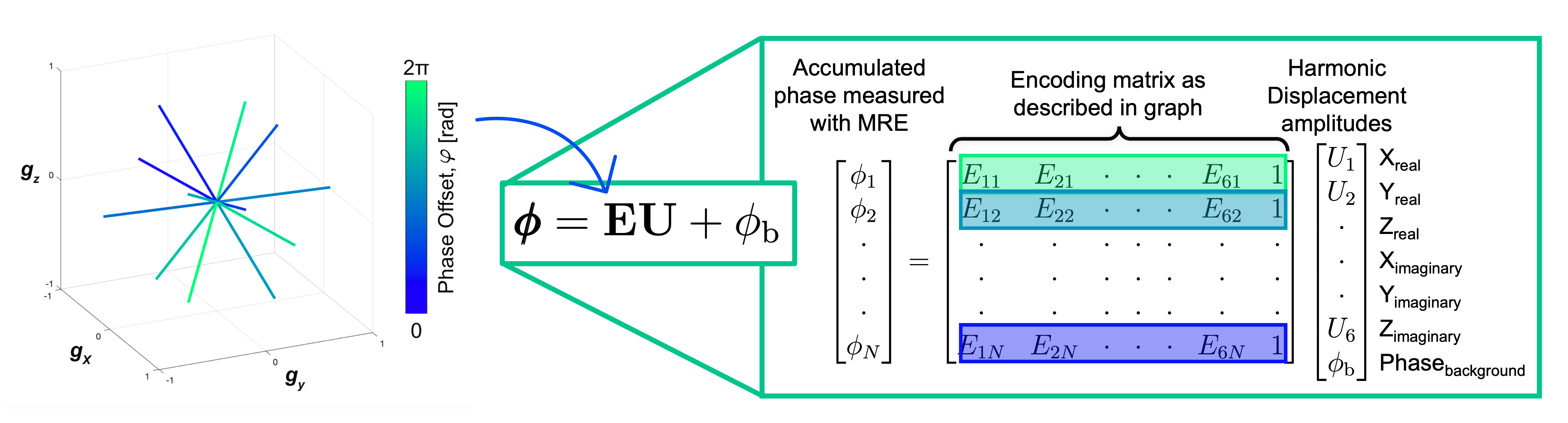
Traditional MRE requires 24-48 images, separately sampling displacement along three orthogonal directions, with positive and negative gradient polarities, and 4-8 phase offsets to capture wave propagation with time2,4. A temporal Fourier transform is used to extract complex displacement fields from these images. This total number of images is greater than the theoretical minimum required for MRE of just 7, accounting for real and imaginary components of three directions of motion, plus background phase5. We propose a generalized encoding scheme (Fig.1) using a set of directions distributed uniformly across a sphere’s surface, as is common in DTI6, with each sample acquired at a different phase offset spread across one period of motion. Thus, each sample includes information from all three components of the displacement vector, both real and imaginary. We formulate the estimation of motion from these samples as an optimization problem using a general encoding matrix and minimizing the objective function, following analogous examples in field mapping by Fessler et al7,8, in an approach similar to work by Trzasko et al9–11. EDGE minimizes distortion from potential subject motion by shortening scan time and introducing the option to retrospectively reject distorted images.We tested EDGE performance with realistic simulated brain data12 and proved its feasibility with real data acquired using phantoms and human subjects. We used Ndirs=24 phase images as the fully sampled EDGE condition, mimicking the number of images typical of traditional MRE, and Ndirs=20,14,10,7 for EDGE undersampled conditions. Final encoding matrices for each number of directions were optimized using the minimum condition number of the matrix. Traditional EPI MRE and EDGE with Ndirs=24,20,14,10,7 were acquired at 2.5mm isotropic resolution with FOV of 240x240x100mm3. Traditional MRE and fully sampled EDGE had acquisition time (TA) of 2min19s, and EDGE with the minimum sampling had TA of 58s. Stiffness maps were generated using a nonlinear inversion algorithm13 and compared with traditional MRE data.
To demonstrate how EDGE can enhance motion robustness, we retrospectively deleted images from the Ndirs=24 case prior to estimating displacement fields, simulating rejection of volumes deemed corrupted by motion.
Results
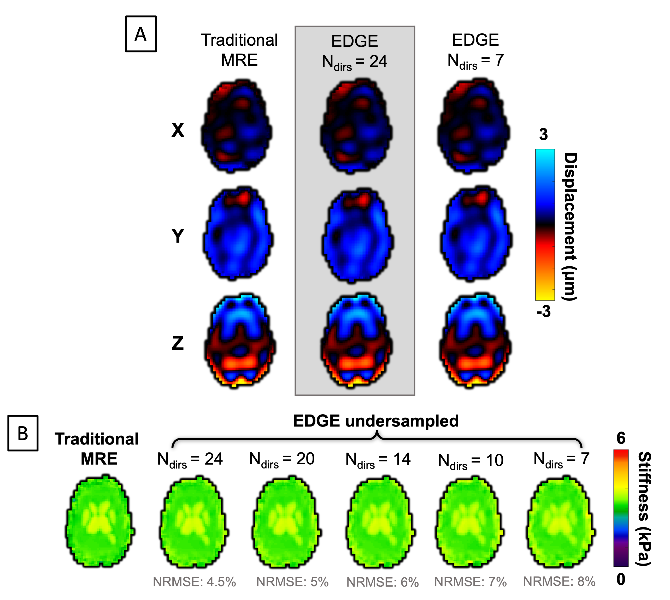
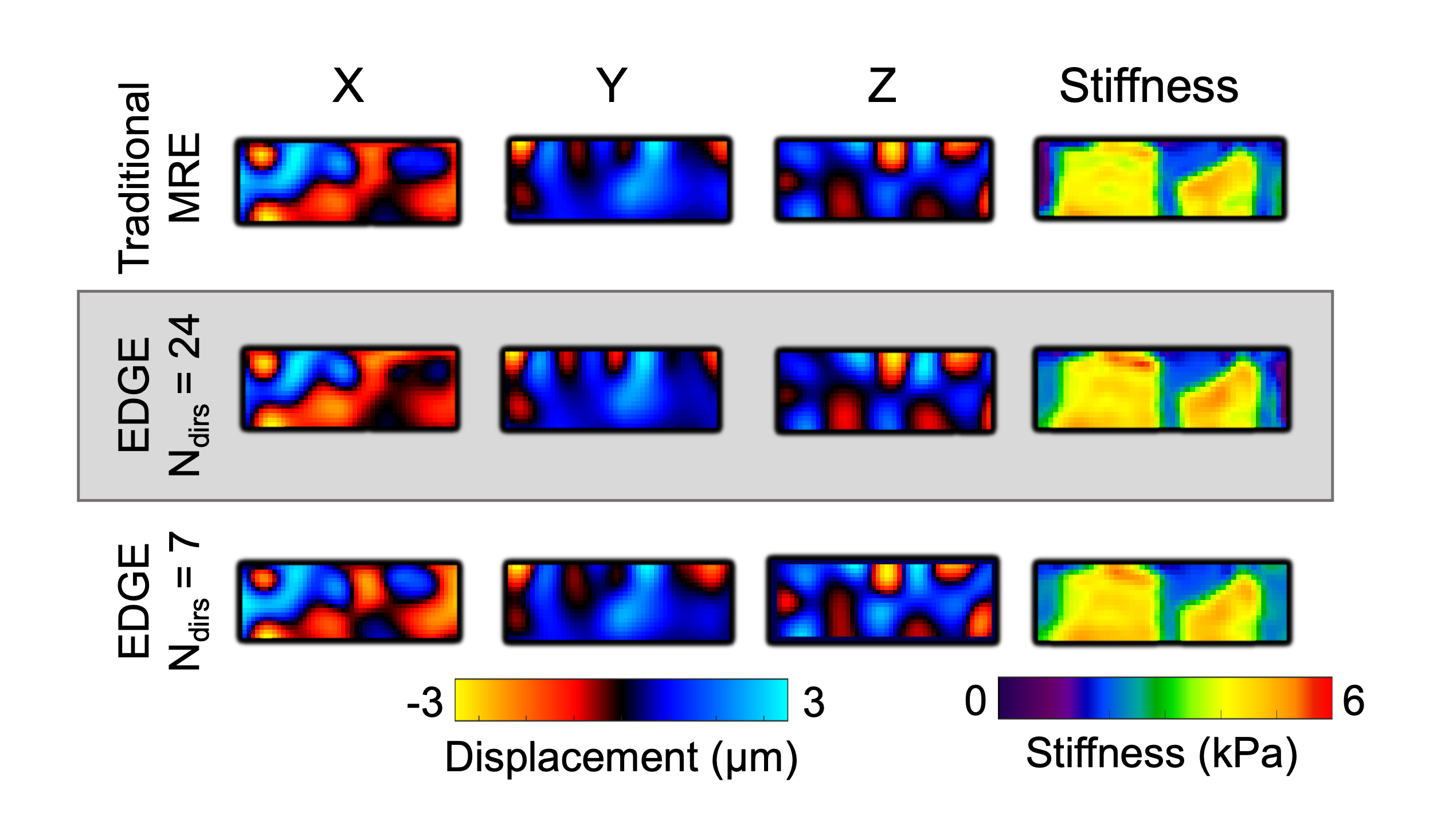
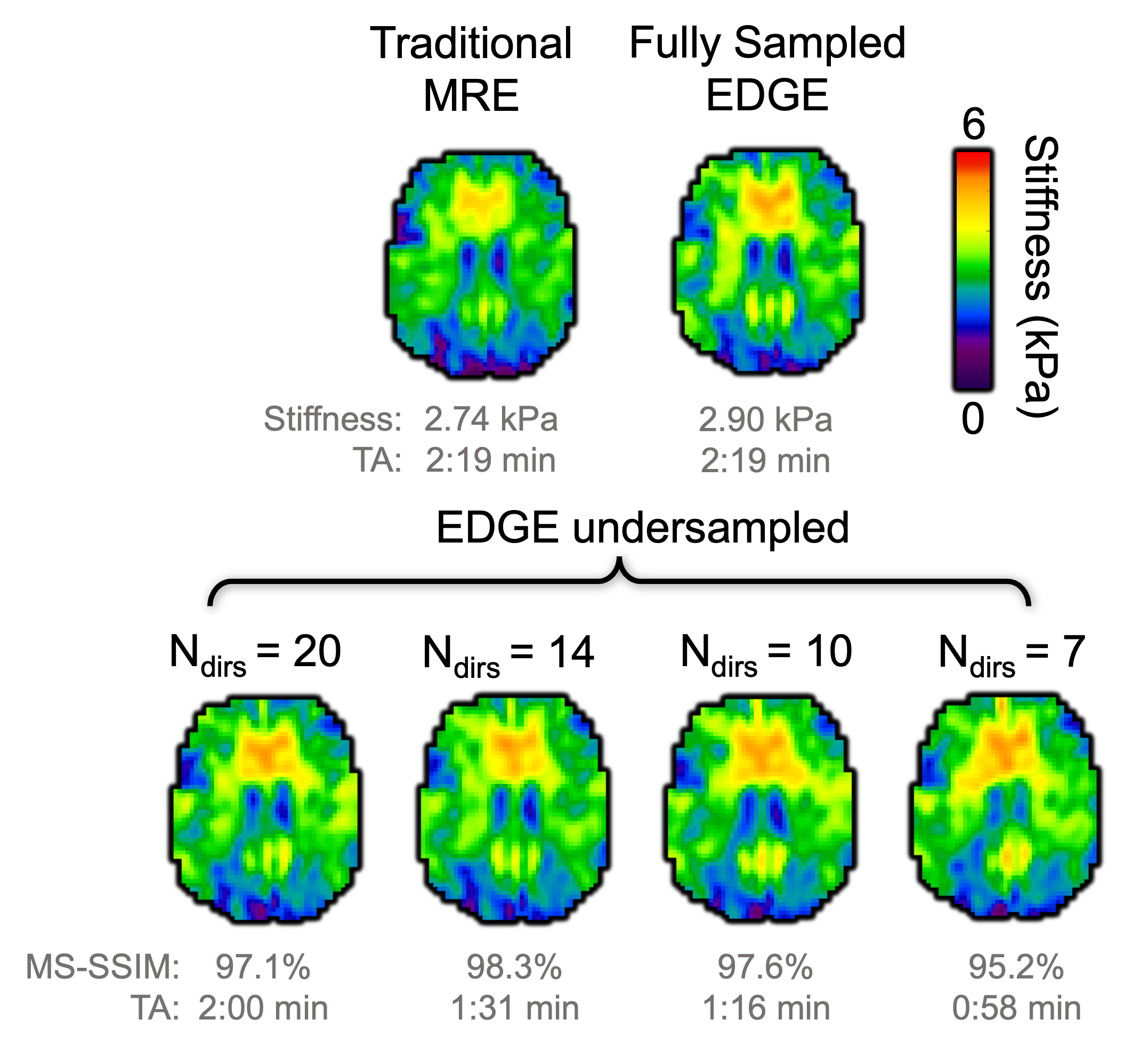
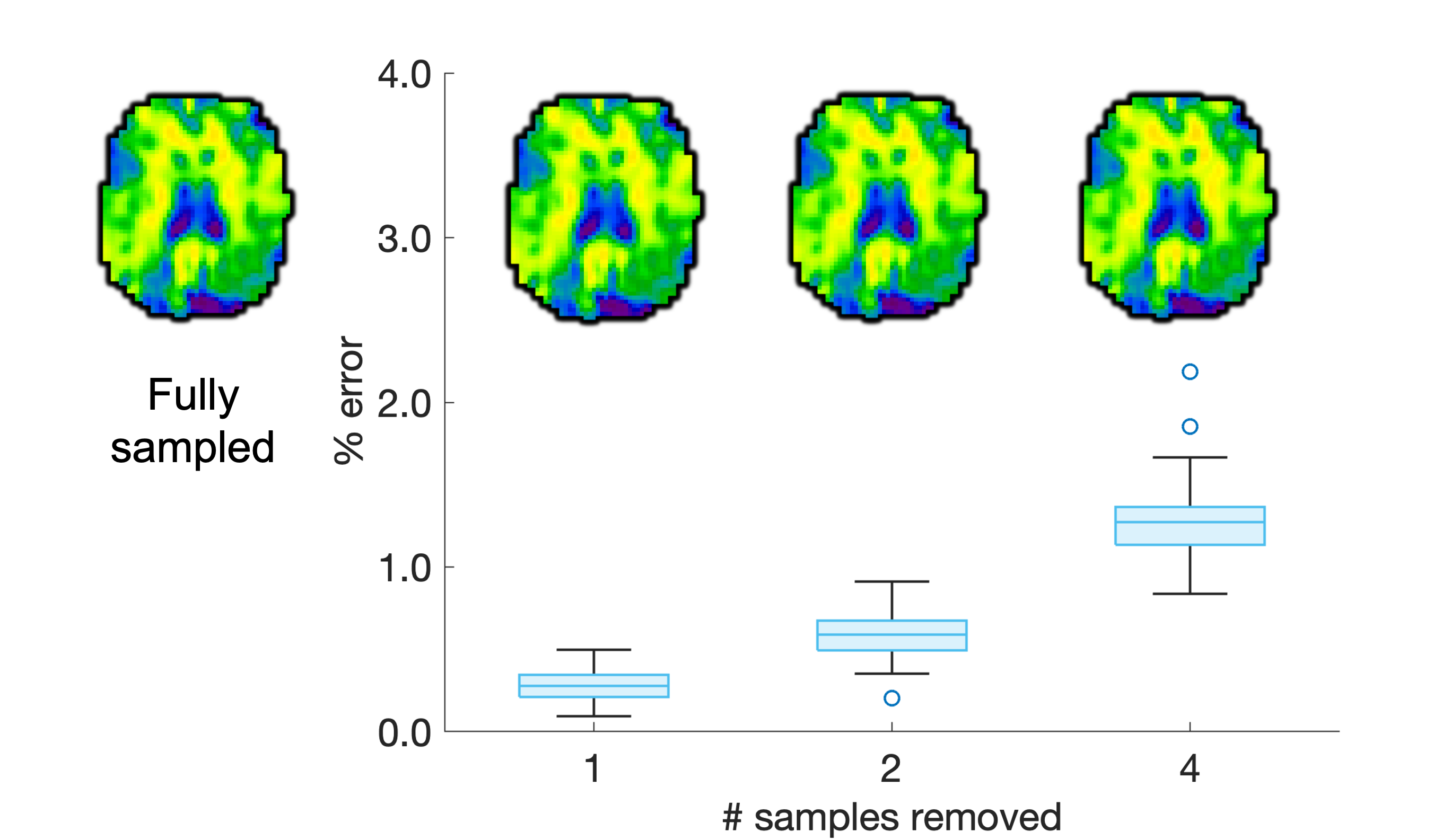
Stiffness maps and displacement fields generated with EDGE data showed good agreement with traditional MRE methods, even after maximum undersampling. With simulations, maximum undersampling with EDGE resulted in a normalized root mean square error (NRMSE) of 8% compared to traditional MRE (Fig.2). With phantom data, multiscale structural similarity (MS-SSIM)14 of 97.4% was found with fully sampled EDGE, and 96% using Ndirs=7 providing 2.3x acceleration (Fig.3). In human data, MS-SSIM of 97.1% was found for fully sampled EDGE data, and 95.2% was found with Ndirs=7 (Fig.4). Furthermore, we showed retrospective rejection of data was possible while maintaining whole-image error of less than 1.5% in stiffness maps reconstructed with 4 samples randomly removed from the fully sampled EDGE case (Fig.5).Discussion and Conclusions
EDGE sampling and displacement estimation allows robust MRE results to be acquired with reduced TA. While the algorithm is similar to work by Trzasko et al9–11, using a distributed set of encoding directions overcomes the major limitation of previous work. Traditional MRE encoding results in infinite local minima when phase wrapping occurs in the brain, which is unwrappable due to bulk motion15. This can only be overcome by including scans with two different motion sensitivities (gradient strengths)16,17, but EDGE sampling effectively includes high and low sensitivity motion for each component automatically, resulting in a solution space with one global minimum, without additional scans. Future improvements to EDGE include optimization of parameters such as a regularization term to stabilize results and minimize effects of noise. Implementation of EDGE will enable prospective acceleration or retrospective volume rejection, significantly advancing the adoptability of MRE.Acknowledgements
Funding for this work provided by F31-AG086036, R01-AG058853, R01-EB027577, and U01-NS112120.References
1. Muthupillai, R. et al. Magnetic resonance elastography by direct visualization of propagating acoustic strain waves. Science. 269, 1854–1857 (1995).
2. Manduca, A. et al. Magnetic resonance elastography: Non-invasive mapping of tissue elasticity. Med. Image Anal. 5, 237–254 (2001).
3. Hiscox, L. V., Schwarb, H., McGarry, M. D. J. & Johnson, C. L. Aging brain mechanics: Progress and promise of magnetic resonance elastography. Neuroimage 232, (2021).
4. Nir, G., Sahebjavaher, R. S., Sinkus, R. & Salcudean, S. E. A framework for optimization-based design of motion encoding in magnetic resonance elastography. Magn. Reson. Med. 73, 1514–1525 (2015).
5. Trzasko, J. D. Robust Harmonic Estimation for MR Elastography: Application to Brain. in International Society of Magnetic Resonance in Medicine 2518 (2015).
6. Jones, D. K., Horsfield, M. A. & Simmons, A. Optimal strategies for measuring diffusion in anisotropic systems by magnetic resonance imaging. Magn. Reson. Med. 42, 515–525 (1999).
7. Funai, A. K., Fessler, J. A., Yeo, D. T. B., Noll, D. C. & Olafsson, V. T. Regularized field map estimation in MRI. IEEE Trans. Med. Imaging 27, 1484–1494 (2008).
8. Lin, C. Y. & Fessler, J. A. Efficient Regularized Field Map Estimation in 3D MRI. IEEE Trans. Comput. Imaging 6, 1451–1458 (2020).
9. Trzasko, J. D. & Manduca, A. Regularized Harmonic Estimation for Steady-State MR Elastography. Proc. Int. Soc. Magn. Reson. Med. 20, 3425 (2012).
10. Trzasko, J. D. et al. A graph cut approach to regularized harmonic estimation for steady state MRE. Proc. Int. Soc. Magn. Reson. Med. 22, 4268 (2014).
11. Trzasko, J. D. et al. Robust Harmonic Estimation for MR Elastography: Application to Brain. Proc. Int. Soc. Magn. Reson. Med. 24, 1962 (2016).
12. McGarry, M. D. J. et al. A heterogenous, time harmonic, nearly incompressible transverse isotropic finite element brain simulation platform for MR elastography. Phys. Med. Biol. 66, 055029 (2021).
13. McGarry, M. D. J. et al. Multiresolution MR elastography using nonlinear inversion. Med. Phys. 39, 6388–6396 (2012).
14. Wang, Z., Simoncelli, E. P. & Bovik, A. C. Multi-Scale Structural Similarity for Image Quality Assessment. Thirty-Seventh Asilomar Conf. Signals, Syst. Comput. 1398–1402 (2003).
15. Badachhape, A. A. et al. The Relationship of Three-Dimensional Human Skull Motion to Brain Tissue Deformation in Magnetic Resonance Elastography Studies. J. Biomech. Eng. 139, 1–12 (2017).
16. Herthum, H. et al. Multiple motion encoding in phase-contrast MRI: A general theory and application to elastography imaging. Med. Image Anal. 78, 102416 (2022).
17. Yin, Z. et al. In vivo characterization of 3D skull and brain motion during dynamic head vibration using magnetic resonance elastography. Magn. Reson. Med. 80, 2573–2585 (2018).
Figures