0554
Magnetic resonance imaging of macrophage response to radiation therapy1University of Kentucky, Lexington, KY, United States, 2University of California San Diego, San Diego, CA, United States
Synopsis
Keywords: Biomarkers, Contrast Agent
Motivation: Cellular response to cancer treatment is difficult to track in real time. Standard practice involves immunostaining of a biopsied tumor, but this is severely limited by a number of factors.
Goal(s): Non-invasive imaging methods such as MRI could obviate the need for biopsies and serve as a biomarker of radiation therapy efficacy.
Approach: This study aimed to observe macrophage response in a mouse model by use of a fluorine nanoemulsion and 19F MRI.
Results: It was shown that macrophage recruitment can be quantified through MRI. Moreover, the findings suggest that macrophage response to radiation therapy is dependent on several factors including tumor origin.
Impact: Our results demonstrate the potential of 19F MRI to non-invasively track macrophages during radiation therapy and its prognostic value with regards to tumor growth. This technique will be extremely beneficial in future analysis of inflammation’s role in tumor recurrence.
Introduction
Surgery, chemotherapy, and radiotherapy (RT) are all mainstay modalities for treating cancer. The body responds to these therapies through immune responses and distinguishing between antitumor and tumor promoting immune responses can be difficult. Standard practice of macrophage quantification currently involves immunostaining of a biopsied tumor. However, this technique is fraught with numerous limitations, including the invasive nature of the test, heterogeneous tumor associated macrophage (TAM) distribution in primary and metastatic lesions, and lack of a clear cut-off point for positive staining. An imaging biomarker capable of non-invasively depicting and quantifying tumor associated macrophages and de novo macrophages infiltrating the tumor in vivo could obviate the need for such invasive means.Methods
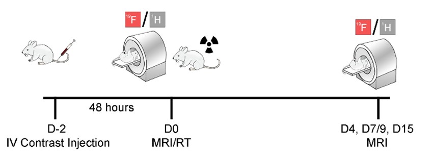
In this study, a fluorine nanoemulsion (perfluoro-crown-ether formulated with egg yolk phospholipid) was intravenously delivered to mice bearing breast or colorectal tumors to label and subsequently track macrophage dynamics in response to radiation therapy1-3. Murine colon cancer cell line MC38 and murine breast cancer cell line 4T1 were selected due to their high worldwide incidence and frequent treatment with radiation therapy (RT). Ten Balb/c mice received unilateral mammary fat pad injection of 5 × 10⁵ 4T1 cells in 100 µL PBS. Ten C57BL/6J mice received unilateral mammary fat pad injection of 5 × 10⁵ MC38 cells in 100 µL PBS. Tumors we allowed to grow for 4-5 days prior to radiation therapy start. Two days prior to radiation (D-2, Fig.1), all mice received intravenous injection of fluorine nanoemulsion (200 µL, C = 150 mg/ml). After baseline MR imaging, five mice in each tumor model were irradiated with a single 8 Gy dose over an approximately four-minute interval. Mice were monitored for two weeks for tumor growth and 19F signal using a 7T Bruker MR Scanner with a dual‐tune 1H/19F volume coil. To gauge the power of our imaging method, we ran correlation tests between the fluorine signal detected and tumor burden irrespective of the timepoint.Results
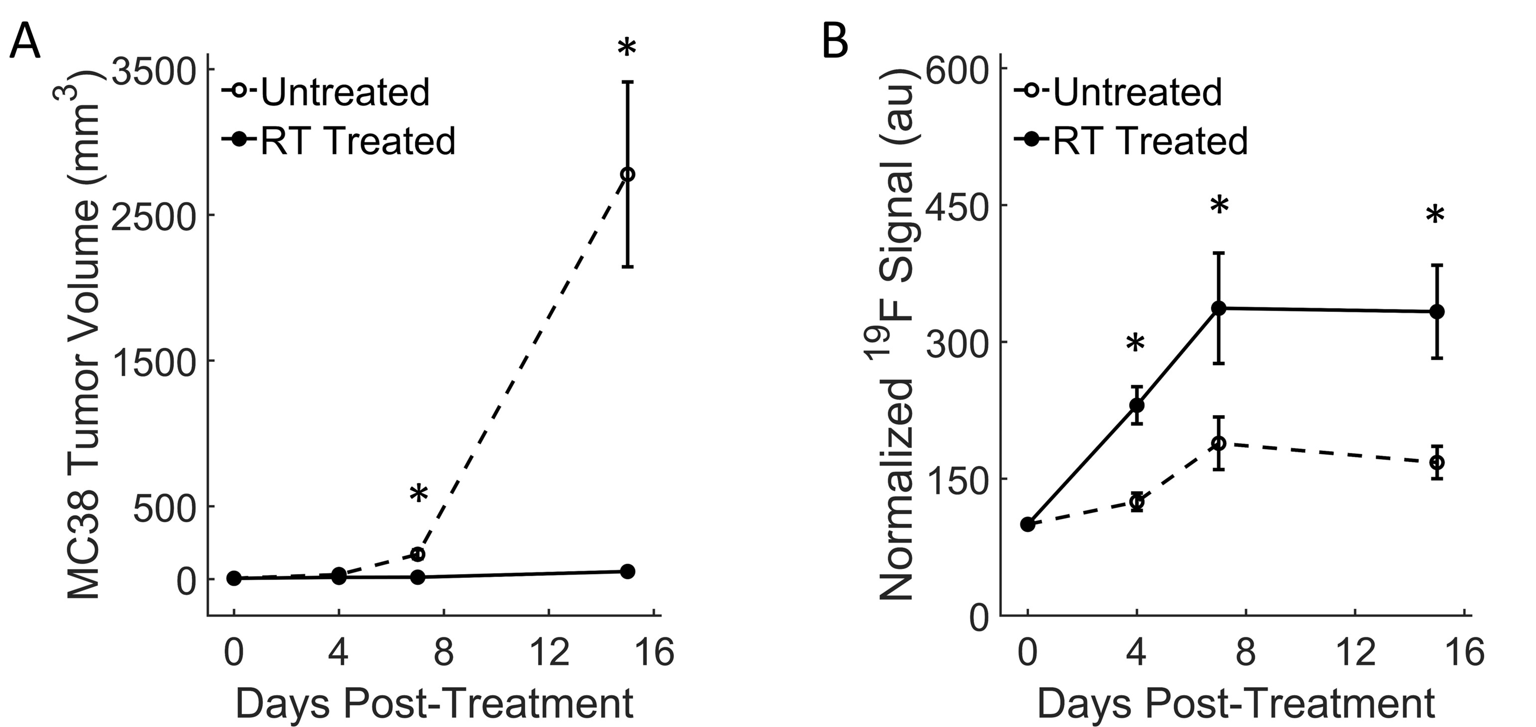
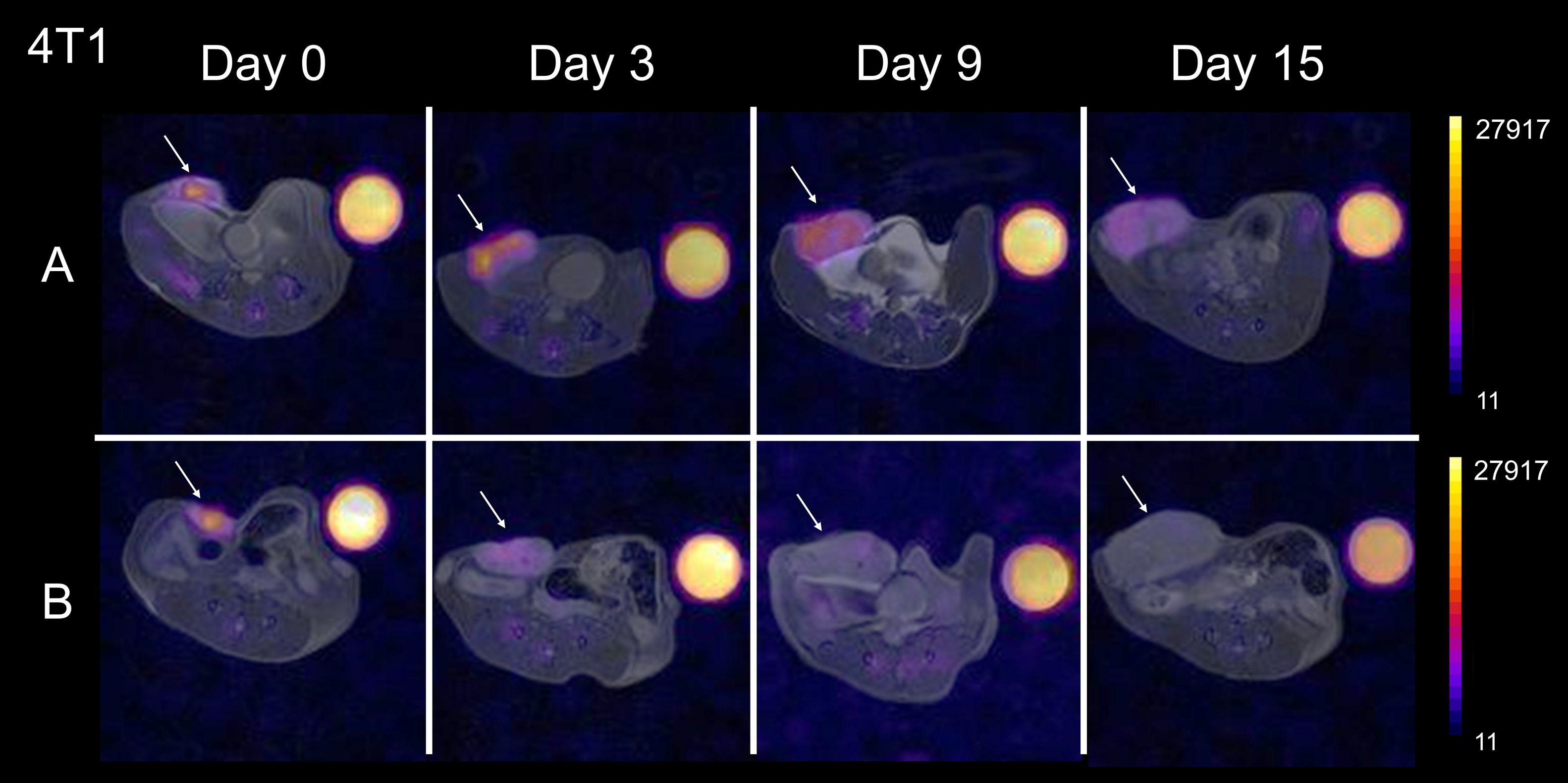
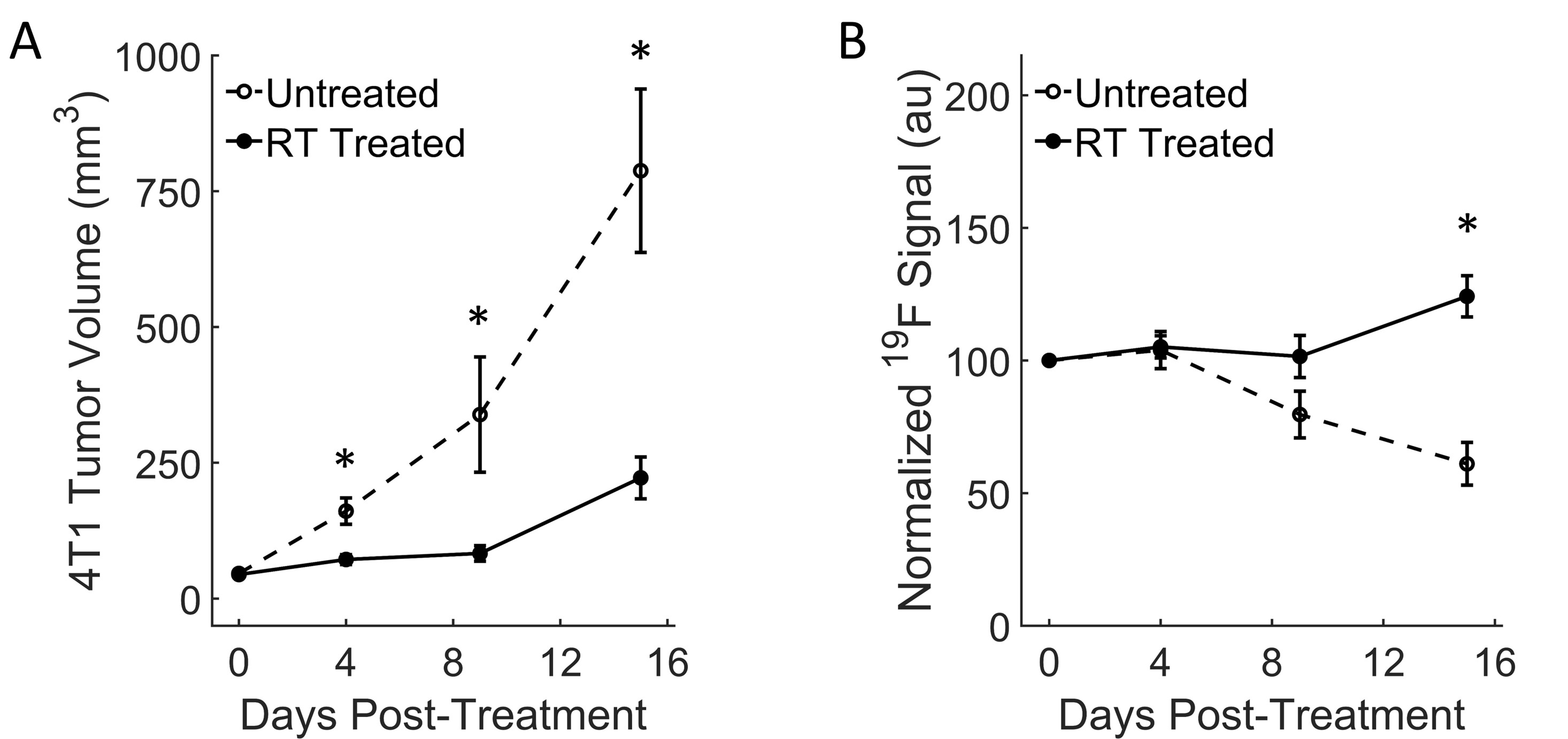
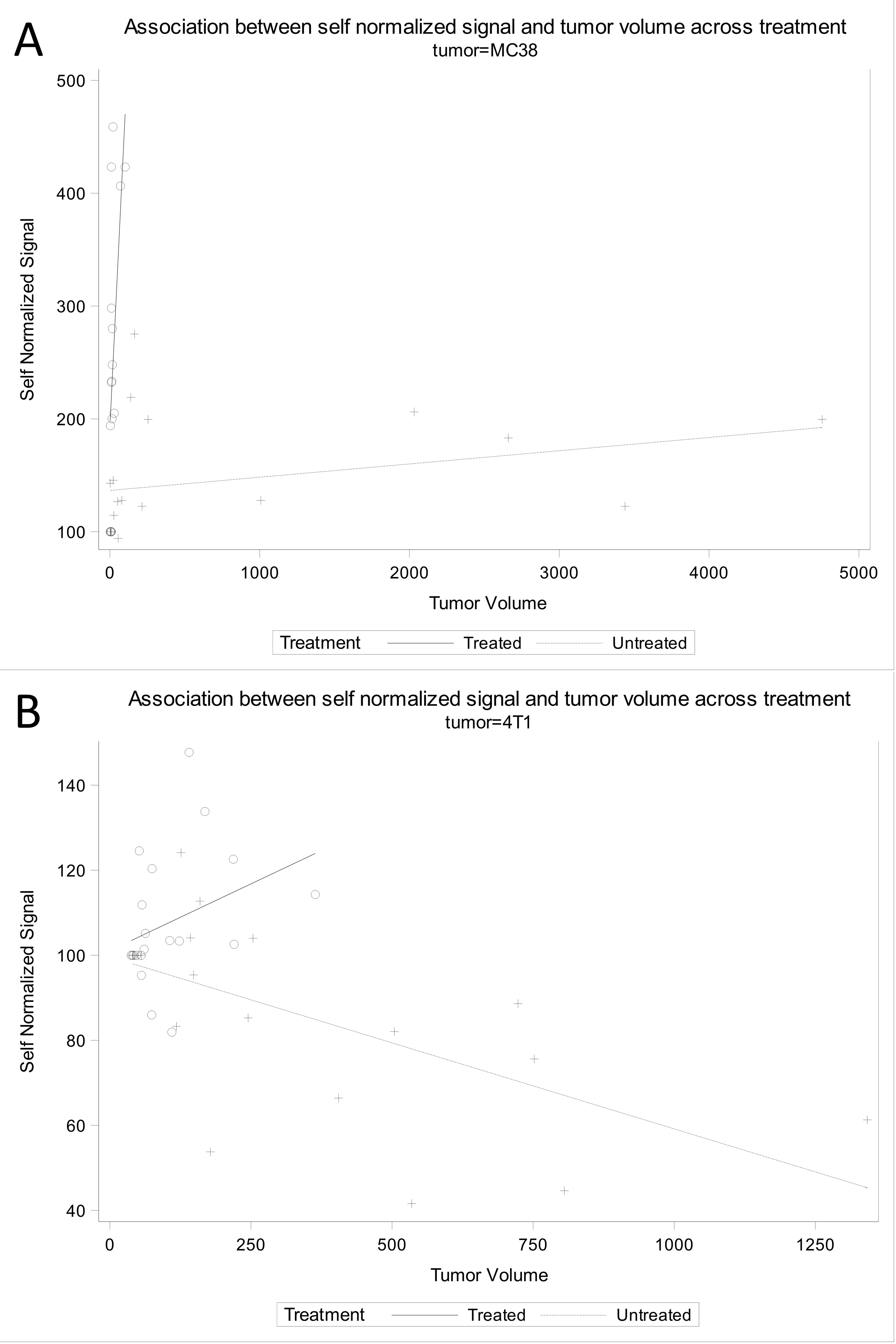
Colon tumors displayed significant tumor growth reduction following high dose radiation therapy as early as 7 days post-RT (*p=0.01, Fig. 2A). Normalized fluorine signal measurements in tumors increased significantly in the RT treated group as early as day 4 post-treatment (*p=0.01, Fig. 2B) and continued to remain significantly higher than the untreated group at days 7 and 15 (*p = 0.05 and *p=0.02, respectively). This indicates significant macrophage influx in the treated tumors, which concurs with previous literature4, 5. In the breast cancer model, baseline (day 0) fluorine signal was higher for all mice compared to the colon cancer model (Fig. 3). In the treated group, signal intensity appeared to persist or increase with respect to tumor growth (Fig. 3A), whereas signal decreased in the control group (Fig. 3B). Conversely to the colon cancer model, all mice bearing 4T1 tumors displayed slower growth but persistent growth, even after therapy. Quantitatively, tumor growth reduction in the RT-treated mice was significant as early as day 4 post-therapy (*p=0.009, Fig. 4A) and remained significantly different throughout the experiment. In 4T1 tumors, macrophage influx seemed slower compared to the MC38 model, as significant signal change only occurred 15 days post-RT (*p=0.0005, Figs. 2B&4B). Spearman’s correlation coefficients were significant for all treatment groups (p<0.03, Fig. 5).Discussion
This study is the first to correlate macrophage signal change to tumor volume and a first step towards accurate non-invasive assessment of RT efficacy. A significant change in 19F signal was found between radiotherapy treated and untreated groups for both the breast and colon cancer models two weeks after irradiation. Treated groups in both cohorts also displayed significant tumor regression. Fluorine signal change correlated to tumor volumes, indicating that MR imaging of macrophage recruitment in tumors may be a valid biomarker of RT efficacy. Additionally, tumors possess apparent mechanistic differences depending on their origin and have therefore varied responses to RT. Therefore, it was imperative to analyze more than one tumor model to acquire a better understanding of differences in radiation-induced TAM involvement.Conclusion
Our findings support the potential therapeutic benefit of incorporating MRI of macrophage dynamics in RT workflow. Non-invasive imaging tools are becoming increasingly necessary to provide safe and accurate tumor prognosis. Finding ways to increase probe specificity to distinguish macrophage phenotypes will be the next challenge but ultimately can dramatically better patient outcomes.Acknowledgements
This study was supported by the National Center for Research Resources and the National Center for Advancing Translational Sciences, National Institutes of Health, through Grant UL1TR001998, by grant # IRG-19-140-31 from the American Cancer Society and by the Shared Resource Facilities of the University of Kentucky Markey Cancer Center P20CA177558. H. Y. received support from the Commonwealth Undergraduate Research Experience (CURE) Fellowship for this project. The content is solely the responsibility of the authors and does not necessarily represent the official views of the NIH.References
(1) Yang, R.; Sarkar, S.; Yong, V. W.; Dunn, J. F. In Vivo MR Imaging of Tumor-Associated Macrophages: The Next Frontier in Cancer Imaging. Magn Reson Insights 2018, 11, 1178623X18771974. DOI: 10.1177/1178623X18771974 From NLM PubMed-not-MEDLINE.
(2) Daldrup-Link, H. E.; Golovko, D.; Ruffell, B.; Denardo, D. G.; Castaneda, R.; Ansari, C.; Rao, J.; Tikhomirov, G. A.; Wendland, M. F.; Corot, C.; et al. MRI of tumor-associated macrophages with clinically applicable iron oxide nanoparticles. Clin Cancer Res 2011, 17 (17), 5695-5704. DOI: 10.1158/1078-0432.CCR-10-3420.
(3) Khurana, A.; Chapelin, F.; Xu, H.; Acevedo, J. R.; Molinolo, A.; Nguyen, Q.; Ahrens, E. T. Visualization of macrophage recruitment in head and neck carcinoma model using fluorine-19 magnetic resonance imaging. Magnetic resonance in medicine 2017. DOI: 10.1002/mrm.26854.
(4) Croci, D.; Santalla Mendez, R.; Temme, S.; Soukup, K.; Fournier, N.; Zomer, A.; Colotti, R.; Wischnewski, V.; Flogel, U.; van Heeswijk, R. B.; et al. Multispectral fluorine-19 MRI enables longitudinal and noninvasive monitoring of tumor-associated macrophages. Sci Transl Med 2022, 14 (667), eabo2952. DOI: 10.1126/scitranslmed.abo2952 From NLM Medline.
(5) Beach, C.; MacLean, D.; Majorova, D.; Arnold, J. N.; Olcina, M. M. The effects of radiation therapy on the macrophage response in cancer. Front Oncol 2022, 12, 1020606. DOI: 10.3389/fonc.2022.1020606 From NLM PubMed-not-MEDLINE.
Figures