0553
Assessing pathological prognostic factors of resectable rectal cancer: comparison of a monoexponential model and multiple advanced DWI models1Department of Radiology, Sichuan Provincial People's Hospital, University of Electronic Science and Technology of China, Chengdu, China, 2MR Research Collaboration, Siemens Healthineers, Chengdu, China, 3MRI clinical application, Customer Service Department, Siemens Digital Medical Technology Co., LTD, Shanghai, China
Synopsis
Keywords: Biomarkers, Diffusion/other diffusion imaging techniques
Motivation: Diffusion-weighted imaging (DWI) can help to predicti rectal cancer prognosis; however, inconsistencies in its effectiveness necessitate exploring advanced DWI models to improve diagnostic accuracy.
Goal(s): We investigated the value of the stretched exponential (SEM), fractional-order calculus (FROC), and continuous-time random-walk (CTRW) models in assessing prognostic factors for rectal cancer.
Approach: This study included 181 rectal cancer patients with using traditional and advanced DWI models to explore correlations between DWI parameters and histopathological indicators.
Results: The parameters of advanced DWI models were significantly correlated with histopathological prognostic factors. CTRW-α was the superior predictor for histological type and pT stage.
Impact: Our study demonstrates the value of CTRW model in predicting prognostic factors for rectal cancer. Utilizing these noninvasive and accurate advanced DWI models before surgery can help physicians to make appropriate surgical plan to help patients achieve better prognosis.
Introduction
Colorectal cancer (CRC) is the third most common cancer worldwide and the second leading cause of cancer-related deaths, of which, approximately one-third are related to rectal cancer. The prognosis is affected by several factors, including tumor-node-metastasis (TNM) stage and histological type, which are widely accepted as main prognostic factors and are recommended by the National Comprehensive Cancer Network1. Accurate preoperative assessments of these prognostic factors are crucial for improving the prognoses of patients with CRC. Magnetic resonance imaging (MRI), specifically diffusion-weighted imaging (DWI), has emerged as a promising non-invasive tool; however, inconsistencies exist regarding its effectiveness2. These inconsistencies arise from technical limitations and the inherent assumptions of DWI. Recently, advanced DWI models based on high b-value DWI have been proposed to better get the complexity of tissue diffusion3,4. While these models have shown potential in various organs, their application in rectal cancer remains poorly studied. Here, we investigated the value of these advanced DWI models, including stretched exponential (SEM), fractional-order calculus (FROC), and continuous-time random-walk (CTRW) models, and traditional DWI parameter such as apparent diffusion coefficient (ADC) in assessing prognostic factors for rectal cancer.Methods
MR imaging: This study included 181 consecutive patients with rectal cancer. MRI scans were conducted on a 3T MR scanner (MAGNETOM Vida; Siemens Healthineers, Erlangen, Germany) with comprehensive protocols, including T2-weighted imaging and DWI sequence. The DWI protocols included eleven b-values (0, 50, 100, 200, 500, 800, 1000, 1500, 2000, 2300, and 2600 s/mm2 in three directions) with a repetition time/echo time of 3100/99 ms, field of view of 226 × 226 mm2, matrix size of 110 × 110, slice thickness of 2 mm, and simultaneous multislice factor of 2. The DWI acquisition time was 6 min and 21 seconds.Reconstruction & Segmentation: Parameters for these DWI models, including ADC, distributed diffusion coefficient (SEM_DDC), intravoxel heterogeneity index (SEM_α), diffusion coefficient (FROC_D), fractional order derivative in space (FROC_β), spatial constant (FROC_µ), anomalous diffusion coefficient (CTRW_D), temporal diffusion heterogeneity (CTRW_α), and spatial diffusion heterogeneity (CTRW_β), were generated using in-house postprocessing software (NeuDiLab) based on the open-resource tool Diffusion Imaging in Python (https://dipy.org). Two experienced radiologists delineated the tumor lesions using MRIcron software. Histopathological assessment followed the 17th edition of the American Joint Committee on Cancer TNM classification system.
Statistical Analysis: Statistical analysis included the Shapiro-Wilk test, independent-samples t-test, Mann-Whitney U test, and receiver operating characteristic (ROC) curves by using SPSS (v 26) and MedCalc (v 16.8) softwares.
Results
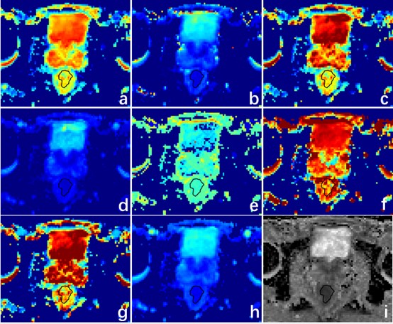
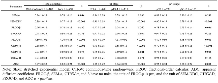
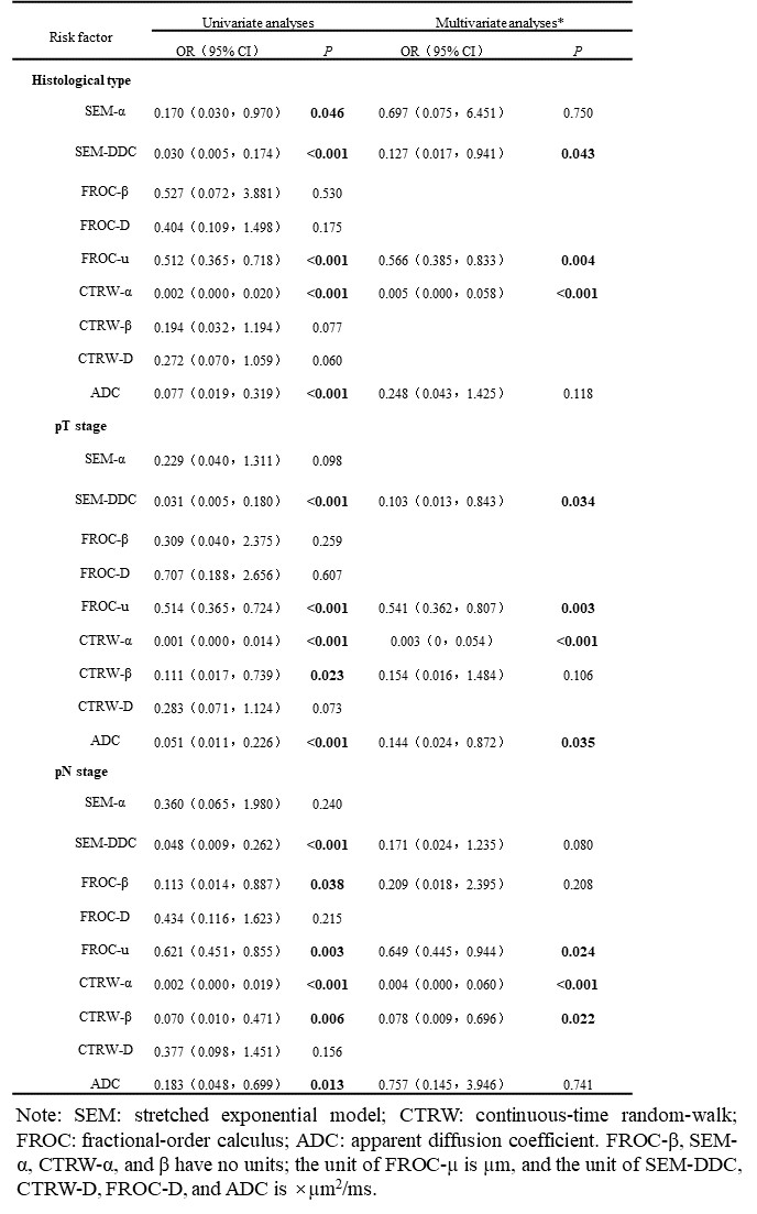
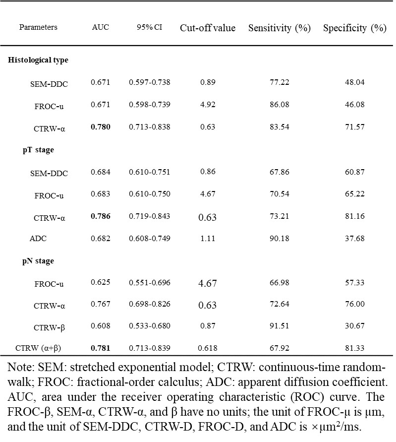
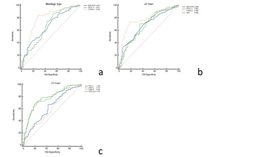
Figure 1 illustrates typical parameters from a patient with moderately differentiated rectal canser. We found significant correlations between specific MRI parameters and histopathological prognostic factors in patients with rectal cancer. SEM-DDC, FROC-u, CTRW-α, and ADC differed significantly across histological differentiation, pT stage, and pN stage (all P<0.05)(Table 1). Among these, SEM-DDC, FROC-u and CTRW-α demonstrated pronounced differences in both tumor depth (pT1-2 vs pT3-4) and nodal involvement (pN0 vs pN1-2)(all P<0.05). Multivariate analysis identified SEM-DDC, FROC-u, and CTRW-α as significant parameters associated with histological type(all P<0.05), and SEM-DDC, FROC-u, CTRW-α, and ADC were notably correlated with pT stage(all P<0.05). FROC-u, CTRW-α, and CTRW-β demonstrated independent associations with pN stage (all P<0.05)(Table 2). Table 3 and Figure 2 show the diagnostic effectiveness of each parameter. CTRW-α demonstrated a notably higher area under the curve (AUC) in predicting histological type and pT stage than did SEM-DDC, FROC-u, and ADC, exhibiting its enhanced predictive ability for these categories. For predicting pN stage, both CTRW-α and CTRW(α+β) outperformed FROC-u and CTRW-β, with CTRW(α+β) yielding the best AUC.Discussion
Integrating advanced DWI models in high b-value DWI offers a promising method in rectal cancer diagnosis. Among the parameters, CTRW-α stood out for its superior predictive ability, particularly in determining histological type and pT stage, challenging traditional ADC values and emphasizing a potential shift towards these advanced models for a more accurate prognosis5.Conclusion
CTRW-α showed the potential to be a highly valuable parameter in DWI-based rectal cancer prognosis. Its enhanced predictive capability compared with conventional ADC value highlights the importance of adopting these advanced models to ensure more accurate patient assessment and therapeutic interventions.Acknowledgements
No acknowledgement found.References
1. Boras Z, Kondza G, Sisljagić V, et al. Prognostic factors of local recurrence and survival after curative rectal cancer surgery: a single institution experience. Collegium antropologicum. 2012;36:1355-1361.
2. Curvo-Semedo L, Lambregts D M, Maas M, et al.Diffusion-weighted MRI in rectal cancer: apparent diffusion coefficient as a potential noninvasive marker of tumor aggressiveness. J Magn Reson Imaging. 2012;35:1365-1371.
3. Magin R L, Hall M G, Karaman M M, et al. Fractional Calculus Models of Magnetic Resonance Phenomena: Relaxation and Diffusion. Critical reviews in biomedical engineering. 2020;48:285-326.
4. Guo H, Liu J, Hu J, et al. Diagnostic performance of gliomas grading and IDH status decoding A comparison between 3D amide proton transfer APT and four diffusion-weighted MRI models. J Magn Reson Imaging. 2022;56:1834-1844.
5. Mazaheri Y, Afaq A, Rowe D B, et al. Diffusion-weighted magnetic resonance imaging of the prostate: improved robustness with stretched exponential modeling. J Comput Assist Tomogr. 2012;36:695-703.
Figures