0549
Imaging glutamine utilization in a prostate cancer xenograft model1Johns Hopkins University, Baltimore, MD, United States, 2Spelman College, Atlanta, GA, United States, 3Kennedy Krieger Institute, Baltimore, MD, United States
Synopsis
Keywords: Biomarkers, Cancer, Prostate, Metabolism
Motivation: MRI agents that can monitor abnormal glutamine utilization, a hallmark of many aggressive cancers, are not clinically available.
Goal(s): We investigated the utility of glutamine as a chemical exchange saturation transfer (CEST) agent to evaluate its utilization.
Approach: Phantoms revealed contrast differences between glutamine and its metabolic products. Dynamic CEST images (in vivo) were acquired in 2 prostate cancer xenograft models after intravenous injection of glutamine. MALDI (matrix-assisted laser desorption/ionization) images of the same tumors (ex vivo) were collected for validation.
Results: CEST and MALDI images could distinguish prostate tumors derived from cell lines with known differences in glutamine utilization.
Impact: The potential of CEST (chemical exchange saturation transfer) MRI to evaluate glutamine utilization was evaluated in preclinical prostate cancer models (DU-145 and LNCaP). CEST enhancement images upon infusion of glutamine could distinguish tumors that differed in glutamine utilization.
Introduction
Glutamine is an attractive target for cancer therapy and imaging because of its multifaceted roles in the disease. The physiological roles of glutamine—a source of energy1, a source of nitrogen and carbon for biosynthesis1, and a signaling molecule2, 3—are often upregulated in cancers1. In the preclinical stage, multiple drugs that target a range of proteins involved in glutamine utilization are currently in development. In the clinical stage, seven drugs designed to target glutamine utilization have been tested to treat cancer4-7. Clinically-available imaging agents that can evaluate glutamine utilization require radiolabeling8 or isotopic substitution9 for visualization. We investigated the utility of the chemical exchange saturation transfer (CEST) method to evaluate utilization glutamine using magnetic resonance imaging.Methods
Prostate cancer model: DU-145 (2-3x10⁶) or LNCaP (3-5x10⁶) cells were injected subcutaneously into 6-10 week old Rag2-/- mice. Phantom CEST MRI: Phantoms containing metabolites or cells were imaged using a Bruker 11.7T vertical bore spectrometer and a 20 mm TR coil. Z-spectra were collected over a range from ±8.0 ppm relative to the water frequency with a 0.1 ppm step-size, B1 = 3.6 μT, and tsat = 3 s. CEST maps of these phantoms were generated from Z-spectral signal (1-S/S0). in vivo CEST MRI: Tumors were imaged using a Bruker 11.7T horizontal bore spectrometer and an 8-channel TR coil. Dynamic CEST images were collected using the following saturation parameters—ω = +3.1 ppm, B1 = 3.6 μT, and tsat = 3 s—prior to intravenous glutamine injection (6 mmole/kg) to establish a baseline (1-2 min) and then over a period of 60 minutes after injection. CEST enhancement (ΔS(t)/S0) was quantified by subtracting the signal (S(t)/S0) at time t from the average baseline signal obtained prior to injection. Injected glutamine also contained rare, stable isotopes (13C5,15N2) in order to track its fate using MALDI imaging without affecting the CEST signal in these studies, which is generated through the exchange of protons (1H). MALDI imaging: Seventy-five minutes after injection of 13C5,15N2-glutamine, tumors were excised for imaging. Tumors were cryosectioned (10 µm thick) onto indium tin oxide slides. Four passes of 10mg/10mL 1,5-diaminophthlene in 70% acetonitrile with 0.1% were applied. MALDI images were obtained using a Bruker Rapiflex MALDI-TOF/TOF mass spectrometer in reflection negative mode with a 100x200 μm raster. Statistics: An ANOVA or Student’s t-test was performed, as appropriate, with p<0.05 considered significant.Results
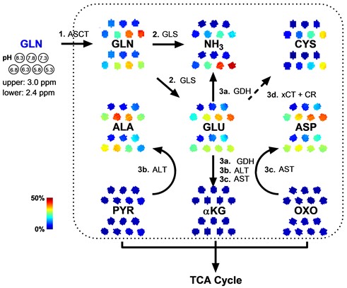
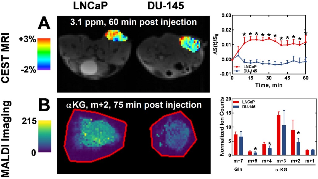
We first evaluated the potential sensitivity of CEST to detect the uptake and metabolism of glutamine. Initially, we screened the CEST signal generated by glutamine and its metabolic products at cancer-relevant pH (5.3-8.3) in phantoms (20 mM in PBS; Figure 1). For much of this range, glutamic acid, aspartic acid, and alanine generated a higher CEST signal at the 3.0 ppm frequency compared to glutamine, while ammonia generated a higher CEST signal at the 2.4 ppm frequency. In contrast, more downstream products typically had lower CEST signal at these frequencies compared to glutamine. We subsequently investigated CEST signal enhancement when glutamine (6 mmol/kg) was administered IV (Figure 2A). When glutamine was administered into mice bearing DU-145 tumors (N=4), CEST signal at the +3.1 ppm frequency remained near baseline levels throughout the scan. When the same amount of glutamine was administered into mice bearing LNCaP tumors, CEST signal at the +3.1 ppm frequency was enhanced throughout the 60-minute scan (N=5). CEST signal was significantly higher in LNCaP tumors compared to DU-145 tumors at 60 minutes (p < 0.05). CEST enhancement maps were heterogenous, which prompted our use of MALDI imaging to evaluated the uptake and metabolism of injected glutamine for CEST image validation. Lastly, we investigated the presence of isotopically-substituted metabolic products ex vivo using MALDI imaging after infusion of 13C5,15N2-glutamine (6 mmol/kg) (Figure 2b). The presence of 13C5,15N2-glutamine trended higher in LNCaP tumors compared to DU-145 tumors at 75 minutes, but was not significant (p > 0.05). Still, the presence of 13C-containing alpha-ketoglutarate was significantly higher in LNCaP tumors compared to DU-145 tumors (p < 0.05). Metabolite maps were also heterogenous, but their patterns appeared similar to that observed in CEST enhancement maps.Discussion and Conclusion
CEST MRI was able to distinguish tumors with differing levels of glutamine utilization, as confirmed with MALDI imaging. Higher CEST enhancement upon glutamine injection was observed in LNCaP versus DU-145 tumors, in line with reports of higher uptake of 14C-containing glutamine in vitro10. This work preliminarily demonstrates the potential of CEST MRI to evaluate glutamine utilization, a prognostic marker of many cancers and an active target for cancer therapy.Acknowledgements
Funding for this work was provided by NIH: K01 EB030612, P41 EB024495, and R01 EB030376.References
1. Rajagopalan KN, DeBerardinis RJ. Role of Glutamine in Cancer: Therapeutic and Imaging Implications. Journal of Nuclear Medicine. 2011;52(7):1005-8. doi: 10.2967/jnumed.110.084244.
2. Meng D, Yang Q, Wang H, Melick CH, Navlani R, Frank AR, Jewell JL. Glutamine and asparagine activate mTORC1 independently of Rag GTPases. Journal of Biological Chemistry. 2020;295(10):2890-9. doi: https://doi.org/10.1074/jbc.AC119.011578.
3. Jewell JL, Kim YC, Russell RC, Yu F-X, Park HW, Plouffe SW, Tagliabracci VS, Guan K-L. Differential regulation of mTORC1 by leucine and glutamine. Science. 2015;347(6218):194-8. doi: doi:10.1126/science.1259472.
4. Taylor SA, Crowley J, Pollock TW, Eyre HJ, Jaeckle C, Hynes HE, Stephens RL. Objective antitumor activity of acivicin in patients with recurrent CNS malignancies: a Southwest Oncology Group trial. Journal of Clinical Oncology. 1991;9(8):1476-9. doi: 10.1200/jco.1991.9.8.1476. PubMed PMID: 1649269.
5. Schroeder JM, Ansfield FJ, Curreri AR, LePage GA. Toxicity and Clinical Trial of Azaserine and 6-Thioguanine in Advanced Solid Malignant Neoplasms. British Journal of Cancer. 1964;18(3):449-58. doi: 10.1038/bjc.1964.51.
6. Pettit GR, Nelson PS. Antineoplastic agents. 114. Synthesis of azotomycin. The Journal of Organic Chemistry. 1986;51(8):1282-6. doi: 10.1021/jo00369a020.
7. Rahman A, Smith FP, Luc PT, Woolley PV. Phase I study and clinical pharmacology of 6-diazo-5-oxo-L-norleucine (DON). Invest New Drugs. 1985;3(4):369-74. doi: 10.1007/bf00170760. PubMed PMID: 4086244.
8. Cabella C, Karlsson M, Canapè C, Catanzaro G, Colombo Serra S, Miragoli L, Poggi L, Uggeri F, Venturi L, Jensen PR, Lerche MH, Tedoldi F. In vivo and in vitro liver cancer metabolism observed with hyperpolarized [5-13C]glutamine. Journal of Magnetic Resonance. 2013;232:45-52. doi: https://doi.org/10.1016/j.jmr.2013.04.010.
9. Gallagher FA, Kettunen MI, Day SE, Lerche M, Brindle KM. 13C MR spectroscopy measurements of glutaminase activity in human hepatocellular carcinoma cells using hyperpolarized 13C-labeled glutamine. Magnetic Resonance in Medicine. 2008;60(2):253-7. doi: https://doi.org/10.1002/mrm.21650.
10. Okudaira H, Oka S, Ono M, Nakanishi T, Schuster DM, Kobayashi M, Goodman MM, Tamai I, Kawai K, Shirakami Y. Accumulation of Trans-1-Amino-3-[18F]Fluorocyclobutanecarboxylic Acid in Prostate Cancer due to Androgen-Induced Expression of Amino Acid Transporters. Molecular Imaging and Biology. 2014;16(6):756-64. doi: 10.1007/s11307-014-0756-x.
Figures