0532
The application of multi-modal fMRI to investigate coma induction induced by Endothelin-driven brainstem injury1Athinoula A. Martinos Center for Biomedical Imaging, Massachusetts General Hospital, Charlestown, MA, United States, 2Department of Neurosurgery, Beijing Tsinghua Changgung Hospital, School of Clinical Medicine, Tsinghua University, Beijing, China
Synopsis
Keywords: Functional Connectivity, Brain Connectivity
Motivation: This study aimed to create a reproducible coma animal model and develop neuroimaging techniques to illustrate underlying mechanisms during coma induction.
Goal(s): To investigate brain dynamic changes during coma induction and to identify the key regulatory brain regions involved in the process.
Approach: We induced a brainstem coma in rats, optimizing surgical procedures and utilizing multi-modal fMRI techniques. We recorded Glu signals and BOLD fMRI simultaneously during coma induction.
Results: The study revealed specific Glu-oscillations before coma induction and identified certain subcortical nuclei as potential key regulatory brain regions for inducing coma. These findings could enhance our understanding of coma-related brain state changes.
Impact: The optimized coma model and multi-modal imaging techniques in this study offer a deeper understanding of coma dynamics. The identification of specific regulatory brain regions and Glutamate oscillations may pave the way for improved clinical strategies and patient outcomes.
Introduction
Brain coma leads to dramatic brain dynamic changes, of which the reemergence, in particular, from a vegetative or minimal consciousness states, is seldom understood in clinical practice given the lack of reliable therapeutic strategies to awake the unweakened brain. The ongoing challenges to study brain coma can be summarized in two aspects: i. there is lack of a reproducible and recoverable coma animal model for systematic analysis. ii. There are missing multi-modal neuroimaging tools to map the cross-scale brain functional changes to bridge the molecular/cellular mechanism and global spatiotemporal dynamic patterns. We have established a brainstem injury induced rat coma model [1]. Here, we optimized surgical procedure to enable the real-time coma induction with whole brain fMRI and fiber photometry-based Glutamate recordings, aiming to specify the underlying brain dynamic changes during the coma induction.Materials and Methods
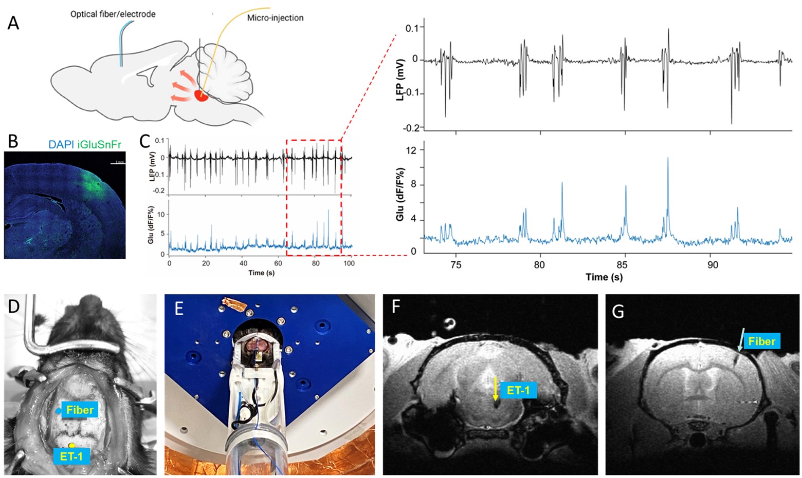
Animal Model: Female Long Evans rats (~250g) were induced with 5% and maintained at 1%-2% isoflurane. The rats’ rectal temperature was maintained at around 37oC. The genetically encoded reporter iGluSnFR was expressed by the AAV9 virus in the barrel cortex. 3 weeks post-surgery, optical fiber (250 µm) was inserted into the barrel cortex expressing the fluorescent biosensors for recording. For come induction, a peek tubing (150μm) was implemented into the brainstem tegmentum through the ventral surface of the brain to inject ET-1. Isoflurane is withdrawn after the coma induction. Animals were incubated during scanning. MR Techniques: All images were acquired with a 14 T/ 13 mm horizontal bore magnet (Magnex), interfaced to a Bruker AV-Neo console (Bruker), and equipped with a 6 cm gradient set, capable of providing 1.2 T/m (Resonance Research). A transceiver surface coil was used to acquire fMRI images. fMRI scans with block design were performed using 3D Echo planar imaging sequence: TR=1.5s, TE=7.5ms, FOV=2.4X2.4X2.0 cm3, 96X96X40 matrix.Results
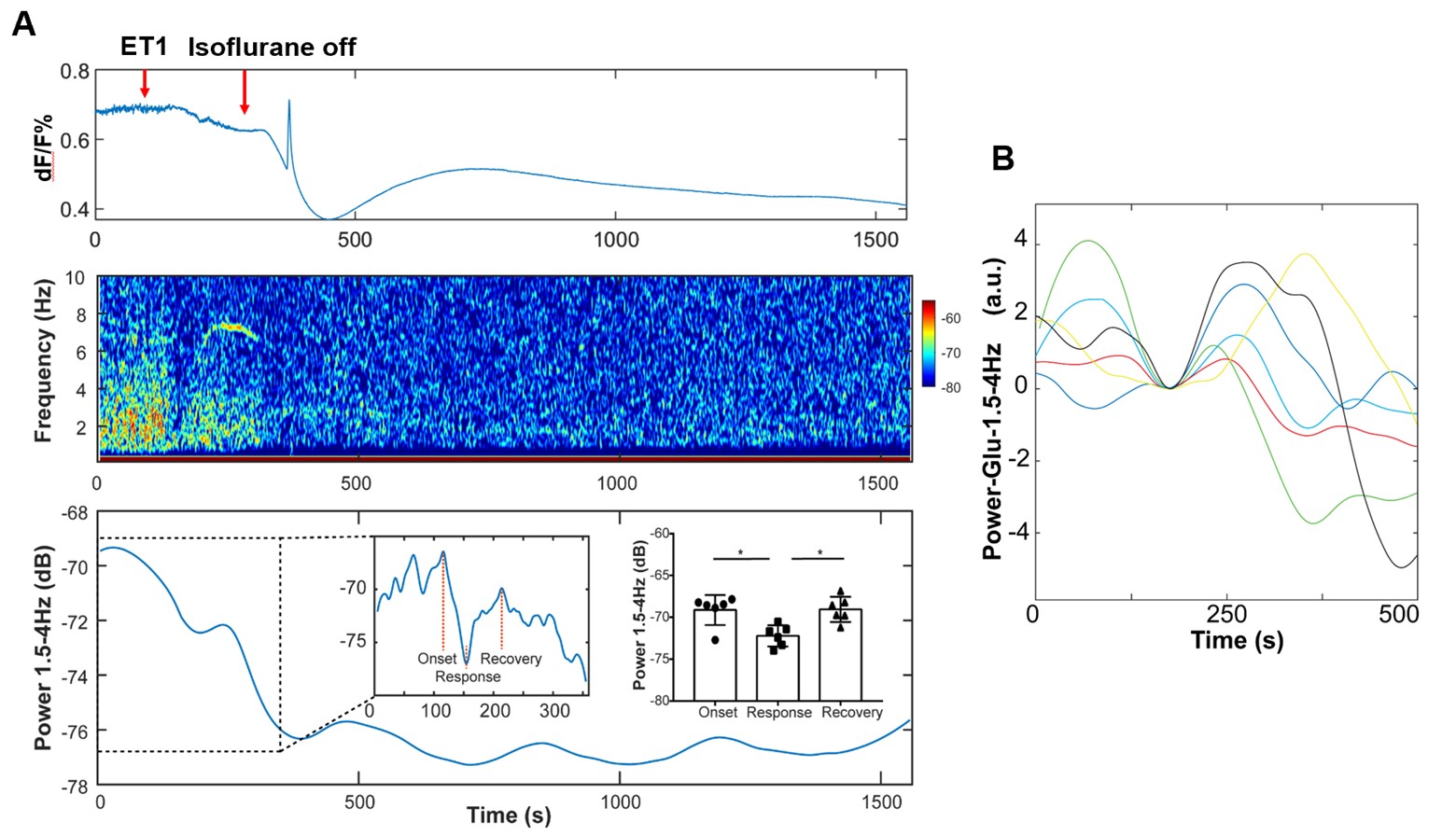
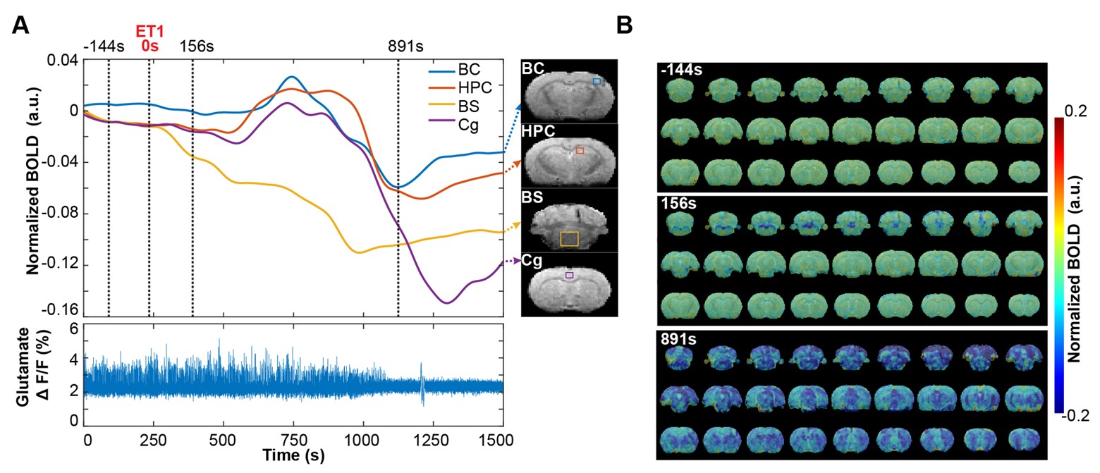
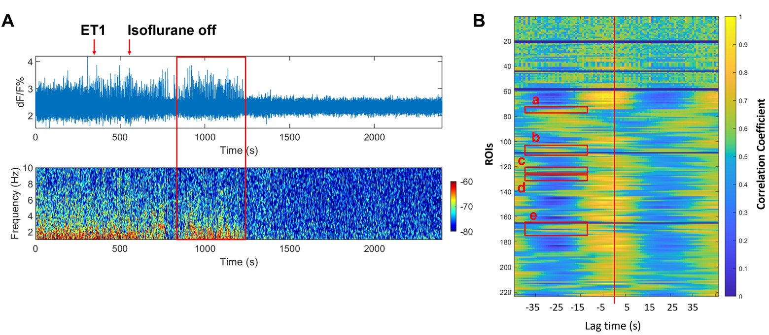
We recorded the cell type-specific Glu signal simultaneously with the local field potential (LFP) in barrel cortex. The spontaneous Glu signal from neurons matched well with the LFP for each spike (Fig1A-C). By combining the Glu signal recording and whole brain fMRI, we can continuously measure the neuronal activities in specific brain area and the whole brain functional dynamics in various condition, including during coma induction (Fig1 D-G). By using the optimized coma model and the multi-modal fMRI methodology, we detected the induction of iso-electric line in 5-20 mins after injecting the ET-1 into brainstem tegmentum, with a 150-300 seconds specific Glu-oscillation feature in advance (Fig 2). Also, during ET-1 microinjection, the BOLD fMRI responses can trace the vasoconstriction in the brainstem injection site (Fig 3, negative BOLD, blue dot). And in 8-15 min, we observed reproducible global negative fMRI responses, also with a similar Bold-oscillation feature in advance in some specific cortical and subcortical regions, e.g., barrel cortex, cingulate cortex, hippocampus, etc. Therefore, it is implied that the specific oscillation feature could be a driving factor for coma induction, and the origins of this oscillation feature could be the key regulatory brain regions for coma-related brain state change. To illustrate the Glu-oscillation feature related brain regions, a ROI based cross-correlation heatmap was conducted with the power of the Glu (0.8-4Hz) and the BOLD signal (Figure4). It is noteworthy that there is an earlier response separating some specific nuclei (e.g., hippocampus, ventral cochlear nucleus, basal ganglia, thalamus, As shown with red box) from the other anatomical subdivisions, highly implying that these specific nuclei are the key regulatory brain regions for coma induction.Discussion
With the optimized brainstem coma induction model and the multi-modal fMRI platform, we observed a specific Glu-oscillation feature before coma induction. The specific Glu-oscillation feature could originate from some specific subcortical nuclei (e.g., hippocampus, ventral cochlear nucleus, basal ganglia, thalamus), which might be the key regulatory brain regions for coma induction.Conclusions
We optimized brainstem coma induction model and established a multi-modal fMRI platform. Some subcortical nuclei derived specific Glu-oscillation feature could be the key regulatory factor for coma induction.Acknowledgements
This research was funded by NIH funding (RF1NS113278, RF1NS124778, R01NS122904, R01NS120594), NSF grant 2123971, and the S10 instrument grant (S10 MH124733–01) to Martino’s Center)References
1 Pais-Roldan, P. et al. Multimodal assessment of recovery from coma in a rat model of diffuse brainstem tegmentum injury. Neuroimage 189, 615-630, doi:10.1016/j.neuroimage.2019.01.060 (2019).Figures