0529
Design of an Open-transmit / 24-channel flexible receiver head coil for MRI/fMRI of somatosensory and motor cortex at 5T1Lauterbur Imaging Research Center, Shenzhen Institutes of Advanced Technology, Chinese Academy of Sciences, Shenzhen, China, 2Shanghai United Imaging Healthcare, Shanghai, China, 3Key Laboratory for Magnetic Resonance and Multimodality Imaging of Guangdong Province, Shenzhen, China, 4Research Center for Medical AI, Shenzhen Institutes of Advanced Technology, Chinese Academy of Sciences, Shenzhen, China, 5Department of Biomedical Engineering, State University of New York at Buffalo, Buffalo, NY, United States
Synopsis
Keywords: High-Field MRI, High-Field MRI
Motivation: Functional magnetic resonance imaging (fMRI) is a non-invasive in vivo functional mapping technique, which afforded a high-quality glimpse of the cortex.
Goal(s): Sampling brain activity across cortical layers by using proposed RF coil
Approach: The Open-Face birdcage coil was designed by removing and adjusting the legs of the 16-rung high-pass birdcage coil. The 24ch flexible receive array was designed for high spatiotemporal-resolution MRI/fMRI at cortical region.
Results: In this study, we designed and constructed an open-transmit and 24 channel flexible receiver head coil assembly for human somatosensory and motor cortex in vivo cortical imaging on a whole body 5T scanner.
Impact: SNR maps, T2* weighted images and fMRI images were acquired with the proposed coil assembly, which were compared with those using a quadrature birdcage transmit/48-channel receiver coil assembly.
Introduction
Functional magnetic resonance imaging (fMRI) is a non-invasive in vivo functional mapping technique, which afforded a high-quality glimpse of the cortex. Current fMRI studies are starting to map out the functional neural circuits underlying the somatosensory and motor cortex. The CBV-fMRI method is developed for sampling brain activity across cortical layers and can measure laminar activity with higher accuracy than BOLD-fMRI [1].Ultra-high field MRI provides significant intrinsic gains in both imaging signal-to-noise ratio (SNR) and functional signal contrast-to-noise ratio (CNR), which can achieve high spatial resolution for the visualization of submillimeter cortical columns and subnuclei structures in fMRI. Local birdcage coils are widely used as transmit coils for ultra-high field MRI systems. RF shielding is indispensable to reduce radiation loss. However, the closed structure is not friendly for cognitive functional MRI experiment, especially in the visual fMRI. For the RF receive coils design in human brain studies, a denser number of coil elements are laid out on top of a head shell to improve SNR and parallel acceleration capacity. The channel number has been increased from 32 to 64, or even 128[2-3]. These designs are based on a rigid shell, of which coil elements locate at a certain distance from the imaging position. The flexible head coils show some advantages in the aspects of adaptability and SNR.
In this study, we designed and constructed an open-transmit and 24 channel flexible receiver head coil assembly for human somatosensory and motor cortex in vivo cortical imaging on a whole body 5T scanner (Shanghai United Imaging Healthcare, Shanghai, China). SNR maps, T2* weighted images and fMRI images were acquired with the proposed coil assembly, which were compared with those using a quadrature birdcage transmit/48-channel receiver coil assembly [4].
Methods
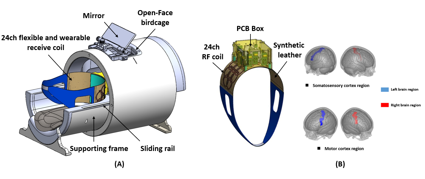
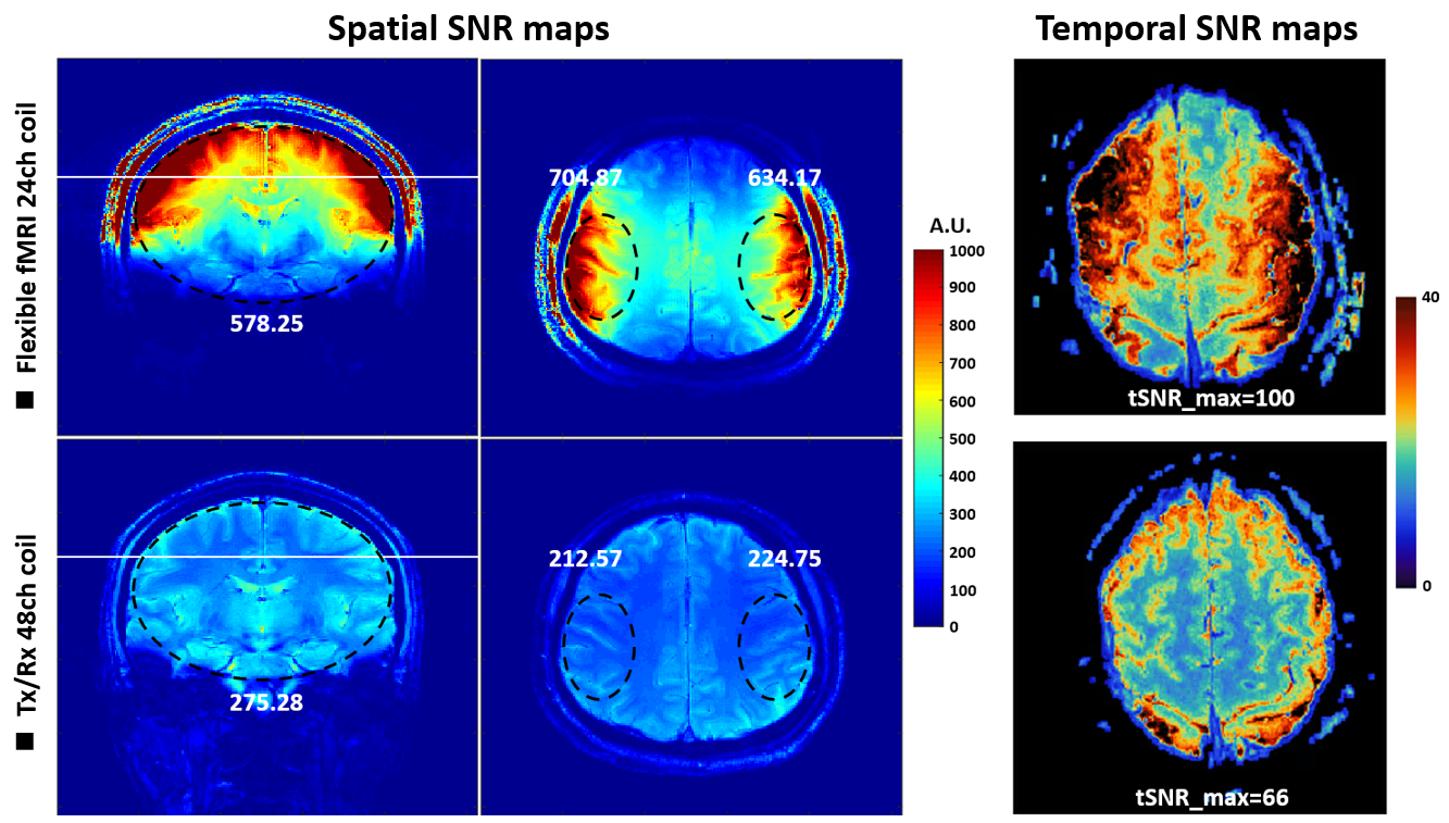
Figure 1 shows the layout of the Open-Face birdcage coil assembled with flexible 24-channel receiver head coil. The Open-Face birdcage coil was designed by removing and adjusting the legs of the 16-rung high-pass birdcage coil [5]. The customized surface coil array was designed for high spatiotemporal-resolution MRI/fMRI at cortical region using24 closely spaced loops with a diameter of 40 mm to maximize SNR and parallel imaging performance. The receiver coil elements were arranged on a flexible cushion. The overlap decoupling method was applied to cancel the inductive coupling between neighboring loops.For spatial-SNR comparisons, a two-dimensional (2D) gradient echo (GRE) sequence was applied for signal acquisition with following parameters: TR/TE=1000/15ms, bandwidth (BW)=130Hz/pixel, slice thickness=5mm, FOV=200x200mm2, matrix size=256x256. Noise images were acquired by setting the flip angle to 0. SNR maps were calculated using the sum-of-squares method and were then normalized by the sine value of flip angle. The flip angle maps were obtained using the dual refocusing echo acquisition mode (DREAM) sequence. For temporal-SNR comparisons, single-shot GRE-EPI images were acquired with following parameters: TR/TE=3000/26.6ms, Flip angle=900, FOV=100x50mm2, matrix size=128x64, resolution:0.8x0.8x0.8mm3. The tSNR was calculated for each voxel as the mean value divided by temporal standard deviation.
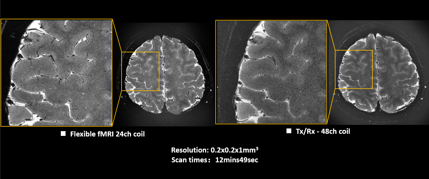
To evaluate the coil capability of high-resolution imaging, 2D GRE T2* weighted sequence was applied with the following parameters: TR/TE=1671/34ms, Flip angle=300, FOV=180x180mm2, matrix size=896, spatial resolution:0.2x0.2x1mm3, receiver bandwidth=30Hz/pixel, scan time=12mins49sec.
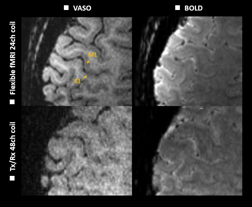
The finger-tapping task and Slice-Selective Slab-Inversion VASO sequence were performed for layer-specific study. The post-processing of the fMRI data was performed using the FSL toolbox(Analysis Group, FMRIB, Oxford, UK), AFNI (NIMH Scientific and Statistical Computing Core, NIH, Maryland, US) and LANII (software suite for layer-fMRI).
Results
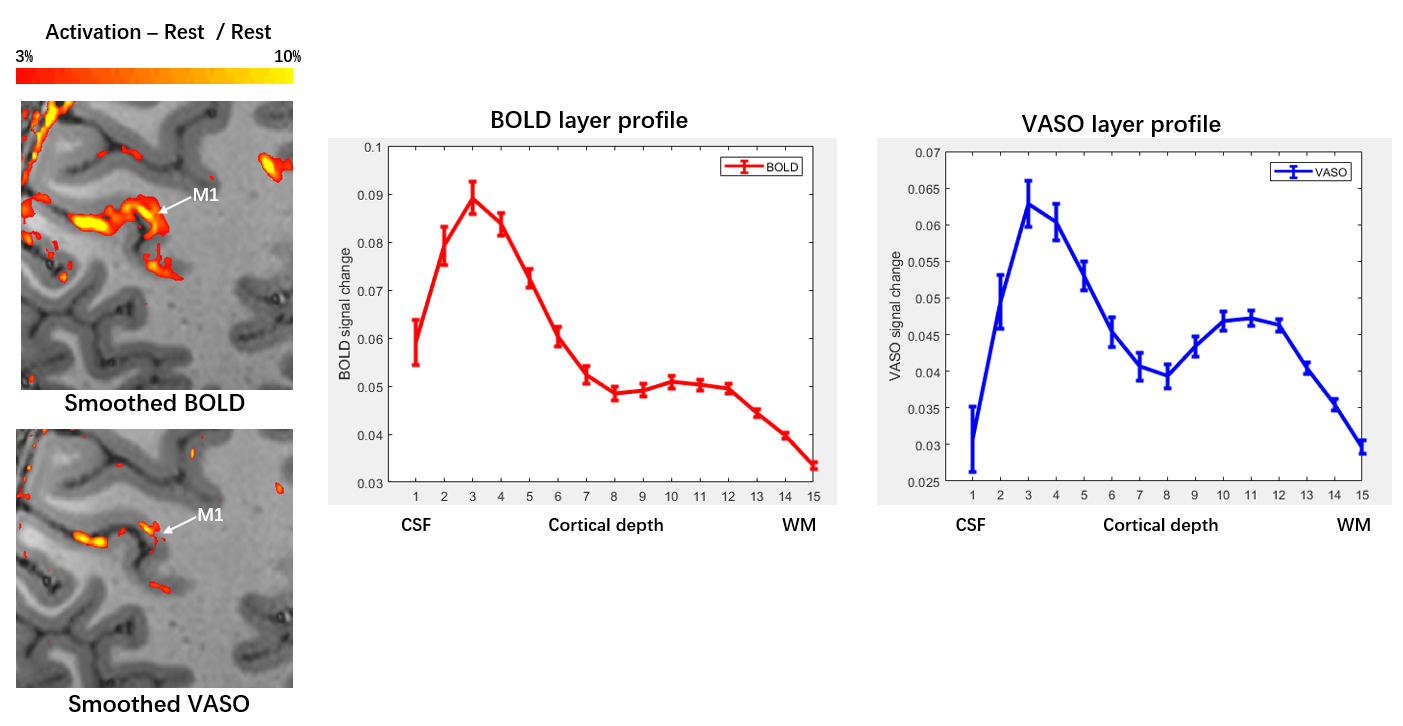
Figure 2 shows the spatial-SNR maps with correction in flip angle and temporal-SNR maps. The mean values of SNR in the ROI are depicted in the maps. The proposed receiver coil can provide SNR improvement up to 3-fold in the cortex region and better temporal-SNR value than the 48-channel head coil. Figure 3 shows the T2*-weighted brain images with a resolution of 0.2x0.2x1mm3 using the proposed coil assembly and quadrature birdcage transmit/48-channel receiver coil assembly, respectively. As shown in Figure 4, the 24-channel fMRI head coil can provide better VASO and BOLD image quality for finger-tapping task. Figure 5 shows the layer-specific results by using proposed coil, which indicated that similar activation maps and signal change in M1 cortex as well as Huber’s 7T fMRI results [1].Discussions/Conclusion
The proposed Open-Face birdcage coil assembled with 24-channel flexible receiver head coil provides better clinical feasibility and SNR performance. In the future work, more functional MRI studies and layer-specific studies using the proposed coil will be performed at 5T MRI.Acknowledgements
This work was supported in part by the Project on Global Common Challenges of Chinese Academy of Sciences (No. 321GJHZ2022081GC), the NSFC grant (81627901, U22A20344), the Key Laboratory Project of Guangdong Province, China (2020B1212060052), the Funding Program of Shenzhen, China (RCYX20200714114735123), the Chinese Academy of Sciences Youth Innovation Promotion Association funded project (Y2021098), Shenzhen Science and Technology Program (GJHZ20210705141405016).References
[1] Huber L, Handwerker DA, Jangraw DC, Chen G, Hall A, Stüber C, Gonzalez-Castillo J, Ivanov D, Marrett S, Guidi M, Goense J, Poser BA, Bandettini PA. High-Resolution CBV-fMRI Allows Mapping of Laminar Activity and Connectivity of Cortical Input and Output in Human M1. Neuron. 2017,96(6):1253-1263.e7.
[2] Kraff O, Quick HH. Radiofrequency Coils for 7 Tesla MRI. Top Magn Reson Imaging. 2019, 28(3):145-158.
[3] Gruber B, Stockmann JP, Mareyam A, Keil B, Bilgic B, Chang Y, Kazemivalipour E, Beckett AJS, Vu AT, Feinberg DA, Wald LL. A 128-channel receive array for cortical brain imaging at 7 T. Magn Reson Med. 2023, 90(6):2592-2607.
[4] Wei Z, Chen Q, Han S, Zhang S, Zhang N, Zhang L, Wang H, He Q, Cao P, Zhang X, Liang D, Liu X, Li Y, Zheng H. 5T magnetic resonance imaging: radio frequency hardware and initial brain imaging. Quant Imaging Med Surg. 2023, 13(5):3222-3240.
[5] Wei Z, Chen Q, He Q, Zhang X, Liu X, Zheng H, Li Y. Design of an improved Open-Face birdcage for human whole brain imaging at 5T. Proc. 31th Annual Meeting of ISMRM, Toronto, 2023, p.4226.
Figures