0527
Remarkably enhanced B1 and SNR with uHDC ceramics integrated with novel RF transreceiver array for 2H, 17O, and 1H imaging of human brain at 10.5T1Center of Magnetic Resonance Research (CMRR), Department of Radiology, University of Minnesota, Minneapolis, MN, United States, 2Department of Biomedical Engineering, University of Minnesota, Minneapolis, MN, United States
Synopsis
Keywords: RF Arrays & Systems, Parallel Transmit & Multiband, High Permittivity Material (HPM), ultrahigh Dielectric Constant (uHDC) Material, MRSI, UHF, Ultrahigh field, Broadband RF Coil
Motivation: 2H and 17O MRSI are useful to study brain energy metabolism, however, have low imaging sensitivity.
Goal(s): To develop a RF coil engineering solution offering superior performance for human brain 1H MRI, 2H and 17O MRSI at UHF.
Approach: We constructed a novel RF transreceiver array coil, which can operate at 1H, 2H and 17O resonant frequencies at 10.5T. The integration of ultrahigh dielectric constant (uHDC) ceramics enhanced RF transmission field (B1+) and SNR.
Results: The new array coil functioned well for performing multinuclear imaging, and the integrated uHDC technology remarkably enhanced B1+ and SNR for performing 2H and 17O MRSI.
Impact: In this study, we introduce and demonstrate an advanced RF array coil integrated with the uHDC material enabling imaging of three important nuclei (1H, 2H and 17O) signals with superior performance aiming for human brain applications at UHF of 10.5T.
Introduction
X-nuclear imaging methods are highly valuable in studying metabolism, energetics, and physiopathology in the human brain. Among them, 17O and 2H MRSI enable noninvasive imaging of cerebral glucose and oxygen metabolism and rates under normal and diseased state1-4. However, the low natural abundance of X-nuclei and low intrinsic signal-to-noise ratio (iSNR) of X-nuclear imaging present challenges to obtaining high-quality and high-resolution X-nuclear images. Increasing the field strength and improving the radiofrequency transmission field (B1) and imaging iSNR using high or ultrahigh dielectric constant (HDC, uHDC) material and metamaterial5-7 would partially surmount the challenges. In this study, we introduce the advanced broadband RF coil system with low-loss uHDC ceramic disks for 2H and 17O imaging applications at 10.5T.Methods
A 4-channel triple-frequency-tunable loop coil (Snowman coil)array was designed and built (FIG. 1A), based on the recently introduced dual-frequency loop coil concept8,9. The coil array could be tuned and matched to either 2H (68.6MHz) or 17O (60.6MHz) resonant frequency while being tuned to 1H resonant frequency (447MHz) at 10.5T. Adjacent coil loops were decoupled with conventional overlaps; and the modified inductive decoupling method, which was previously introduced in 7T 4-channel 2H coil array9 was applied to decouple loops in parallel. Broadband preamplifiers and TR switches, a 4-way power divider, and phase shifters in the frequency range between 60MHz and 70MHz were designed and assembled (FIG. 1B) to operate 2H and 17O imaging at 10.5T. 1H imaging was utilized to acquire high-quality structural MRI and perform B0-shim. the circular uHDC ceramic disks (εr ≈ 5400, 8.5cm diameter and 1.6cm thickness, FIG. 1C) were positioned between the coil and a square bottled water phantom (56mM Na+) to demonstrate the effect of uHDC disks for improving 2H and 17O imaging performance. To acquire the RF transmission and reception field (B1+/-) maps for 2H and 17O imaging, the sinusoidal curve fitting method was implemented to process multiple volumes of CSI with various RF pulse voltages. Comparison between control (without uHDC disks) and uHDC conditions was conducted by registering the center of the phantom in the same position and calculating the ratio of B1 field maps and SNR maps between the two conditions.Results
Figure 1A shows the prototype of the 4-channel triple-frequency tunable RF coil array, which was connected to the 4-channel 1H interface and the 4-channel broadband interface (Fig. 1B) for 2H and 17O operation. The tuned and matched coils have reflection coefficients below -13dB and transmission coefficients below -12dB at all resonant frequencies.3D 17O and 2H CSIs with various RF pulse voltages were acquired, and B1 field maps were estimated. Based on the estimated B1 maps, the effect of uHDC ceramic disks was illustrated by assessing the ratio of B1 and SNR maps compared to control for each nucleus. In B1 field maps of 17O CSI (Fig. 2), uHDC disks could enhance transmission efficiency (proportional to B1+), reception sensitivity (proportional to B1-), and SNR by 35%, 47%, and 46%, respectively. Like the 17O results, estimated B1- and SNR maps of 2H CSI (Fig. 3) exhibited an increase of 32% and 30%, respectively, when using uHDC disks, and in parallel improved the transmission efficiency by more than a twofold increase.
Discussion
Following the relationship between optimal uHDC permittivity and MR imaging operation frequency from the previous study5, the permittivity of uHDC disks (εr ≈ 5400) implemented in this study was close to the optimal permittivity of 2H at 10.5T (εr = 5212). This could explain the substantial improvement of B1+ fields in 2H imaging. Additionally, 17O imaging presented significant improvement in RF fields and SNR even though the permittivity was less optimized for 17O imaging at 10.5T, which has the optimal permittivity of εr = 6888. Considering the remarkable enhancement in B1+/- fields and SNR in both 17O and 2H imaging, the application of uHDC disks is a promising technology to enhance SNR and transmission efficiency.Conclusion
In conclusion, we have developed a high-fidelity, simple-structured, and triple-frequency tunable human head array coil showing excellent performance for 2H, 17O, and 1H imaging at 10.5T, and demonstrated remarkable improvements using the uHDC technology for 2H and 17O imaging. The integrated RF coil approach opens new opportunities in studying brain energy metabolisms in normal and diseased human brains at UHF.Acknowledgements
This work was supported, in part, by NIH grants: U01 EB026978, R01CA240953, R01NS133006, S10 RR029672, and P41EB027061.References
- De Feyter, H. M., Behar, K. L., Corbin, Z. A., Fulbright, R. K., Brown, P. B., McIntyre, S., Nixon, T. W., Rothman, D. L., & de Graaf, R. A. (2018). Deuterium metabolic imaging (DMI) for MRI-based 3D mapping of metabolism in vivo. Sci Adv, 4(8), eaat7314. https://doi.org/10.1126/sciadv.aat7314
- Li, Y., Zhao, Y., Guo, R., Wang, T., Zhang, Y., Chrostek, M., Low, W. C., Zhu, X. H., Liang, Z. P., & Chen, W. (2021). Machine Learning-Enabled High-Resolution Dynamic Deuterium MR Spectroscopic Imaging. IEEE Trans Med Imaging, 40(12), 3879-3890. https://doi.org/10.1109/TMI.2021.3101149
- Lu, M., Zhu, X. H., Zhang, Y., Mateescu, G., & Chen, W. (2017). Quantitative assessment of brain glucose metabolic rates using in vivo deuterium magnetic resonance spectroscopy. J Cereb Blood Flow Metab, 37(11), 3518-3530. https://doi.org/10.1177/0271678X17706444
- Zhu, X. H., Lu, M., & Chen, W. (2018). Quantitative imaging of brain energy metabolisms and neuroenergetics using in vivo X-nuclear 2H, 17O and 31P MRS at ultra-high field. J Magn Reson, 292, 155-170. https://doi.org/10.1016/j.jmr.2018.05.005
- Chen, W., Lee, B. Y., Zhu, X. H., Wiesner, H. M., Sarkarat, M., Gandji, N. P., Rupprecht, S., Yang, Q. X., & Lanagan, M. T. (2020). Tunable Ultrahigh Dielectric Constant (tuHDC) Ceramic Technique to Largely Improve RF Coil Efficiency and MR Imaging Performance. IEEE Trans Med Imaging, 39(10), 3187-3197. https://doi.org/10.1109/TMI.2020.2988834
- Lee, B. Y., Zhu, X. H., Rupprecht, S., Lanagan, M. T., Yang, Q. X., & Chen, W. (2017). Large improvement of RF transmission efficiency and reception sensitivity for human in vivo 31P MRS imaging using ultrahigh dielectric constant materials at 7T. Magn Reson Imaging, 42, 158-163. https://doi.org/10.1016/j.mri.2017.07.019
- Soon, S., et al. (2023). Significant signal-to-noise ratio (SNR) enhancement with ultrahigh dielectric constant (uHDC) ceramic disks for 2H MRSI application at 7T. Proceedings of the Annual Meeting of ISMRM, Toronto, Canada. p. 4083.
- Soon, S., et al. (2022). Development of 8-Channel 1H-2H Dual-Frequency loop coil array with LC tanks for 1H MRI and 2H MRS imaging of human brain at 7 Tesla. Proceedings of the 31st Annual Meeting of ISMRM, London, UK. p. 1542.
- Li, X., et al. (2022). A multinuclear 4-channel 2H loop and 4-channel 1H microstrip array coil for human head MRS/MRI at 7T. Proceedings of the 31st Annual Meeting of ISMRM, London, UK. p. 4510.
Figures
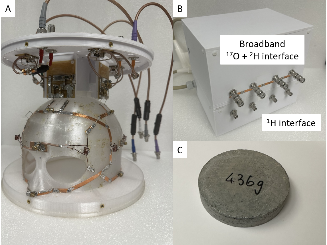
Figure 1. (A) The Prototype of 4-channel triple-frequency-tunable head coil (Snowman Coil) array with LC tank circuits to enable multi-frequency tuning and matching. Adjacent coils were decoupled with overlaps, and coils in parallel consist of modified inductive decoupling method. (B) Broadband interfaces implemented in the study. (C) An ultrahigh dielectric constant (uHDC) ceramic disk was applied on each side of the square bottled water phantom with Na+ concentration of 56mM.
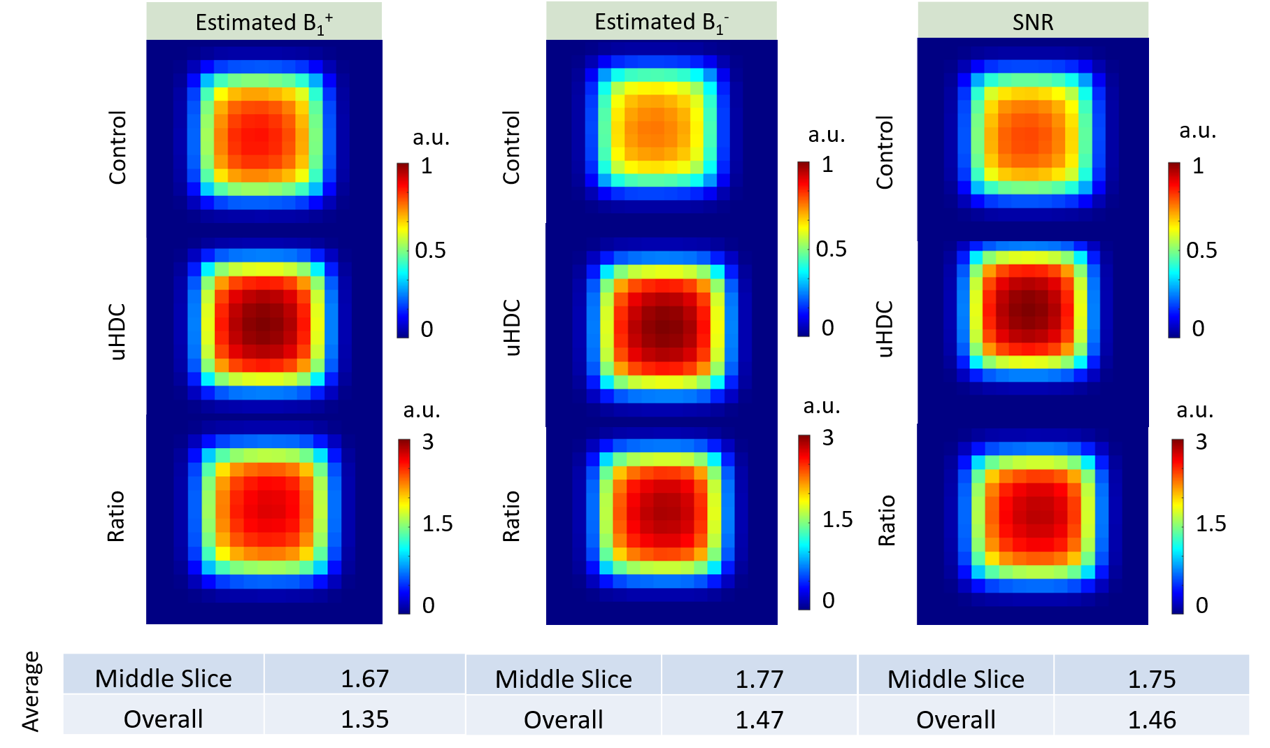
Figure 2. Normalized estimated B1+/- field maps of 17O CSIs in the middle slices where the uHDC disks are placed. The sinusoidal curve fitting method was employed to approximate radiofrequency field maps. In the 17O B1 field maps, the overall enhancement of transmission (B1+) and reception (B1-) fields in the volume where the uHDC disks covered the phantom were elevated by 35% and 47%, respectively. The comparison of normalized SNR maps demonstrates an averaged improvement of 46% with the uHDC disks.
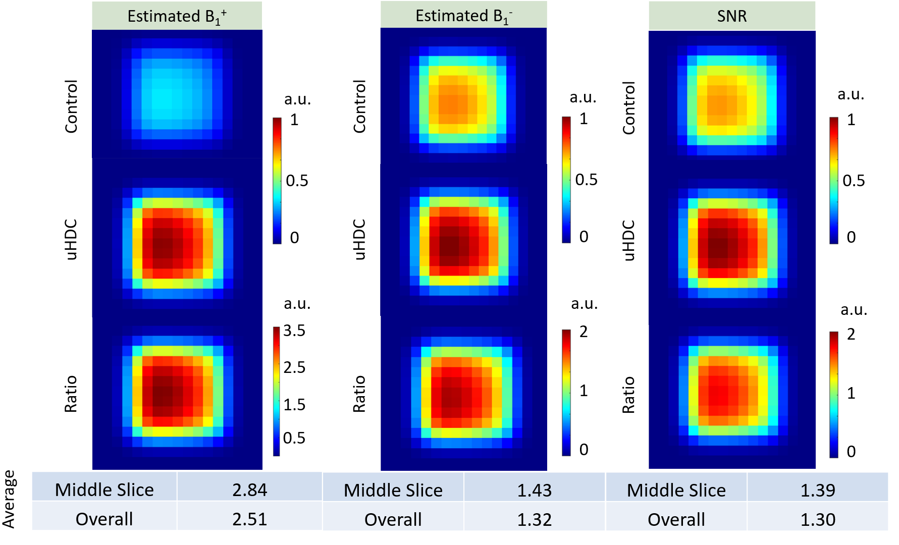
Figure 3. Normalized estimated B1+/- field maps of 2H CSIs in the middle slices where the uHDC disks are placed. Overall averaged SNR gain and B1- gain with the uHDC disks were 32% and 30%, respectively. Overall averaged B1+ was improved by 2.5 times higher with uHDC disks than without uHDC disks.