0526
Motion-robust high-resolution multi-echo 3D gradient echo imaging of the human brain at 10.5 tesla using 80 receive channels1Advanced Imaging Research Center, UT Southwestern Medical Center, Dallas, TX, United States, 2Radiology, UT Southwestern Medical Center, Dallas, TX, United States, 3Advanced MRI section, NINDS, NIH, Bethesda, MD, United States, 4CMRR, Radiology, Medical School, University of Minnesota, Minneapolis, MN, United States
Synopsis
Keywords: High-Field MRI, High-Field MRI, multi-parametric mapping; motion and field correction
Motivation: There is an increasing interest in T2*-related contrast at ultrahigh field for increased signal-to-noise and contrast-to-noise ratios.
Goal(s): To demonstrate the feasibility and utility of high-resolution T2*-weighted brain MRI at 10.5 tesla by combining a motion-robust multi-echo gradient-echo method with a high-channel-count RF coil.
Approach: Images were collected at 0.5-mm isotropic resolution using a custom 80-channel receive (80Rx) coil and used for quantitative R2* and susceptibility mapping.
Results: Our method effectively eliminated artifacts from motion, producing quality images and multi-parametric maps. Parallel imaging performance was improved using the 80Rx coil relative to the commercial 7-tesla Nova 32Rx coil.
Impact: The demonstrated feasibility and utility of motion-robust high-resolution multi-echo gradient echo imaging in humans at 10.5 tesla may shed light on future optimal implementation of anatomic T2*-weighted brain MRI at ultrahigh field, paving the way for many neuroscience applications.
Introduction
Pushing the resolution limit for in vivo MRI motivates the development of ultrahigh field (UHF) technologies. Significant progress has been made in human MRI beyond the clinically approved 7 tesla (7 T), including initial human studies at 10.5 T1,2. To fully realize the promise of high resolution at UHF, it is important to address subject motion while developing radiofrequency (RF) detectors with high parallel imaging performance. Here, we demonstrated the benefit of combining the motion-robust data acquisition and image reconstruction strategies3 for the multi-echo 3D gradient-recalled echo (GRE) contrast and a custom 16-channel transmit, 80-channel receive (16Tx80Rx) RF coil4 in producing high-quality, high-resolution T2*-weighted (T2*w) images and associated quantitative R2* and susceptibility (χ) maps in human at 10.5 T.Methods
MRI experimentsExperiments were performed on a Siemens 10.5 T Dotplus MR scanner (Siemens, Erlangen, Germany) (Fig. 1) capable of 16-channel parallel transmission and 128-channel signal reception and equipped with a whole-body gradient (80 mT/m maximum strength and 200 T/m/s maximum slew rate). One healthy adult was scanned with a signed consent form approved by the local Institutional Review Board. The custom 16Tx/80Rx coil was operated in its circularly polarized mode for excitation.
T2*w MRI data at three echo times (TE) were acquired using a 3D multi-echo GRE sequence. In the same sequence, volumetric navigator images covering the same field of view (FOV) were simultaneously acquired using multi-shot 3D echo planar imaging (EPI) at a short TE, for motion and B0 fluctuation correction3. Data were obtained with the following parameters: isotropic resolution=0.5 mm, FOV=256×192×48 mm3, slice oversampling=50%, TE1/TE2/TE3=13.2/23.6/34.0 ms, TR=50 ms, nominal flip angle=17°, bandwidth=98 Hz/pixel, 2D acceleration rate=3×2 with controlled aliasing in parallel imaging (CAIPI)5 and scan time=7.7 min.
The navigators were obtained with spatial resolution of 5.3×6×6 mm3 and temporal resolution of 0.3 s. To estimate the receive sensitivity maps for parallel imaging reconstruction, a reference scan was conducted using a multi-slice 2D GRE sequence, with an isotropic resolution of 4 mm, FOV=256×192×160 mm3 and scan time=12 s.
Data processing and analysis
Image reconstruction was performed using a custom MATLAB software incorporating estimated sensitivity maps, motion correction and spatially linear B0 correction3. For comparison, reconstruction was also performed without these corrections. The relaxation rate R2* was quantified using mono-exponential fitting of the multi-echo magnitude images. Quantitative susceptibility mapping (QSM) was carried out using the JHU/KKI QSM toolbox, including path-based phase unwrapping6, skull stripping (BET in FSL)7, VSHARP background field removal8, and dipole inversion using structural feature-based regularization9 and a L2-norm data consistency function10.
The g-factor maps, characterizing the spatial noise amplification of parallel imaging reconstruction, were calculated for various 2D acceleration configurations and two FOVs utilizing the estimated sensitivity maps and measured noise covariance matrices. The results were compared to those obtained at 7 T using the commercial Nova 1Tx/32Rx coil (Nova Medical Inc., MA).
Results
The reconstruction with motion and B0 correction led to high image quality (Fig. 2), effectively eliminating image artifacts (especially near the cortex) observed for reconstruction without corrections. The maximum rotation across all three axes was 0.6° and the maximum translation was 1.6 mm measured by the navigator.The quality multi-echo GRE images translated into quality quantitative R2* and χ maps (Fig. 3), delineating rich anatomical details in many brain regions including superficial white matter and intracortical layers.
The 10.5 T 80Rx coil outperformed the 7 T Nova 32Rx coil in parallel imaging (Figs. 4 and 5), reducing g-factor, especially at higher acceleration rates.
Discussion
We have demonstrated the feasibility of high-resolution, high-quality multi-echo T2*w GRE human brain imaging at 10.5 T and its utility for multi-parametric mapping. Critical to the success was a synergistical combination of technical developments including a high-channel-count RF receive coil, and a motion-robust multi-echo 3D GRE imaging method.The g-factor comparison for the 10.5 T 80Rx coil vs. the 7 T Nova 32Rx coil confirmed that the combination of a high-channel-count RF coil and a higher field strength can provide multiplicative gains in parallel imaging, enabling higher acceleration factors.
Given that susceptibility-based MRI and related quantitative maps are important neuroimaging application at UHF11, part of our future studies is to investigate the field dependent susceptibility contrast and to optimize the high-resolution susceptibility imaging methods at 10.5 T.
Conclusion
It is feasible to perform high-quality high-resolution multi-echo T2*-weighted imaging of the human brain at 10.5 T using a high-channel-count RF coil in combination with a motion-robust multi-echo 3D gradient echo imaging method.Acknowledgements
This work was supported in part by Hamon Foundation, Texas Instrument Foundation, NIH grants (NIBIB P41 EB027061, U01 EB025144 and S10 RR029672), and the intramural research program of NINDS/NIH.
References
1. Sadeghi-Tarakameh, A. et al. In vivo human head MRI at 10.5T: A radiofrequency safety study and preliminary imaging results. Magn Reson Med 84, 484–496 (2020).
2. He, X. et al. First in-vivo human imaging at 10.5T: Imaging the body at 447 MHz. Magn Reson Med 84, 289–303 (2020).
3. Liu, J., van Gelderen, P., de Zwart, J. A. & Duyn, J. H. Reducing motion sensitivity in 3D high-resolution T2*-weighted MRI by navigator-based motion and nonlinear magnetic field correction. Neuroimage 206, 116332 (2020).
4. Waks, M. et al. A self-decoupled 16-channel transmit, 80-channel receive array for 10.5 Tesla human head imaging. in Proceedings of the 31st Annual Meeting of ISMRM 211 (2023).
5. Breuer, F. A. et al. Controlled aliasing in volumetric parallel imaging (2D CAIPIRINHA). Magn Reson Med 55, 549–556 (2006).
6. Abdul-Rahman, H., Gdeisat, M., Burton, D. & Lalor, M. Fast three-dimensional phase-unwrapping algorithm based on sorting by reliability following a non-continuous path. in vol. 5856 32–41 (International Society for Optics and Photonics, 2005).
7. Smith, S. M. Fast robust automated brain extraction. Hum Brain Mapp 17, 143–155 (2002).
8. Wu, B., Li, W., Guidon, A. & Liu, C. Whole brain susceptibility mapping using compressed sensing. Magn Reson Med 67, 137–147 (2012).
9. Bao, L., Li, X., Cai, C., Chen, Z. & van Zijl, P. C. M. Quantitative Susceptibility Mapping Using Structural Feature Based Collaborative Reconstruction (SFCR) in the Human Brain. IEEE Trans Med Imaging 35, 2040–2050 (2016).
10. Milovic, C., Bilgic, B., Zhao, B., Acosta-Cabronero, J. & Tejos, C. Fast nonlinear susceptibility inversion with variational regularization. Magnetic Resonance in Medicine 80, 814–821 (2018).
11. Duyn, J. H. et al. High-field MRI of brain cortical substructure based on signal phase. Proc. Natl. Acad. Sci. U.S.A. 104, 11796–11801 (2007).
Figures
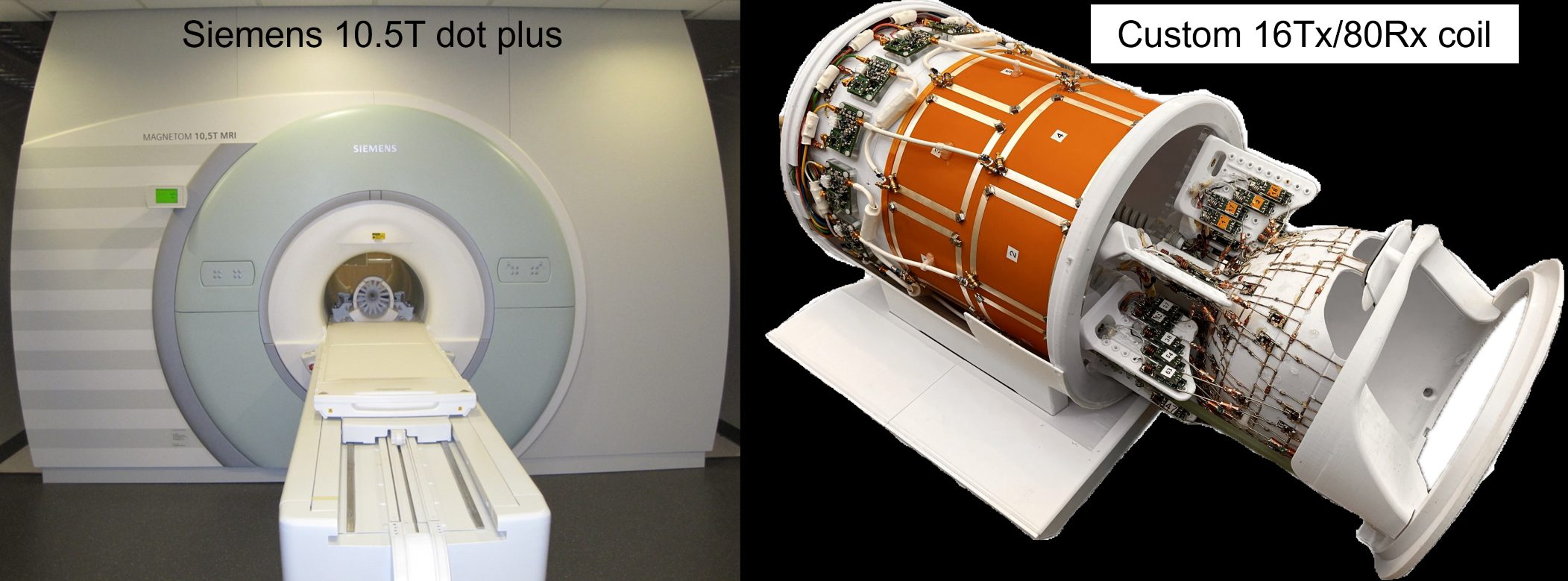
Fig. 1. Experimental setup. Image data were collected in healthy volunteers on a 10.5 tesla (10.5 T) Siemens Magnetom Dotplus scanner (left) using a custom 16-channel transmit and 80-channel receive (16Tx/80Rx) RF coil (right).
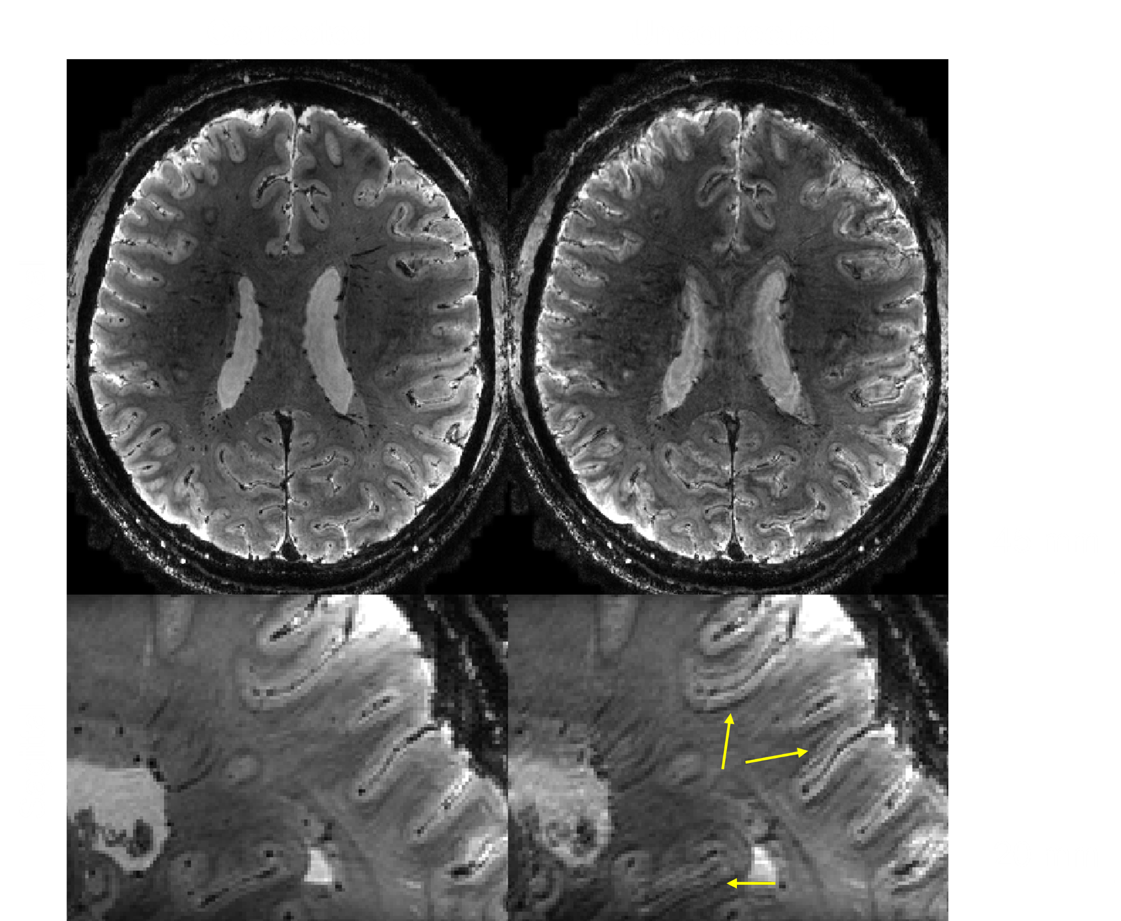
Fig. 2. Importance of simultaneous motion and B0 change corrections in preserving the image quality for the isotropic 0.5 mm resolution T2*-weighted (T2*w) images at 10.5 T. Shown are T2*w echo-averaged magnitude images in axial (top) and sagittal (bottom) views, for reconstruction with (left) and without (right) motion and B0 corrections, respectively. The corrections effectively eliminated image artifacts over the whole field of view (e.g., those near the cortex as highlighted by arrows).
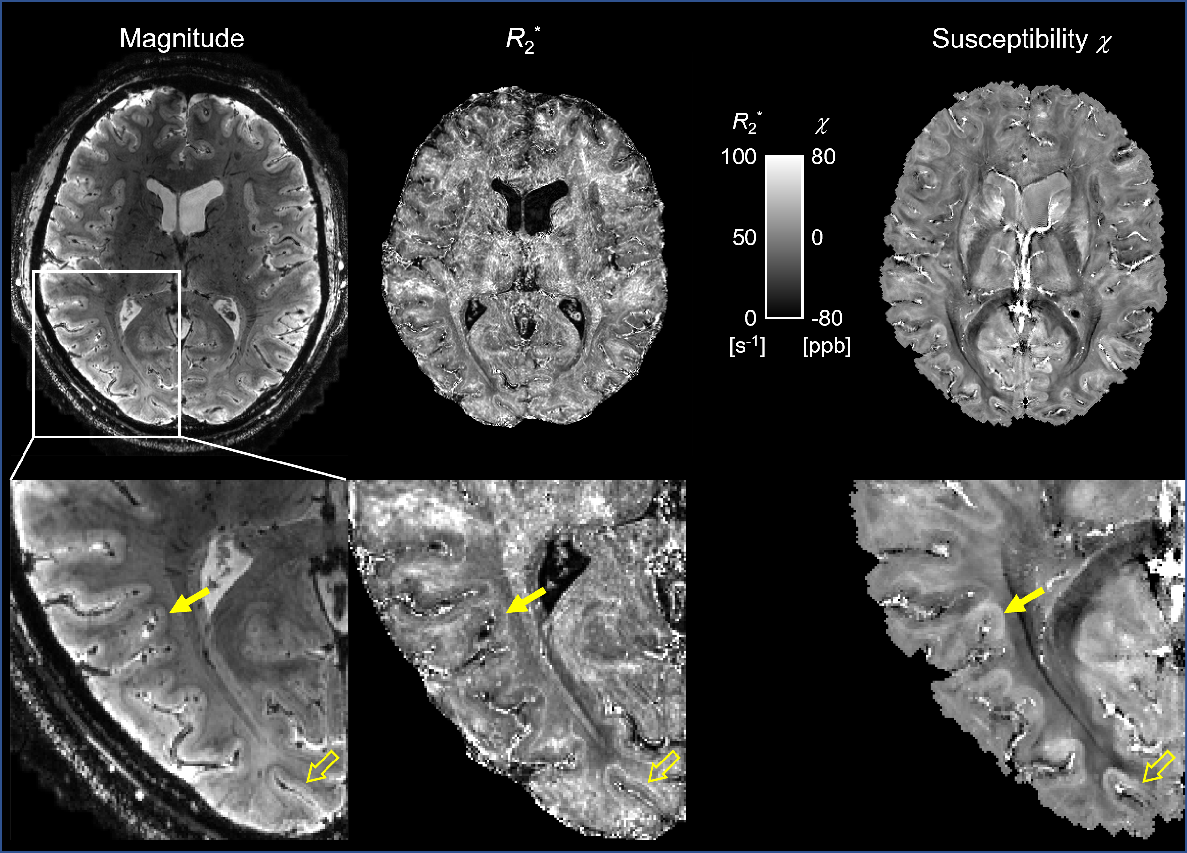
Fig. 3. Utility of motion and B0 corrected multi-echo GRE for multi-parametric mapping at 10.5 T. Shown are a reference T2*w echo-averaged magnitude image (left), alongside quantitative R2* (middle) and susceptibility (right) maps in the same representative slice. The zoomed-in images (bottom) highlight the image quality, delineating fine brain structures, e.g., the superficial white matter as indicated by solid arrows and intracortical structures as indicated by open arrows.
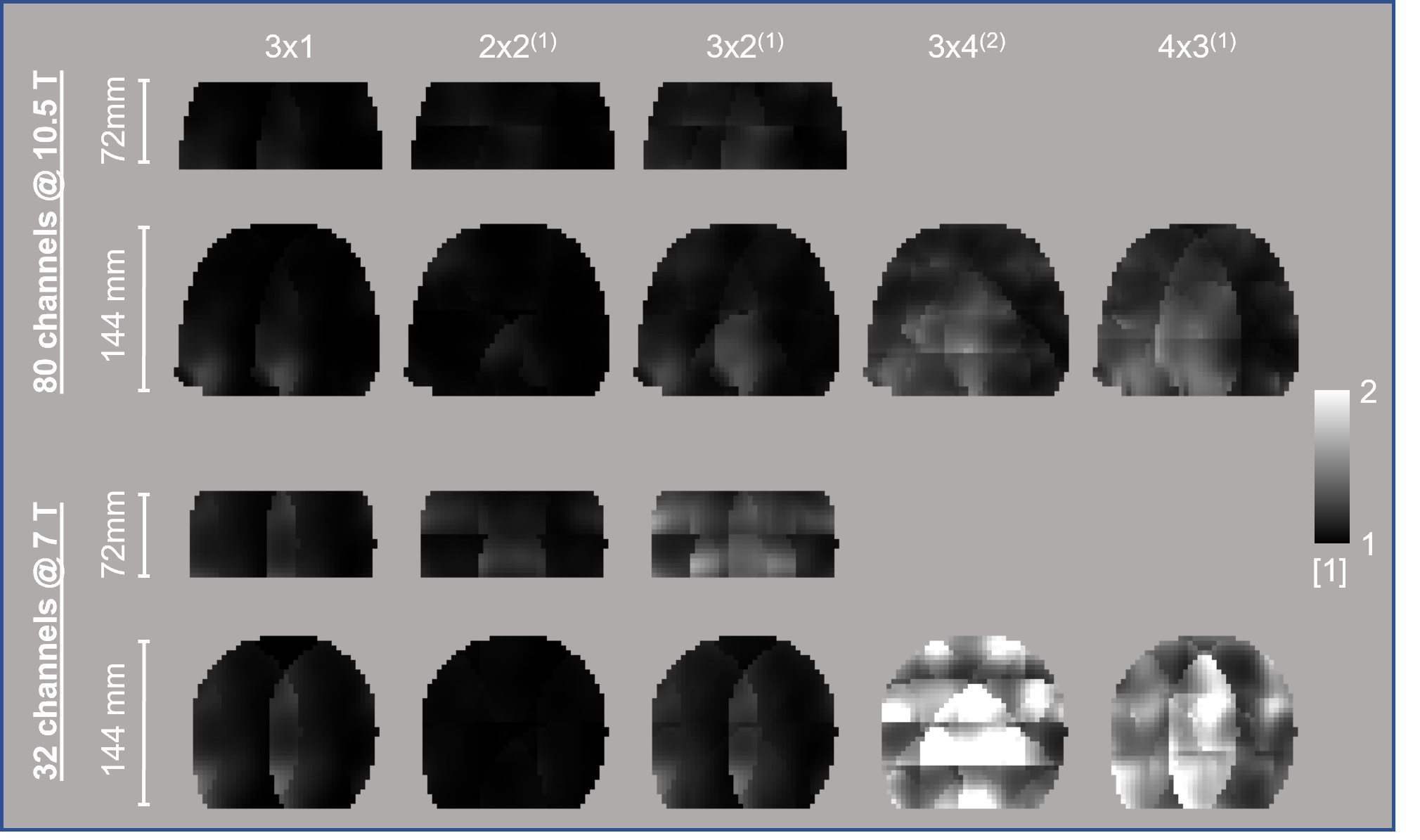
Fig. 4. Comparing the new 10.5 T 80-channel receive coil (top panel) against the commercial 7 T 32-channel Nova coil (bottom panel) in parallel imaging performance. Shown are g-factor maps in representative coronal slices, for different 2D acceleration schemes (Rp×Rs(∆s)) and two fields of view in the slice direction (72 vs. 144 mm). Here, Rp and Rs stand for the acceleration rate in the phase (left-right) and slice (head-foot) directions, respectively; ∆s is the CAIPI shift for controlled aliasing.
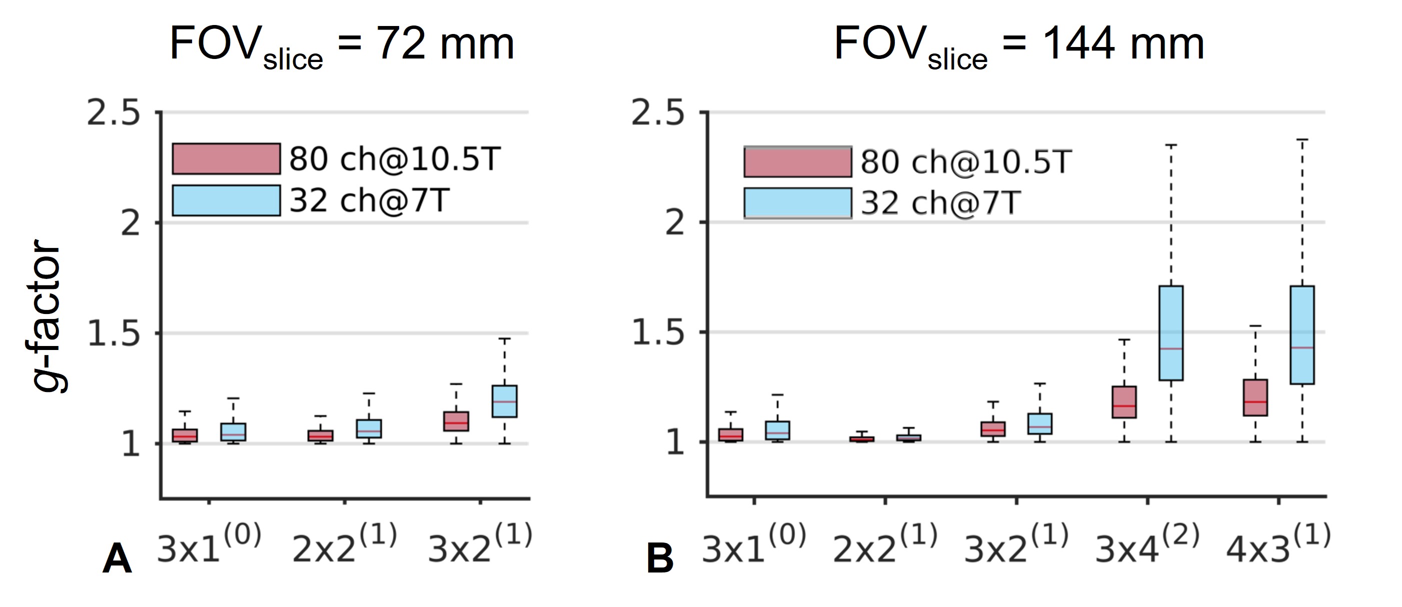
Fig. 5 Comparing statistical distributions of the g-factor across the whole FOV. Shown are boxplots summarizing g-factor values for the two RF coils when used with different acceleration rates and two FOVs in the slice direction. The horizontal bar within each box indicates the median value and the box height represents the 25th and 75th percentile range. Note how the use of our 80-channel receive coil at 10.5 T reduced g-factors, especially at higher acceleration or with shorter slice FOV or both.