0523
Opening new horizons with the first human brain in vivo experiments at 11.7T1University Paris-Saclay, CEA, CNRS, BAOBAB, NeuroSpin, Gif-sur-yvette, France, 2Siemens Healthcare SAS, Courbevoie, France, 3IRFU, CEA, Gif-sur-yvette, France, 4University Paris-Saclay, CEA, Uniact, NeuroSpin, Gif-sur-yvette, France
Synopsis
Keywords: High-Field MRI, High-Field MRI
Motivation: Ultra-high magnetic field MRI offers new opportunities due to its higher SNR and CNR.
Goal(s): After receiving the authorization for scanning 20 adult healthy volunteers on the whole-body Iseult MRI scanner, we performed a preliminary MRI investigation to acquire the first brain images at 11.7T.
Approach: Using a homemade multi-transmit multi-receive head coil and parallel transmission, 3D anatomical images were acquired with multiple sequences and tissue contrasts at high resolution.
Results: In this study, high quality whole brain images acquired at 11.7T on the Iseult MRI scanner are presented for the first time to the MR community.
Impact: Showing for the first time in vivo brain images acquired at 11.7T on the whole-body Iseult MRI scanner, this study reveals first potential and challenges of such systems to the ultra-high field MR community.
Introduction
Pushing the boundaries of field strength in MRI, this study reports the first in vivo human brain images acquired at 11.7T on the whole-body Iseult MRI scanner1. Approval to scan in vivo 20 adult healthy volunteers (18-40 years old) at 11.7T was obtained from the French National Regulatory Body (ANSM) and the local ethics committee in Feb 2023. This first protocol consisted mostly of evaluating the safety for subjects exposed to the main magnetic field, for which results will be communicated elsewhere, and perform preliminary MRI testing and investigations with 1h30 long exams.Methods
Informed consent was obtained from all volunteers. The 90-minute exam included the following sequences: localizer, B0 mapping and shimming, B0 and B1+ mapping for parallel transmission (pTx) pulse design, 3D multi-echo GRE in pseudo-CP mode (phase-shimming) and pTx, 3D slab-selective T2*-weighted GRE, 3D T1-weighted MP2RAGE and 3D T2-weighted SPACE. The Iseult 11.7T whole-body scanner has been equipped with the SC72 whole-body gradient coil (Siemens Healthcare, Erlangen, Germany). Experiments were also carried out with a homemade pTx RF coil whose design is described in2. SAR was explicitly taken into account in the RF pulse design using first level mode with virtual observation points derived from homemade electromagnetic simulations and using the RF safety management strategy described in3. RF pulses were designed with kT-points4, kT-spokes5 and GRAPE6 depending on the sequences. The complex RF coefficients were optimized simultaneously with the transmit k-space trajectory7.Results
Figure 1 shows B0 maps of one subject after a 2nd-order B0 shimming procedure. The resulting inhomogeneity (standard deviation over the 3D brain) was evaluated around 90Hz. Figure 1 also depicts individual B1+ maps of the transmit coil. In the default pseudo CP-mode, the peak total power necessary to achieve 180° flip angle in 1 ms was found to be 5.2 kW, on average over the 3 dimensional brains and over the subjects. Figure 2 compares a sagittal 3D-GRE multi-echo using the pseudo-CP mode versus a tailored dynamic pTx solution with a 830 µs-long 7-kT points pulse, showing great mitigation of the RF field inhomogeneity problem with dynamic-pTx. Figure 3 highlights the exquisite contrast obtained with a high in-plane resolution T2*-weighted 3D slab-selective GRE with pTx (TE=15 ms, 0.22×0.22×1.2 mm3). High-resolution (0.55 mm isotropic) T1 and T2-weighted 3D anatomical images with pTx are finally provided in Fig.4 and Fig.5, showing again mitigation of the RF field inhomogeneity problem and high SNR despite the high resolutions.Discussion
The increase in B0 inhomogeneity is consistent with a linear trend versus field, reflecting Iseult meeting its field homogeneity specifications. Due to time-constraints, comparisons of pseudo-CP versus pTx could not be systematically conducted for all sequences. The results reported in Fig.2 however highlight the mitigation of the B1+ field inhomogeneity problem we could achieve at 11.7T, which was further confirmed with other sequences, e.g., including refocusing pTx pulses (Fig.5). Due to the current architecture of the transmit RF coil and the increased power losses at 500 MHz, yet it has been difficult so far to achieve complete magnetization inversion everywhere in the brain. More work therefore is under way to tackle this problem with different transmit channels pairing2 or different RF coil designs8, which we expect by the same token will increase SNR. Our results also emphasize the need to further accelerate sequences and develop robust motion correction methods to achieve ultra-high resolutions when motion-induced field fluctuations are accentuated. These topics are currently the subject of current research to unleash the full potential of 11.7T MRI.Conclusion
This work reports the first brain images acquired at 11.7T on adult healthy volunteers. The results remain preliminary given the limited number of subjects scanned so far and the apparent need of always refining protocols and methods for more optimal acquisitions. Given the severity of the RF field inhomogeneity, parallel transmission hereby appears unavoidable. The images however reveal already superb tissue contrast and pave the way for exciting and unique exploration of the human brain at unprecedented field strength.Acknowledgements
AROMA H2020 FET-Open (885876). ANR-21-ESRE-0006 (“Investissements d'avenir"). The authors are greatly indebted to the Irfu department of CEA for designing and commissioning the Iseult magnet. Edouard Chazel is thanked for assembling the RF coil. The authors also thank Siemens Healthineers, especially Felix Koeber, Peter Dietz and Wolfram Ruth (Germany), and Brice Koestel and Xavier Tastet (France) for valuable support. The authors are also very grateful for the valuable support of the NeuroSpin platform for making these experiments possible. Finally, the AROMA consortium is acknowledged for valuable discussions to troubleshoot the scanner during the commissioning stage.
References
1. Boulant N, et al. (2023). Commissioning of the Iseult CEA 11.7 T whole-body MRI: current status, gradient–magnet interaction tests and first imaging experience, Magn Reson Mater Phy 36, 175–189.2. Luong M, et al. (2022). A Compact 16Tx-32Rx Geometrically Decoupled Phased Array for 11.7T MRI, In Proceedings of the 31st Annual Meeting of ISMRM, p. 707.
3. Boulant N, et al. (2018). Workflow proposal for defining SAR safety margins in parallel transmission. In Proceedings of the 27th Annual Meeting of ISMRM, Paris, p. 295.
4. Cloos M A, et al. (2012). kT-points: short three-dimensional tailored RF pulses for flip-angle homogenization over an extended volume. Magn Reson Med. 67(1):72-80.
5. Saïb G, et al. (2018). kT-spokes: combining kT-points with spokes to ease ramp pulse design for TOF slab selection with parallel transmission at 7T. In Proceedings of the 27th Annual Meeting of ISMRM, Paris, p. 2690.
6. Van Damme L, et al. (2021). Universal non-selective excitation and refocusing pulses with improved robustness to off-resonance for Magnetic Resonance Imaging at 7 Tesla with parallel transmission. Magn Reson Med. 85: 678-693.
7. Gras V, et al. (2015). Joint design of kT-points trajectories and RF pulses under explicit SAR and power constraints in the large flip angle regime. JMR. 261:181-189.
8. Chu S, et al. (2023). Electromagnetic and RF pulse design simulation based optimization of an eight-channel loop array for 11.7T brain imaging. Magn Reson Med. 90: 770-783.
Figures
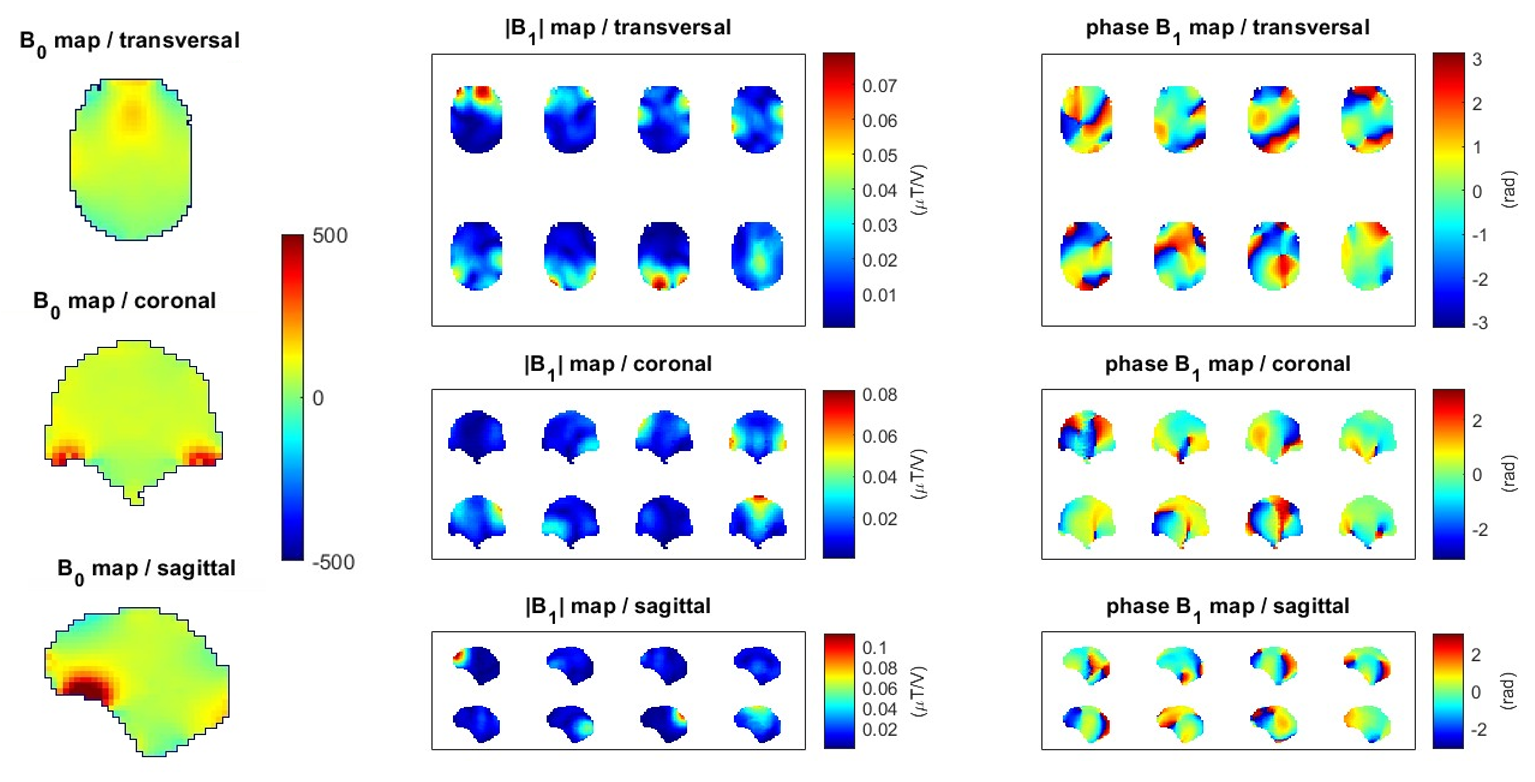
Figure 1. B0 and B1+ maps in the brain of volunteer #8 in transverse, coronal, and sagittal orientations. B0 maps are expressed in Hz and were measured using a 3-echo non-selective 3D-GRE acquired in sagittal (2.5mm isotropic, total acquisition time of 60s). B1+ maps were measured using an interferometric magnetization-prepared 2D turbo-FLASH at a resolution of 5mm isotropic in 4min. Individual B1+ transmit channels are depicted in amplitude and phase, expressed in µT per volt and radians respectively.
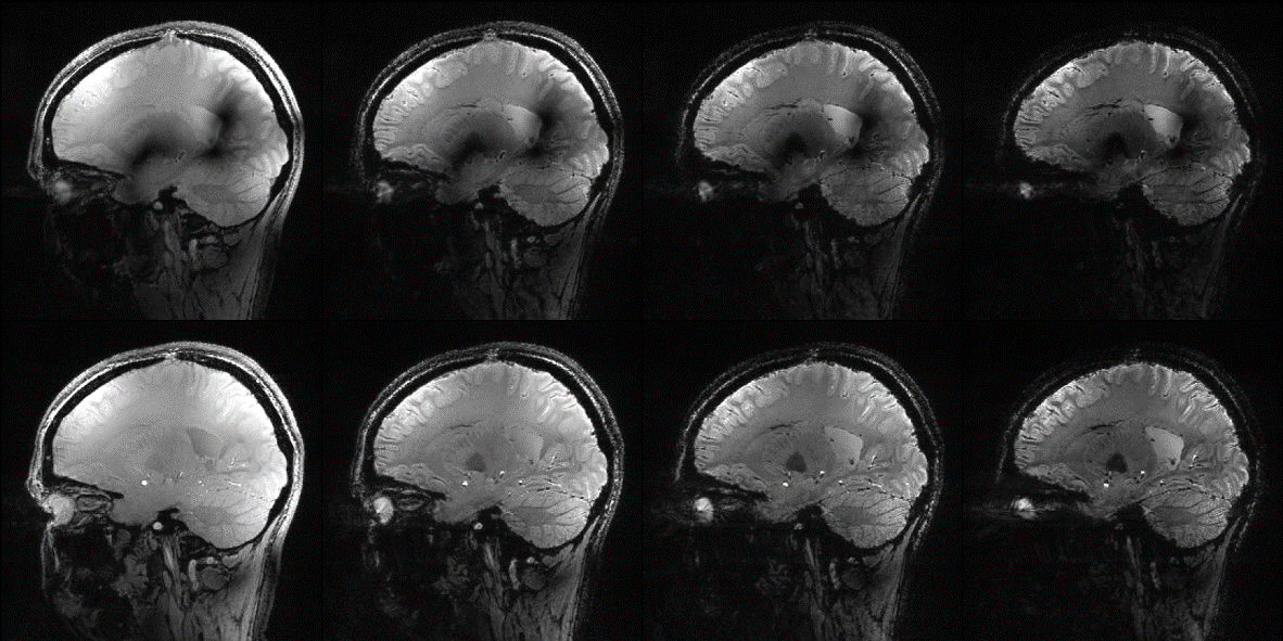
Figure 2. Sagittal views of T2*-weighted isotropic 0.8 mm whole brain peusdo-CP (top row) vs kT-points (bottom row) at different echo times (from left to right, TE = 3.2, 7.2, 11.2 and 15.6 ms) on volunteer #2. Acquisition parameters were 3D non-selective sagittal orientation, TR=25ms, Flip Angle=10°, CAIPIRINHA acceleration 2x2 and a scan time of 6min14.
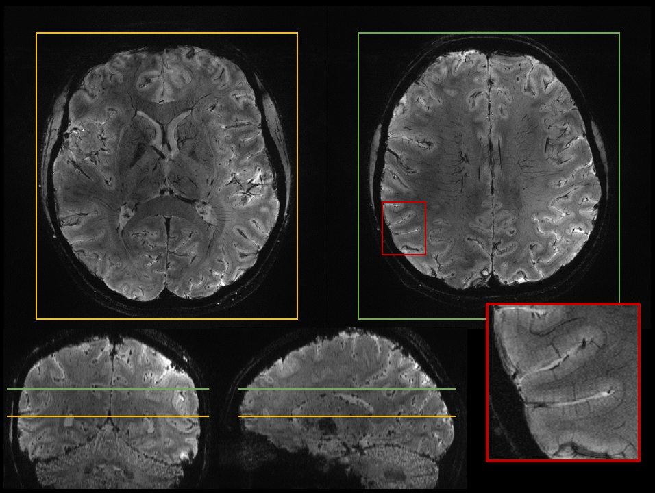
Figure 3. T2*-weighted 3D slab-selective using a 3-spoke pTx pulse with high in-plane resolution (0.22×0.22×1.2 mm3) in axial views (top row) on volunteer #6. Coronal and sagittal views (bottom row) show the slab coverage over the brain. Acquisition parameters were TE=15ms, TR=29ms, a receive bandwidth of 168Hz/pixel, GRAPPA 4 in R>>L phase encode direction and a scan time of 11min31.
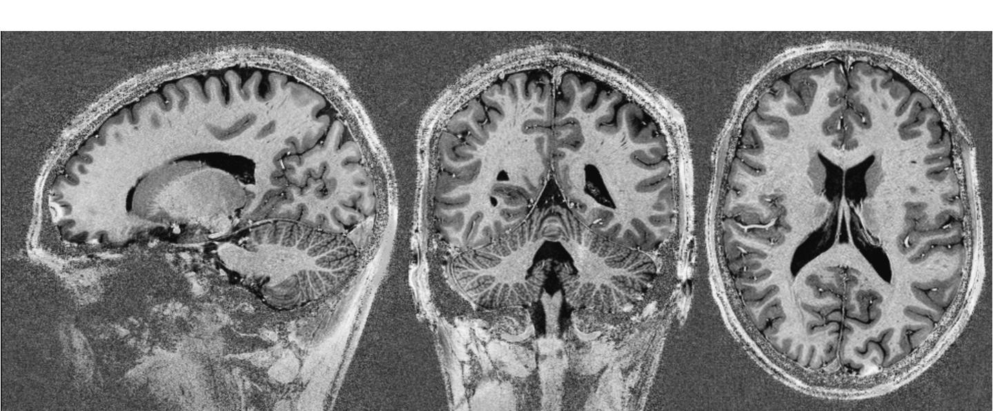
Figure 4. Sagittal, coronal and axial UNI images of a 3D T1-weighted MP2RAGE acquisition at 0.55mm isotropic resolution on volunteer #9. Acquisition parameters were 3D non-selective sagittal orientation, TR=5200ms, TI1=1130ms, TI2=3730ms, flip angles of 4° and 2° in consecutive GRE trains, GRAPPA 4 in A>>P phase encode direction and a scan time of 10min08. Spin inversion and excitation were realized using universal (simultaneous design across 5 B0, B1+ maps) GRAPE pulses of duration 10 ms and 0.3 ms respectively.
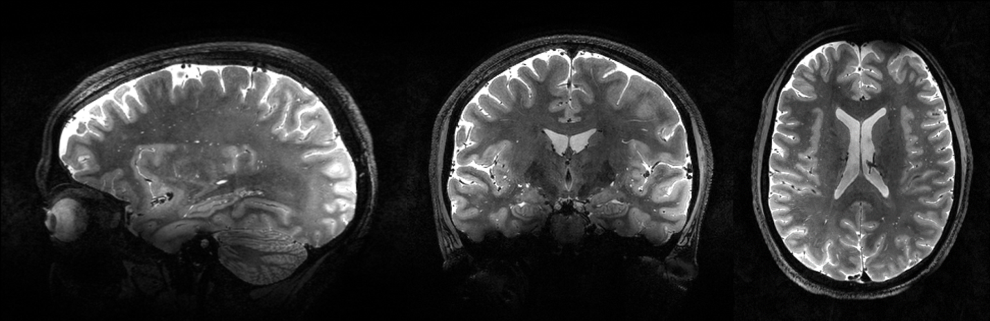
Figure 5. Sagittal, coronal and axial views of a 3D T2-weighted SPACE acquisition at 0.55mm isotropic resolution on volunteer #8. Acquisition parameters were 3D non-selective sagittal orientation, TR=6s, GRAPPA 3x2 in phase and partition directions respectively and a scan time of 13min20. Excitation and refocusing were realized using a single symmetric scalable GRAPE universal pulse of duration 1.4 ms designed across 5 B0, B1+ maps.