0521
Reproducibility of tailored and universal non-selective excitation pulses at 7T for human cardiac body imaging: A 3-year and an inter-day study1Physikalisch-Technische Bundesanstalt (PTB), Berlin, Germany, 2Charité – Universitätsmedizin Berlin, Berlin, Germany, 3Medical Physics in Radiology, German Cancer Research Center (DKFZ), Heidelberg, Germany, 4Center for Magnetic Resonance Research, University of Minnesota, Minneapolis, MN, United States
Synopsis
Keywords: RF Pulse Design & Fields, High-Field MRI, Reproducibility, Universal pulse, 7T, pTx
Motivation: Addressing the issue of reduced spatial variability in flip angle (FA) patterns in ultra-high field 3D imaging of the human heart for inter-year and inter-day studies, ensuring a high level of reproducibility.
Goal(s): Find the correct RF parallel transmission (pTx) excitation scheme that reduces FA spatial variability in 7T 3D imaging of the human heart for long-term and short-term studies.
Approach: Default, tailored pulses (TP), and pre-computed universal pulses (UP) were evaluated to optimize FA homogeneity in B1+ datasets across three years.
Results: The study highlights UPs' robustness in managing FA variations across subjects and coil placements in 3D body imaging at 7T.
Impact: This study confirms pre-computed UPs' suitability for 7T cardiac flip angle homogenization. Different MRI operators maintained consistent RF performance across three years and inter-day tests, with no significative differences in scans at various coil positions for both tests.
Introduction
Ultra-high field (UHF) body MRI faces challenges due to inherent spatial variations in flip angle (FA) patterns1. Subject-specific parallel transmission (pTx) methods, relying on personalized $$$B_1^+$$$-maps, ensure high FA uniformity in the cardiac region2,3,4. In contrast, using cardiac universal pulses (UPs) offers time-saving benefits and robustness for various RF coil placements, while also achieving FA uniformity in surrounding tissues, including the aorta5,6. This study assesses the reproducibility and variability of three excitation methods in six subjects: default excitation, 4kT-points subject-tailored pTx (TP), and calibration-free UP. In addition to a previous study6, three different MRI operators conducted re-scans at one- and two-year intervals, including inter-day scans by two different operators.Methods
MRI was conducted using a Siemens Magnetom 7T with an MRI.TOOLS 32-element body array used in an 8Tx/32Rx mode. A total of 46 channel-wise 3D $$$B_1^+$$$-datasets were acquired during free-breathing4 and subsequently categorized into five groups: i) UP-library (Library: 15M/7F, 21-66y, 19.8-28.3kg/m²), ii) unseen test-cases (Scan 1: 3M/3F, 25-33y, 19.5-35.3kg/m²), iii) re-scans after one year (Scan 2), iv) re-scans after two years (Scan 3), and v) intra-day re-scans of Scan 3 group (Scan 4).A 4kT-point UP was computed based on Library group7 and applied to all subjects and all four scans. TPs were calculated for each subject3 in each Scan and resulting TPs were grouped in four TP configurations, labeled Config. 1 for TPs in Scan 1 to Config. 4 for TPs in Scan 4. Pulses from each Config were then applied for each Scan yielding 16 permutations MRI operators consistently positioned the posterior half of the coil approximately 2 cm from the volunteer's chin, over the volunteer's heart.
The postprocessing encompassed $$$B_1^+$$$-dataset reconstruction4, manual 3D cardiac region selection, RF pulse design, and pulse file generation. Pulse performance was evaluated through FA predictions and coefficient-of-variation (CV) within cardiac volumes. CV cluster points for each scan group were examined using parameters including inertia ($$$I_{CV}$$$), distance from the CV space origin to the cluster centroid ($$$|\bar{x}|$$$), and perpendicular distance from the cluster centroid to the 4D identity line ($$$d_\perp$$$).
Results and Discussion
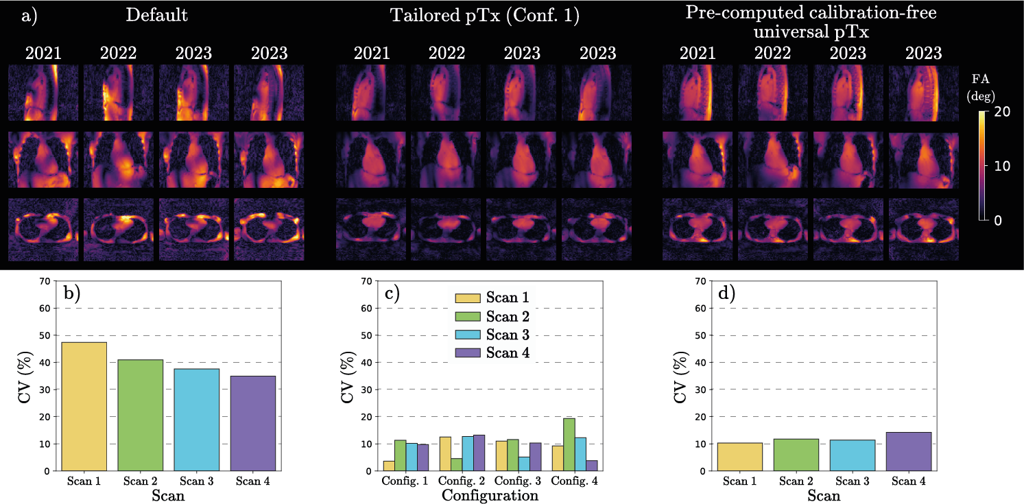
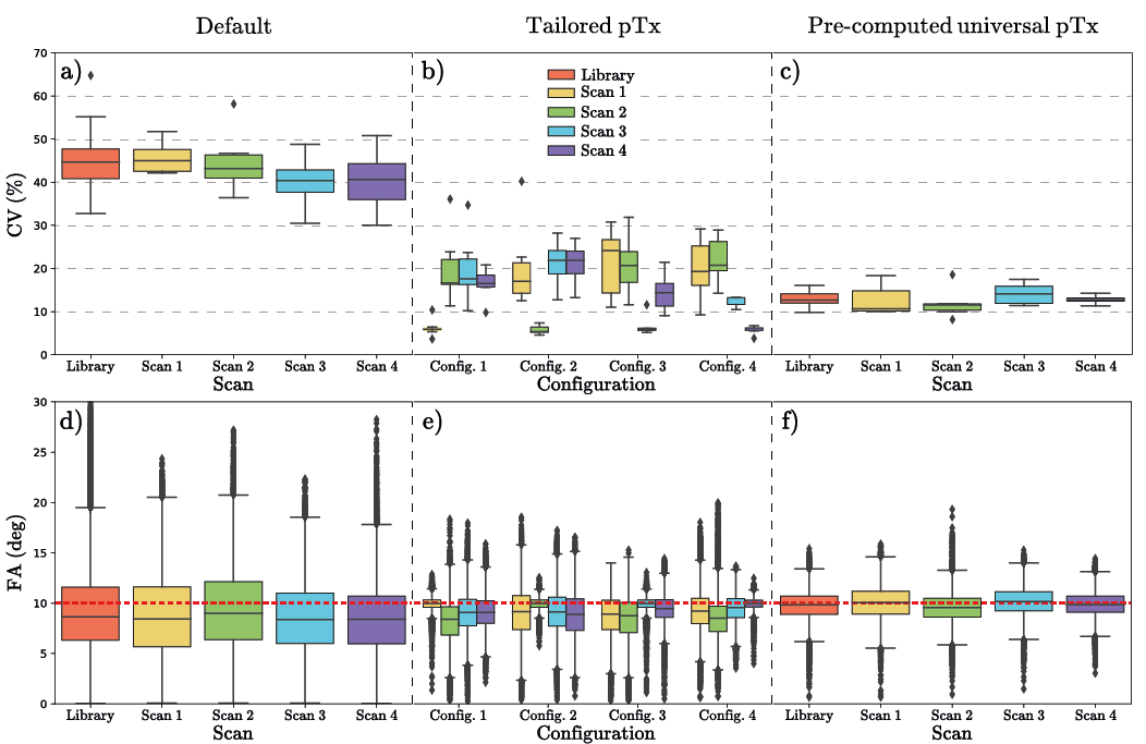
Fig. 1 illustrates FA predictions for three pTx settings in one representative subject. TPs achieved CV values below 5% for Config matching Scan number, but up to 19.6% otherwise, indicating limited reproducibility. In contrast, UP consistently produced similar CV values for all scan groups, demonstrating high reproducibility.Fig. 2 presents predicted CV and FA values for all subjects and excitation methods over the entire cardiac volume. Notably, Config. 3 notably reduces median CV values for Scan 4, contrasting with Scan 1 and Scan 2 groups. This suggests TP's applicability in inter-day studies done by two different operators, but less for inter-year ones. A similar trend is observed for Config. 4. The UP setting maintained median CV values of approximately 10% in all cases, demonstrating reproducibility across all scan groups.
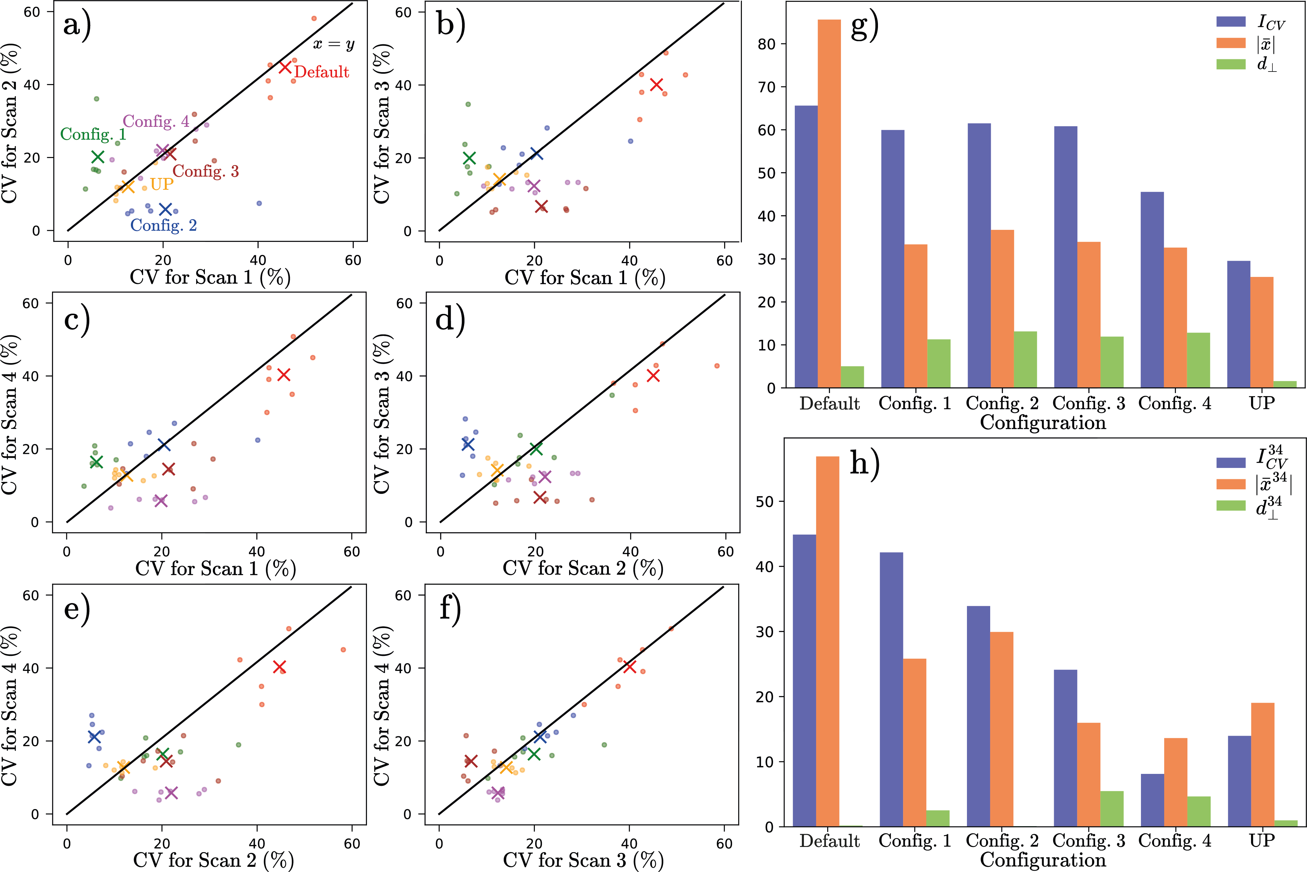
Fig. 3 shows 4D data CV predictions across the cardiac volume for different scan groups. In the inter-year study (see Figure 3g), the UP consistently yields lower $$$I_{CV}$$$, $$$|\bar{x}|$$$, and $$$d_\perp$$$, signifying reduced CVs and strong long-term reproducibility. In the inter-day study (see Figure 3h), UP does not achieve the lowest $$$I_{CV}^{34}$$$, $$$|\bar{x}^{34}|$$$ and $$$d_\perp^{34}$$$ individually, as seen in the inter-year study. Nonetheless, it maintains a $$$d_\perp^{34}$$$ value close to zero while keeping $$$I_{CV}^{34}$$$ and $$$|\bar{x}^{34}|$$$ reasonably low in comparison to other pTx settings, ensuring high reproducibility with low CV values for the inter-day study.
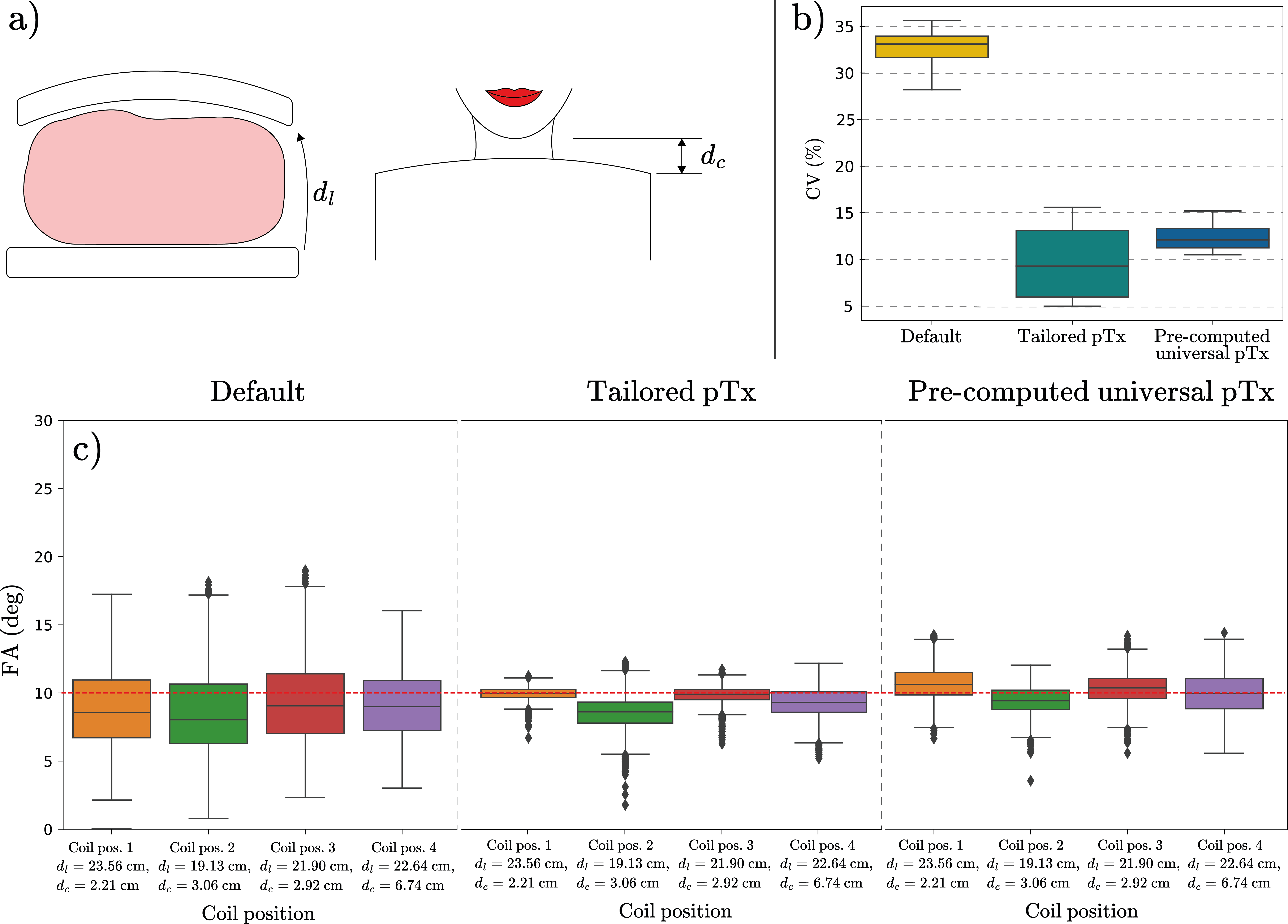
Fig. 4 depicts CV and FA predictions for the three distinct pTx settings explored in this study, obtained from four separate scans conducted on one of the six subjects on the same day, each scan involving different coil positions. TPs were customized for the first scan (Coil. pos 1). In Figure 4b, UP predictions demonstrate exceptional resilience to body coil position variations, as evident from their minimal CV spread. This robustness surpasses other pTx settings for this inter-day study. Figure 4c further accentuates UP's strength, consistently achieving FA values close to the 10° target, outperforming alternative pTx settings in FA accuracy.
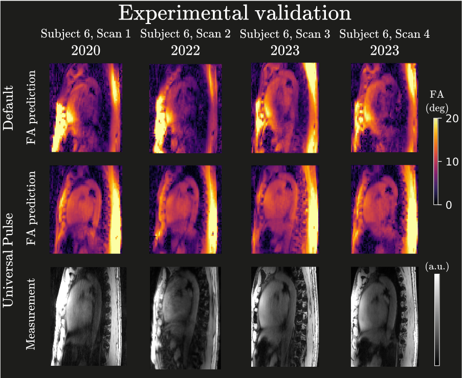
Fig. 5 shows 3D GRE images with UP aligning with $$$B_1^+$$$-predictions, validating coil placement robustness and calibration-free pTx effectiveness in the human heart. Results from 2022 were obtained with a Cartesian sequence, while 2020 and 2023 results used a non-Cartesian sequence5.
Conclusion
This study demonstrates the robustness of pre-computed UPs, which effectively address FA variations among subjects and variations in coil placements in both inter-year and inter-day studies. This property distinguishes UPs from the default pTx setting and TPs, making UPs ideal for 3D body applications at 7T.Acknowledgements
We gratefully acknowledge funding from the German Research Foundation SCHM 2677/2-1, SCHM 2677/4-1 and GRK2260, BIOQIC.References
[1] Ladd, E, Bachert, P, Meyerspeer, M, et al. Pros and cons of ultra-high-field MRI/MRS for human application, Prog. Nuc. Magn. Reson. Spec. 2018; 109:1-50. doi: 10.1016/j.pnmrs.2018.06.001
[2] Padormo, F, Beqiri, A, Hajnal, JV, and Malik, SJ. Parallel transmission for ultrahigh‐field imaging. NMR Biomed. 2016; 29: 1145– 1161. doi: 10.1002/nbm.3313
[3] Aigner, CS, Dietrich, S, Schmitter, S. Three-dimensional static and dynamic parallel transmission of the human heart at 7 T. NMR in Biomedicine. 2021; 34:e4450. doi: 10.1002/nbm.4450
[4] Dietrich, S, Aigner, CS, Kolbitsch, C, et al. 3D Free-breathing multichannel absolute B1+ Mapping in the human body at 7T. Magn Reson Med. 2021; 85: 2552– 2567. doi: 10.1002/mrm.28602
[5] Gras, V., Vignaud, A., Amadon, A., Le Bihan, D. and Boulant, N. (2017), Universal pulses: A new concept for calibration‐free parallel transmission. Magn. Reson. Med., 77: 635-643. doi:10.1002/mrm.26148
[6] Aigner, CS, Bassenge, JP, Dietrich, S, Lutz, M, Krüger, F, and Schmitter, S. Reproducibility and variability of tailored and universal nonselective excitation pulses at 7T for human body imaging: Re-scans after one year. ISMRM 2023, 4590.
[7] Aigner, CS, Dietrich, S, Schaeffter, T, Schmitter, S. Calibration-free pTx of the human heart at 7T via 3D universal pulses. Magn Reson Med. 2021; 87: 70–84. https://doi.org/10.1002/mrm.28952
Figures