0520
Toward Densely Populated Dipole Arrays for Human Prostate Imaging at 7T: 8Tx16Rx Coaxial-End Dipole Array1High-Field MR Center, Max Planck Institute for Biological Cybernetics, Tübingen, Germany, 2Erwin L. Hahn Institute for Magnetic Resonance Imaging, University Duisburg-Essen, Essen, Germany, 3High-Field and Hybrid MR Imaging, University Hospital Essen, Essen, Germany, 4Department for Biomedical Magnetic Resonance, University of Tübingen, Tuebingen, Germany
Synopsis
Keywords: RF Arrays & Systems, Body, Dipoles, Arrays, RF-shimming, SNR, SAR
Motivation: To improve SNR and the SAR-performance in prostate imaging at 7T using a densely populated coaxial-end dipole array.
Goal(s): To numerically optimize and evaluate an 8Tx/16Rx coaxial-end dipole array for prostate imaging at 7T.
Approach: Geometry of a coaxial-end element was optimized to minimize peak SAR and improve coverage. In transmission, 16 coaxial-end dipoles were combined into 8 pairs. This further reduced pSAR. 8-element fractionated dipole and stripline arrays were simulated for comparison.
Results: Optimized 8Tx/16Rx coaxial-end dipole array improved SNR in prostate compared to all other 8-element arrays. SAR-performance of the developed array was better than that of other dipole arrays.
Impact: We demonstrated that densely populated 16-element coaxial-end dipole array improved SNR in the prostate by at least 10% compared to 8-element arrays. In addition, combining 16 elements in 8 pairs during transmission improved SAR-performance in comparison to the 8-channel array.
Introduction
RF arrays of dipole antennas were recently introduced for imaging of deeply located regions of the human body (e.g. the prostate) at ultra-high field (UHF, >7T) (1,2) as a simple and robust alternative to common loop arrays. Due to the large sample size in comparison to the wavelength (~10 cm in the human body at 298 MHz) and the deeply located region of interest (ROI), the RF excitation is more similar to the wave propagation in the lossy media in contrast to the lower-field (1.5, 3T) quasi-static regime. Dipole antennas provide both higher transmit B1+ and SNR compared to loops (2,3). Various eight-element dipole transceiver (TxRx) body arrays have been previously constructed (1,2). Combining eight TxRx-loops and eight TxRx-dipoles has been shown to further improve SNR in the prostate (4). In theory, SNR near the body center can be also improved by increasing the number of dipoles from 8 to 16 (5). To minimize the peak SAR (pSAR) value, coaxial dipoles have been recently proposed for body (6) and head (7) imaging. In addition, combining the dipoles in pairs during transmission to match the number of Tx-channels in typical UHF MR systems allows minimizing pSAR (8). In this work, we numerically optimized and evaluated a densely populated array of 16 coaxial-end dipoles. In transmission, the dipoles were combined in pairs. To our best knowledge, this is the first example of evaluating a16-element single-row dipole array for prostate imaging at 7T.Methods
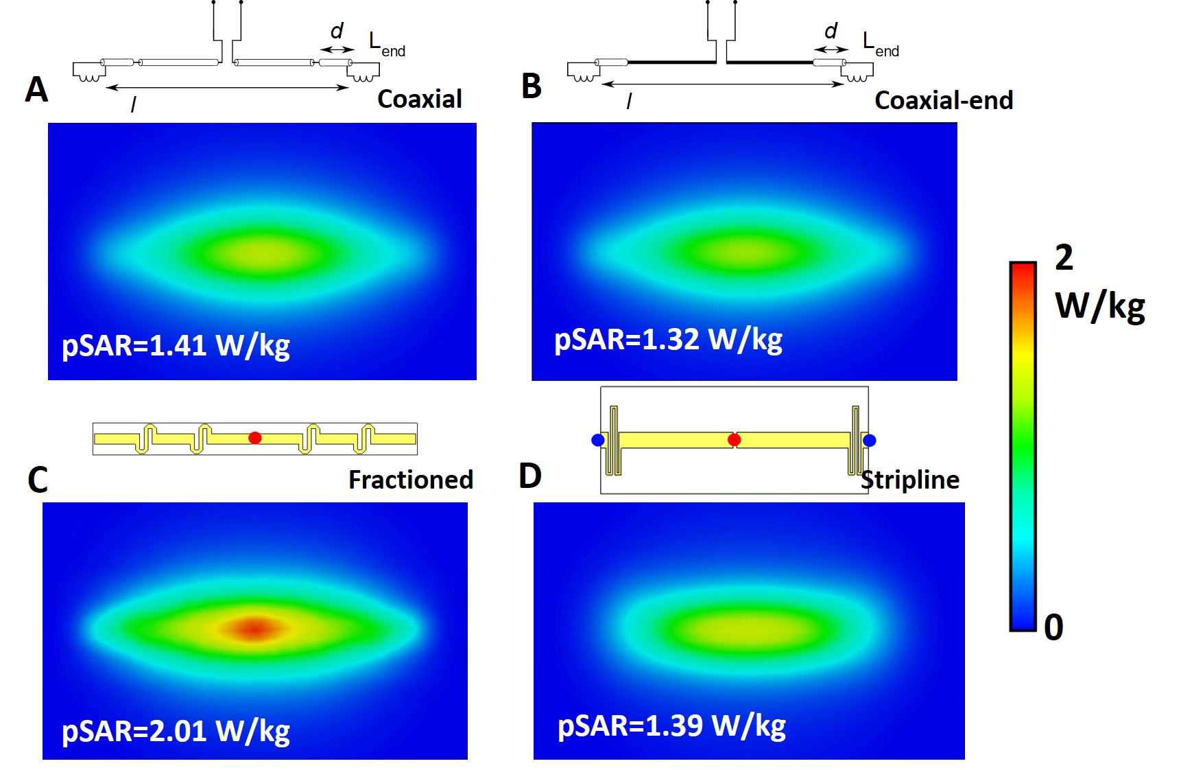
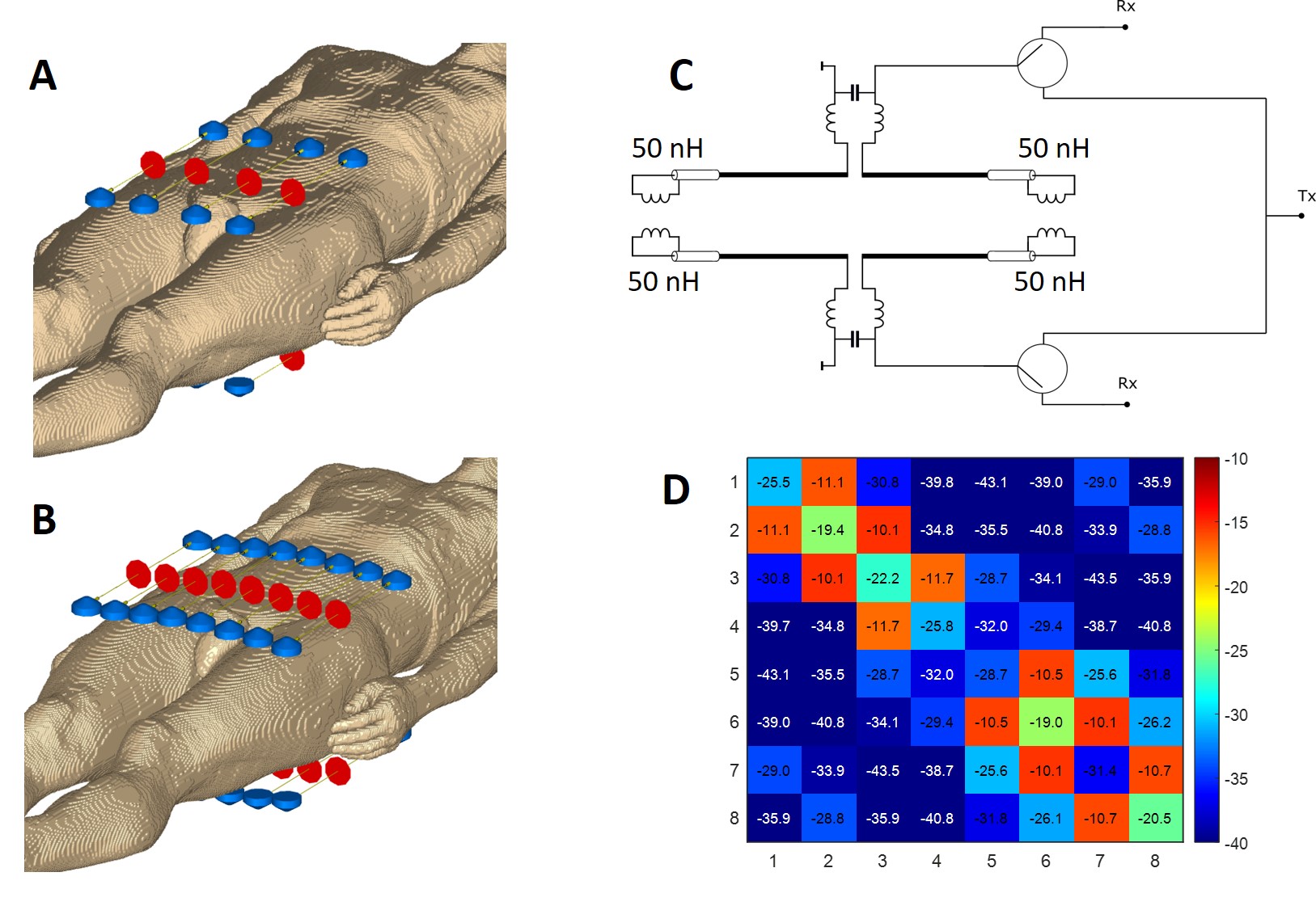
The single coaxial dipole element was very similar to the previously described (8). In addition, we found that the dipole design can be simplified by replacing the central part of the coaxial cable with a wire without a significant change in the dipole performance (Figs. 1A,B), i.e. the coaxial-end dipole. Coaxial-end dipole parameters were optimized to reach a compromise between the best SAR-efficiency (B1+/√pSAR10g) and uniformity of B1+ along the longitudinal z-axis. During optimization, we varied the following parameters (Fig. 1B), i.e. the total dipole length, l, inductance, Lend, and the coaxial part length, d. All dipoles were placed at a 2-cm distance from the phantom. All dipole elements were loaded to the pelvis shaped phantom (ε=34, σ=0.45 S/m). For comparison, we also simulated the fractionated dipole (2) and stripline (9). After finding the optimal coaxial-end dipole configuration, we have simulated four different arrays all loaded by the Duke (Zurich MedTech, Zurich, Switzerland) human body voxel model: 8 coaxial-end dipoles (Fig. 2A), 16 coaxial-end dipoles (Fig. 2B), 8 fractionated dipoles, and 8 striplines. In transmit, 16 dipoles were combined into 8 channels by combining adjacent dipoles in phase (Fig. 2C). Phase-only RF shimming for maximization of efficiency was performed for all arrays with the prostate chosen as ROI. Optimization was performed using CST studio Suite 2021. SAR10g was evaluated using the CST Legacy averaging method. SNR was calculated in MatLab as described in (10) using the imported noise-correlation matrix and individual B1- distributions.Results and Discussion
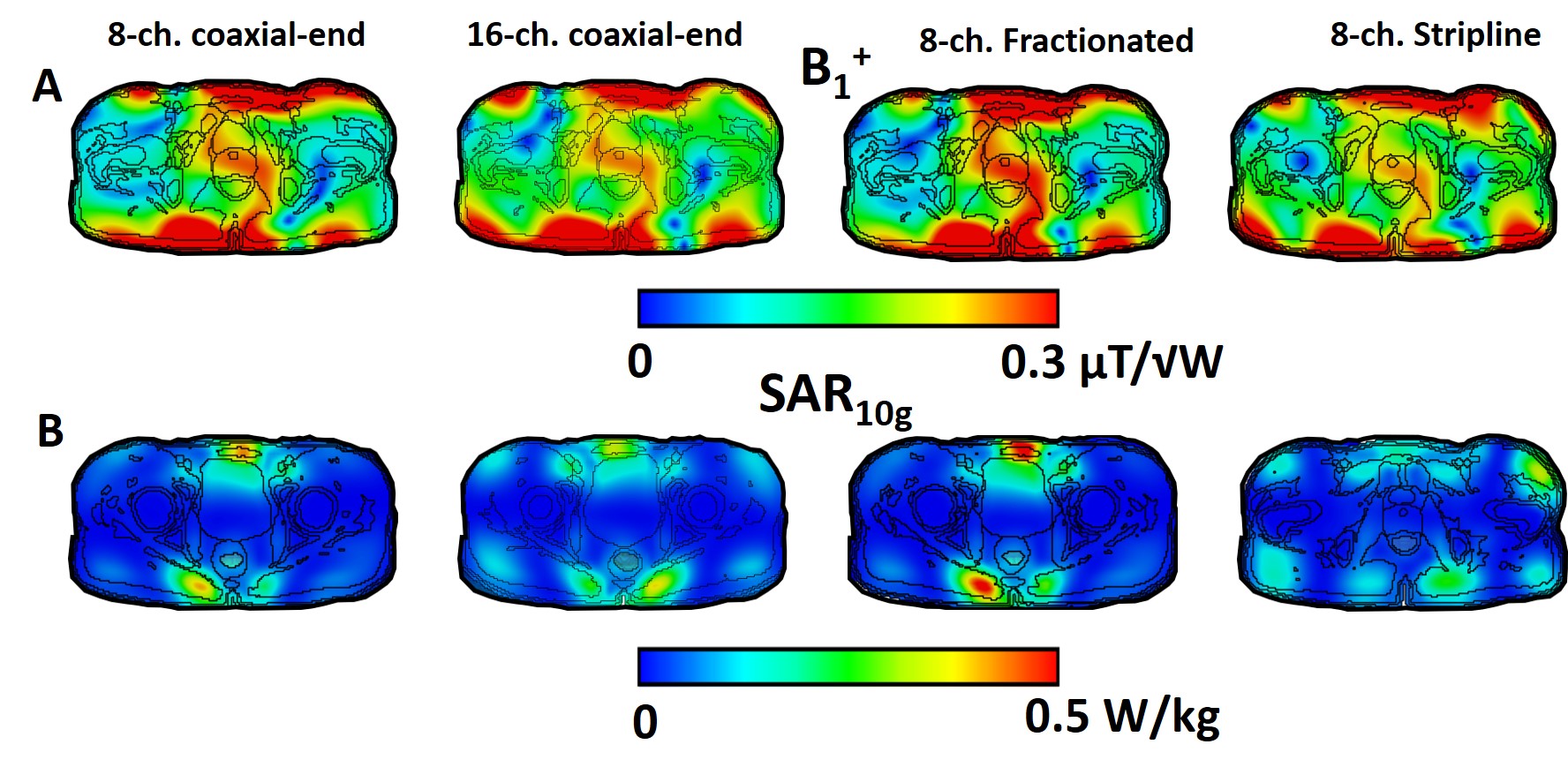
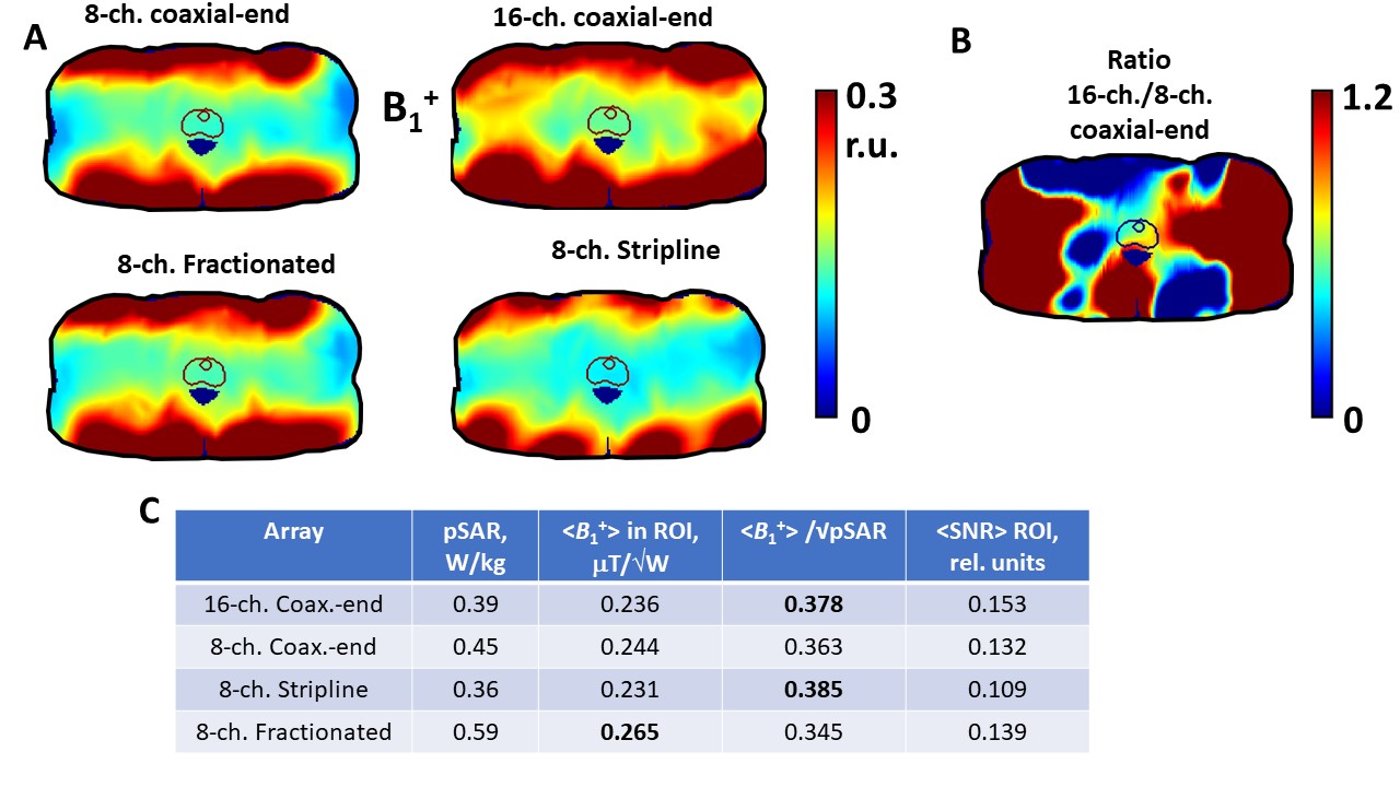
Figs. 1 and 3A show the SAR10g distribution at the top of the homogenous phantom and SAR-efficiency for the different single array elements, respectively. SAR efficiency at 7-cm depth inside the phantom along the z-axis is presented in Fig. 3B. As seen in Fig. 3B, SAR-efficiencies of the coaxial, coaxial-end, and stripline elements are very similar. The SAR-efficiency of the fractionated dipole was approximately 20% lower. Fig. 4 shows transversal B1+ maps cut through the prostate and axial SAR10g maps cut through pSAR for all four arrays. Figs. 5A and 5B present transversal SNR maps of all arrays as well as the SNR ratio of 16-channel and 8-channel coaxial-end dipole arrays. Fig. 5B summarizes all data including <B1+> in prostate, pSAR10g, SAR-efficiency and <SNR>. As seen in the table, using the 16-channel coaxial-end dipole array allows decreasing the pSAR value by 13% and 44% comparing to the 8-channel coaxial-end dipole and fractionated dipole arrays, respectively. Also, the SAR-efficiency of the 16-channel coaxial-end dipole array was 4% and 9% higher than that of the 8-channel coaxial-end dipole and fractionated dipole arrays, respectively. SAR-efficiency and SAR of both coaxial-end dipole arrays were slightly worse than that of the stripline array. Finally, SNR of the 16-channel coaxial-end dipole array was 13%, 10%, and 28% better than that of the 8-channel coaxial-end dipole, fractionated, and stripline arrays, respectively.Conclusion
We developed and numerically evaluated the performance of the novel densely populated 8Tx/16Rx coaxial-end dipole human body array. The array provided lower pSAR value and higher SNR in comparison to several different 8TxRx dipole arrays.Acknowledgements
This work was supported by the Max-Planck-Society and European Union (ERC Advanced Grant SpreadMRI, Number: 834940).References
1. Raaijmakers, AJE., et al. "Design of a radiative surface coil array element at 7 T: the single‐side adapted dipole antenna." Magnetic resonance in medicine 66.5 (2011): 1488-1497.
2. Raaijmakers, AJE, et al. "The fractionated dipole antenna: A new antenna for body imaging at 7 Tesla." Magnetic resonance in medicine 75.3 (2016): 1366-1374.
3. Kraff, O. and Quick HH. "Radiofrequency coils for 7 Tesla MRI." Topics in Magnetic Resonance Imaging 28.3 (2019): 145-158.
4. Arcan EM, et al. "A 16‐channel combined loop‐dipole transceiver array for 7 Tesla body MRI." Magnetic resonance in medicine 77.2 (2017): 884-894.
5. Lattanzi R, et al. "Approaching ultimate intrinsic signal‐to‐noise ratio with loop and dipole antennas." Magnetic resonance in medicine 79.3 (2018): 1789-1803.
6. van Leeuwen CC, et al. "The Coax Dipole: A fully flexible coaxial cable dipole antenna with flattened current distribution for body imaging at 7 Tesla." Magnetic Resonance in Medicine 87.1 (2022): 528-540. 7. Solomakha G, et al. "Evaluation of Coaxial Dipole Antennas as Transceiver Elements of Human Head Array for Ultra-High Field MRI at 9.4T. " In 2023 ISMRM & ISMRT Annual Meeting & Exhibition (ISMRM 2023).
8. Solomakha G, et al. "The dual‐mode dipole: A new array element for 7T body imaging with reduced SAR." Magnetic resonance in medicine 81.2 (2019): 1459-1469.
9. Rietsch SHG, et al. "Parallel transmit capability of various RF transmit elements and arrays at 7T MRI." Magnetic Resonance in Medicine 79.2 (2018): 1116-1126. 10. Roemer PB., et al. "The NMR phased array." Magnetic resonance in medicine 16.2 (1990): 192-225.
Figures