0518
B1+ Inhomogeneity Correction with Dielectric Padding for Improved MR T2 and T1ρ Quantification in Knee Cartilage and Meniscus at 7 Tesla1Program of Advanced Musculoskeletal Imaging (PAMI), Cleveland Clinic, Cleveland, OH, United States, 2Department of Biomedical Engineering, Lerner Research Institute, Cleveland Clinic, Cleveland, OH, United States, 3Department of Diagnostic Radiology, Imaging Institute, Cleveland Clinic, Cleveland, OH, United States, 4Imaging Institute, Cleveland Clinic, Cleveland, OH, United States
Synopsis
Keywords: Cartilage, Cartilage, Osteoarthritis, High-Field MRI, Quantitative Imaging, Relaxometry
Motivation: MR T2 and T1ρ mapping showed great promise for reliable detection and follow-up of knee osteoarthritis, however, B1+ inhomogeneities in knee 7T MRI often lead to signal loss and biased quantification.
Goal(s): Present study therefore evaluates the effect of high-permittivity dielectric padding on the B1+ field distribution and the reproducibility of T2 and T1ρ quantification in knee cartilage and meniscus.
Approach: Twelve subjects received scan-rescan at 7T to quantify B0, B1+, T2, T1ρ, and reproducibility changes associated with dielectric padding.
Results: Dielectric pads positioned over tibia showed improved B1+ homogeneity and reproducibility of T2 and T1ρ quantification in cartilage and meniscus at 7T.
Impact: Improved reproducibility of T2 and T1ρ MRI in cartilage and meniscus with dielectric padding at 7T could facilitate its clinical translation at ultra-high field and improve patient’s follow-up for the noninvasive evaluation of new prevention and treatment strategies for osteoarthritis.
Introduction
Cartilage and meniscus degeneration are hallmarks of knee osteoarthritis (OA). MR T2 and T1ρ mapping of cartilage showed its value for the early detection and prediction of future progression of OA [1-3]. Moreover, T2 mapping of meniscus was correlated with the severity of OA degeneration [4]. Although 7T MRI offers higher signal-to-noise ratio and acceleration factors compared to 3T, shorter RF wavelengths often lead to substantial B1+ inhomogeneities resulting in signal loss and biased quantification at 7T [5,6]. Parallel transmit, post-processing algorithms, and high-permittivity dielectric padding [7,8] were proposed to correct for B1+ inhomogeneities. Dielectric pads placed near the region with low B1+ can increase the local B1+ magnitude at the cost of global B1+ [9,10].The aim of this 7T study is to: i) evaluate the effect of dielectric pads on the B1+ field distribution and the quantification of T2 and T1ρ in knee cartilage and meniscus; and ii) calculate the scan-rescan T2 and T1ρ repeatability with and without dielectric padding.
Methods
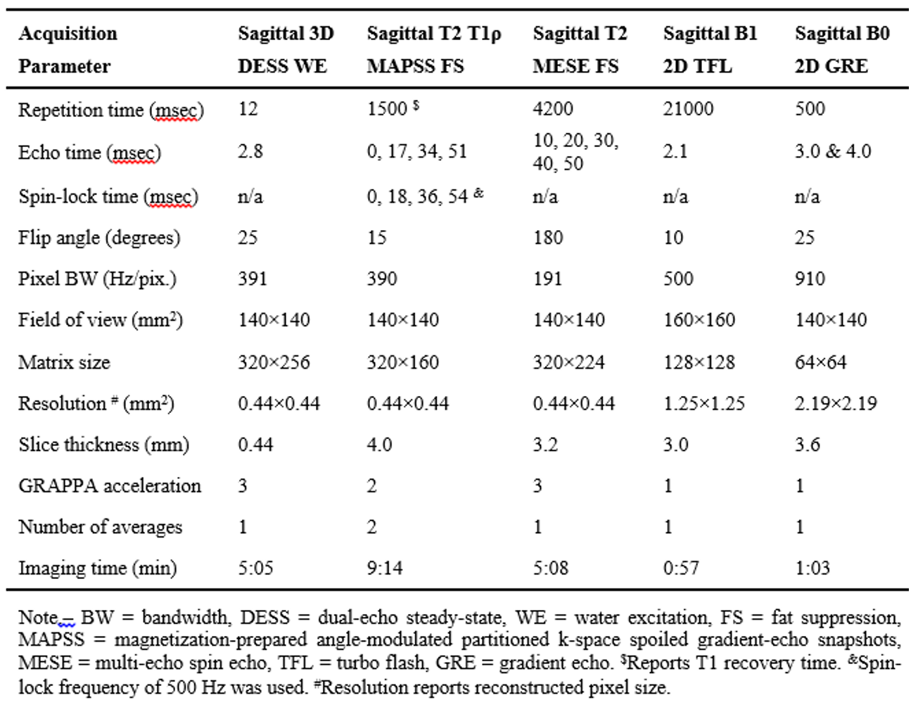
Twelve subjects were (age range, 18-59 years; BMI range, 21.6-34.5 kg/m2; 5 women; 5 right knees) were enrolled in this ongoing IRB-approved study. All subjects were imaged at 7T MRI (Magnetom Terra, Siemens) using a 1-Tx/28-Rx knee coil (Quality Electrodynamics). Morphological turbo-spin echo (TSE), DESS, B0, B1+ [11], MAPSS T2 and T1ρ [12], and multi-echo spin echo (MESE) T2 mapping sequences were acquired (Figure 1). Each subject left the MRI room after first scan and was repositioned for the second scan of the same knee. Repeated scans were measured: i) without dielectric pads (N=4); ii) with dielectric pads (N=3); or iii) one scan with and other scan without dielectric padding (N=5).Two in-house made dielectric pads (155×160 mm2, CaTiO3 suspended in D2O [9]) were placed anteriorly over tibia and over either side of the knee. DESS images from both scans were non-linearly registered in ANTs [13]. Nine cartilage and meniscus compartments were automatically segmented on DESS images using an in-house deep learning algorithm [14], followed by manual corrections in ITK-SNAP as needed. In order to evaluate the same knee location with respect to its position in the magnet, medial compartments in right knees were assigned as left, and medial compartments in left knees were assigned as right (i.e., medial meniscus=right meniscus). B1+ maps were calculated as the ratio of measured and prescribed flip angles. All T2 and T1ρ maps were calculated voxel-wise by fitting a mono-exponential decay to the data using a two-parameter least-square fitting routine. Mean B0, B1+, T2 and T1ρ values were calculated in each compartment. Coefficients of variation (CVs) and Bland-Altman plots were used to evaluate scan-rescan repeatability.
Results
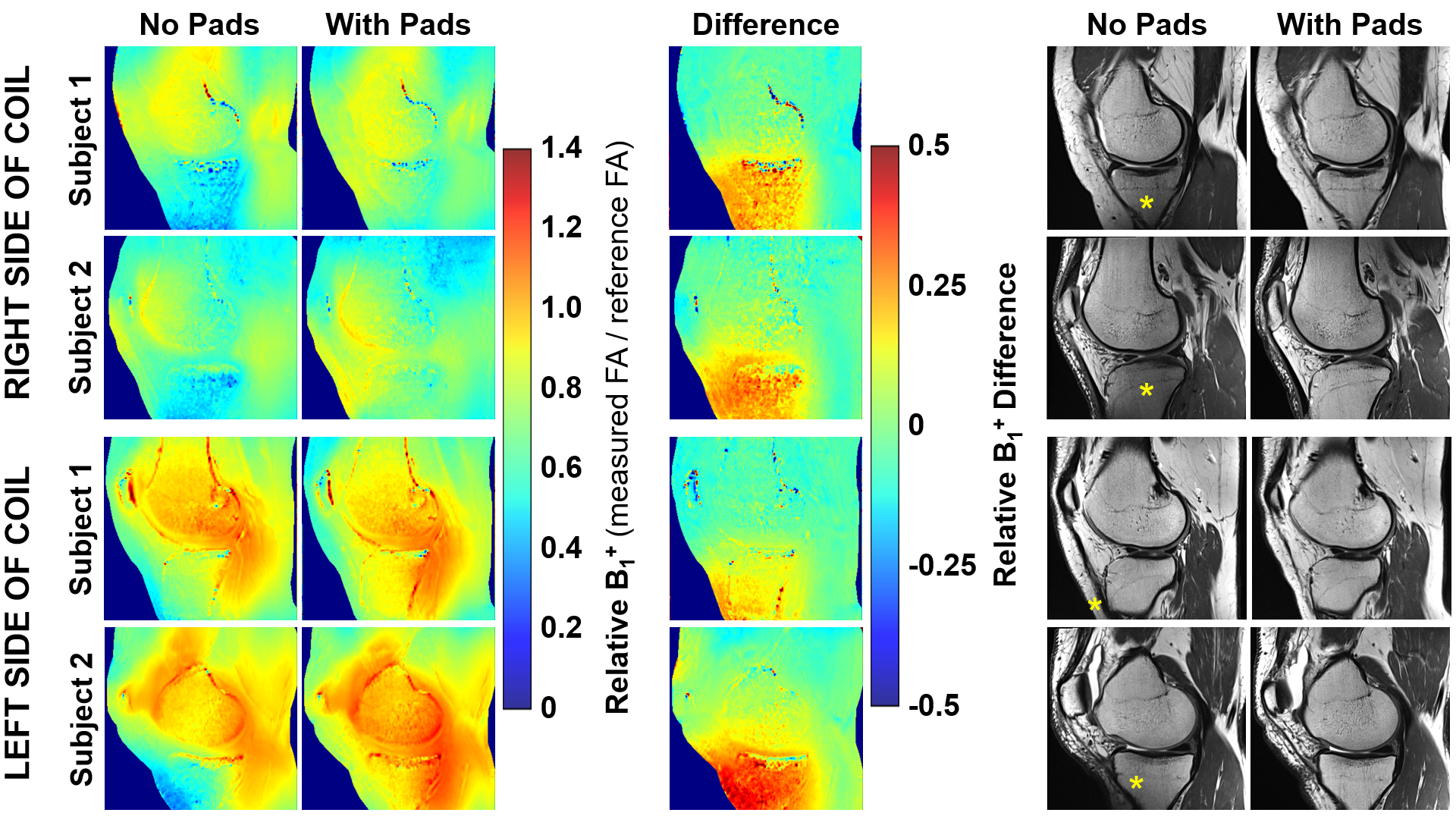
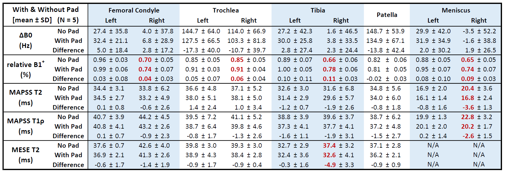
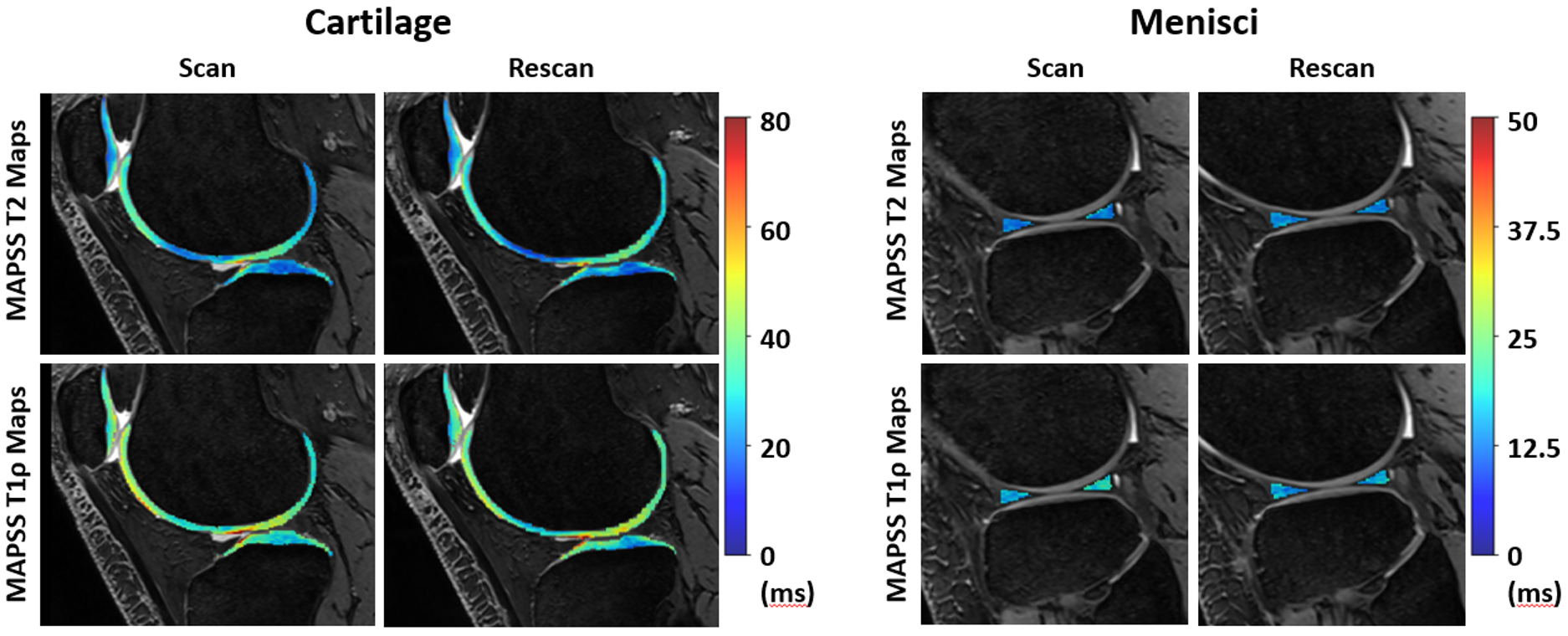
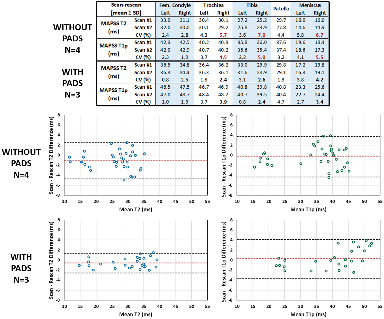
The effect of dielectric pads on morphological TSE images and relative B1+ maps is presented in Figure 2. Application of dielectric pads increased relative B1+ in most regions with statistically significant increase in tibia, meniscus, trochlea and femoral condyle on the right side of the knee coil (Figure 3). Dielectric padding resulted in overall decrease of relaxation times (means: MESE-T2, -1.41 ms; MAPSS-T2, -1.14 ms; MAPSS-T1ρ, -0.71 ms), and in statistically significant decrease in right meniscus (MAPSS) and right tibia (MESE-T2). B0 fields were not significantly affected by padding.Scan-rescan T2 and T1ρ maps acquired without dielectric pads are shown in Figure 4. Scan-rescan CVs for MAPSS T2 and T1ρ were higher in measurements without (range, 1.9-7.0%) than with (range, 0.8-4.7%) dielectric pads (Figure 5). Bland-Altman plots showed more narrow limits of agreement, and thus improved repeatability, with dielectric pads (MAPSS-T2, 3.96 ms; MAPSS-T1ρ, 7.73 ms) than without dielectric pads (MAPSS-T2, 7.23 ms; MAPSS-T1ρ, 8.06 ms) (Figure 5).
Discussion
Dielectric pads positioned over tibia inside the RF knee coil showed improved B1+ homogeneity. Our results agree with the findings of Fagan and colleagues [8]. Insufficient B1+ can lead to imprecise RF pulses and low signal-to-noise ratio resulting in biased T2 and T1ρ quantification. Tibia, meniscus and trochlea compartments on the right side of knee RF coil showed poor scan-rescan T2 and T1ρ repeatability without dielectric padding. Dielectric padding seems to improve the reproducibility of T2 and T1ρ quantification in cartilage and meniscus at 7T, resulting in CVs similar to the previously reported 1.4-4.1% for MAPSS-T2 and 1.6-3.9% for MAPSS-T1ρ in healthy cartilage at 3T [15].Conclusion
Application of dielectric pads reduced B1+ inhomogeneities and improved the quality of morphological images and the reproducibility of T2 and T1ρ quantification in cartilage and meniscus at 7T. Increased reproducibility of quantitative 7T MRI with dielectric padding could help improve patient’s MRI follow-up and the evaluation of new prevention and treatment strategies for OA.Acknowledgements
This study was partially supported by Siemens Healthineers.References
[1] MacKay JW, et al. Systematic review and meta-analysis of the reliability and discriminative validity of cartilage compositional MRI in knee osteoarthritis. Osteoarthritis Cartilage. 2018; 26: 1140-52.[2] Prasad AP, et al. T1ρ and T2 relaxation times predict progression of knee osteoarthritis. Osteoarthritis Cartilage. 2013; 21: 69-76.
[3] Atkinson HF, et al. MRI T2 and T1ρ relaxation in patients at risk for knee osteoarthritis: a systematic review and metaanalysis. BMC Musculoskelet Disord. 2019; 20(1): 182.
[4] Eijgenraam SM, et al. T2 mapping ofthe meniscus is a biomarker for early osteoarthritis. Eur Radiol. 2019; 29(10): 5664-72.
[5] Ladd ME, et al. Pros and cons of ultra-high-field MRI/MRS for human application. Prog Nuc Magn Reson Spec. 2018; 109: 1-50.
[6] Guerin B, et al. The ultimate signal-to-noise ratio in realistic body models. Magn Reson Med. 2017; 78: 1969-80.
[7] Koolstra K, et al. Improved image quality and reduced power deposition in the spine at 3 T using extremely high permittivity materials. Magn Reson Med. 2018; 79: 1192-99.
[8] Fagan AJ, et al. Image artifact management for clinical magnetic resonance imaging on a 7 T scanner
using single-channel radiofrequency transmit mode. Invest Radiol. 2019; 54: 781-91.
[9] Teeuwisse WM, et al. Quantitative assessment of the effects of high-permittivity pads in 7 Tesla MRI of the brain. Magn Reson Med. 2012; 67: 1285-93.
[10] Brink WM, et al. High permittivity dielectric pads improve high spatial resolution magnetic resonance imaging of the inner ear at 7 T. InvestRadiol. 2014; 49: 271-77.
[11] Chung S, et al. Rapid B1+ mapping using a preconditioning RF pulse with TurboFLASH readout. Magn Reson Med. 2010; 64: 439-46.
[12] Li X, et al. Simultaneous Acquisition of T1ρ and T2 Quantification in Knee Cartilage: Repeatability and Diurnal Variation. J Magn Reason Ima. 2014; 39: 1287-93.
[13] Avants BB, et al. Advanced Normalization Tools (ANTS). OR Insight. 2008: 1-35
[14] Gaj S, et al. Automated cartilage and meniscus segmentation of knee MRI with conditional generative adversarial networks. Magn Reson Med. 2019; 84(1): 437-49.
[15] Kim J, et al. Multi-vendor multi-site T1ρ and T2 quantification of knee cartilage. Osteoarthritis Cartilage. 2020; 28: 1539-50.
Figures