0511
TEXTURE ANALYSIS OF CARTILAGE REPAIR TISSUE MATURATION - COMPARISON OF TWO CARTILAGE REPAIR METHODS AND CORRELATION WITH MOCART 2.01High Field MR Centre, Department of Biomedical Imaging and Image-guided Therapy, Medical University of Vienna, Vienna, Austria, 2CD Laboratory for MR Imaging Biomarkers (BIOMAK), Vienna, Austria, 3Institute of Measurement Science, Slovak Academy of Sciences, Bratislava, Slovakia, 4TETEC Tissue Engineering Technologies AG, Reutlingen, Germany, 5Austrian Cluster for Tissue Regeneration, Ludwig Boltzmann Institute for Experimental and Clinical Traumatology, Vienna, Austria, 6Institute for Clinical Molecular MRI in the Musculoskeletal System, Karl Landsteiner Society, Vienna, Austria
Synopsis
Keywords: Cartilage, Cartilage, Repair, Tissue, Maturarion
Motivation: To monitor patients after cartilage repair surgery, non-invasive imaging techniques are needed.
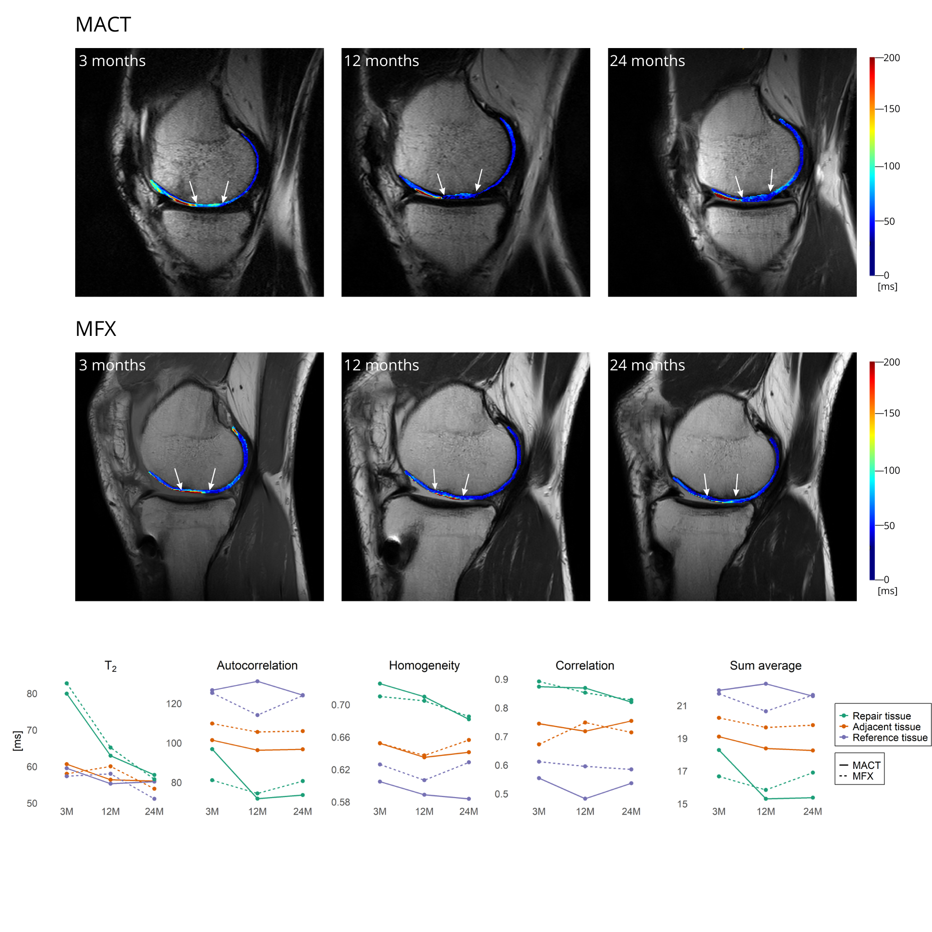
Goal(s): Study aimed to monitor cartilage repair tissue for up to two years post-surgery using GLCM analysis of T2 maps to assess tissue structure and correlate it with the morphological MOCART 2.0 score.
Approach: 37 patients underwent either matrix-associated autologous chondrocyte transplantation or microfracturing. ROI’s were drawn onto T2 maps at three time-points. GLCM features, mean T2 and MOCART 2.0 scores were calculated and analysed.
Results: We found correlations between GLCM features and the MOCART 2.0 score, with significant changes in texture of the repair tissue over time for transplant patients.
Impact: This study reveals a correlation between GLCM and MOCART 2.0 in autologous chondrocyte transplantation and microfracturing. Only chondrocyte transplants showed significant tissue development over time, indicating complex maturation. These findings will impact patient monitoring in clinical trials after cartilage repair.
Introduction
To avoid invasive procedures during monitoring of patients after cartilage repair surgery, gray-level co-occurrence matrix (GLCM) of collagen specific T2 maps is currently being explored as a tool for assessing cartilage repair tissue maturation. Matrix-associated autologous chondrocyte transplantation (MACT) results in repair tissue with more hyaline-like structure in comparison to bone marrow stimulating techniques (MFX)1, which are considered to be the standard repair technique for smaller cartilage lesions. Objective of this study was to assess maturation of MACT grafts up to two years following surgery using GLCM texture analysis of quantitative T2 maps, compare results to the MFX control group and relate these results to the morphological MOCART 2.0 score.Methods
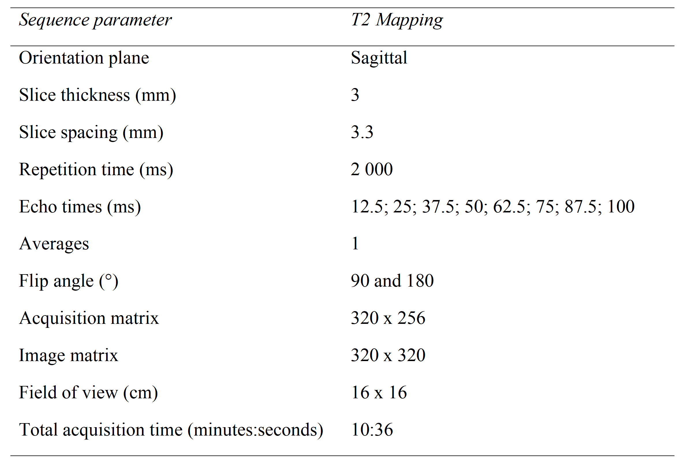
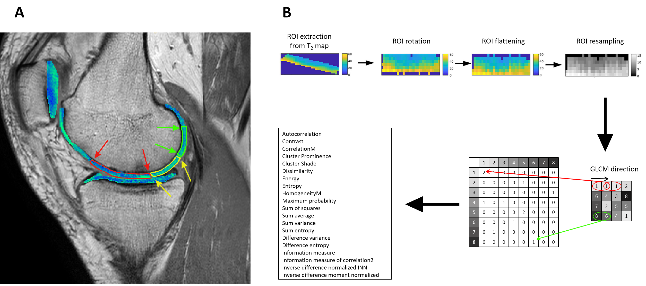
A total of 37 patients were derived from the MRI sub-study of a prospective, multi-center, randomized, controlled, open-label (blinded MRI reading), phase III study comparing the efficacy and safety of MACT using NOVOCART 3D plus (TETEC – Tissue Engineering Technologies AG, Reutlingen, Germany) versus MFX in patients with focal cartilage defects of the knee. For this GLCM analysis, 27 MACT and 10 MFX patients had T2 map available for all three time-points (3, 12 and 24 months). At each time-point, MOCART 2.0 score2 was assessed by an expert reader. T2 mapping sequence parameters at 3T are listed in Table 1. T2 mapping was performed using mono-exponential decay fitting with 2-parameters (M0: ‚zero magnetization‘ and T2: ‚transversal relaxation constant‘). Regions-of-interest (ROI) were defined on two or three consecutive slices on T2 mapping sequence and transferred onto T2 maps using a script written in MATLAB R2019.a (Mathworks, Natick, MA, USA). For each patient, three locations at each time-point were selected: 1) repair tissue; 2) cartilage adjacent to the repair tissue; 3) healthy reference. ROIs were rotated and flattened, quantized into 16 grey levels and GLCM analysis was computed with offset: 0° angle (parallel to cartilage surface) and step of length 1 (considering pixel and its immediate neighbour). Mean T2 value and twenty quantitative features (in Figure 1)3, 4 were extracted averaged through the slices ROI-wise. Spearman coefficient was used to examine the relationship between GLCM features and MOCART 2.0 score. Based on simple comparisons, mean T2, autocorrelation, homogeneity, correlation and sum average were analysed using linear mixed-effects models with random slope and intercept with Kenward-Roger degrees of freedom correction.Results
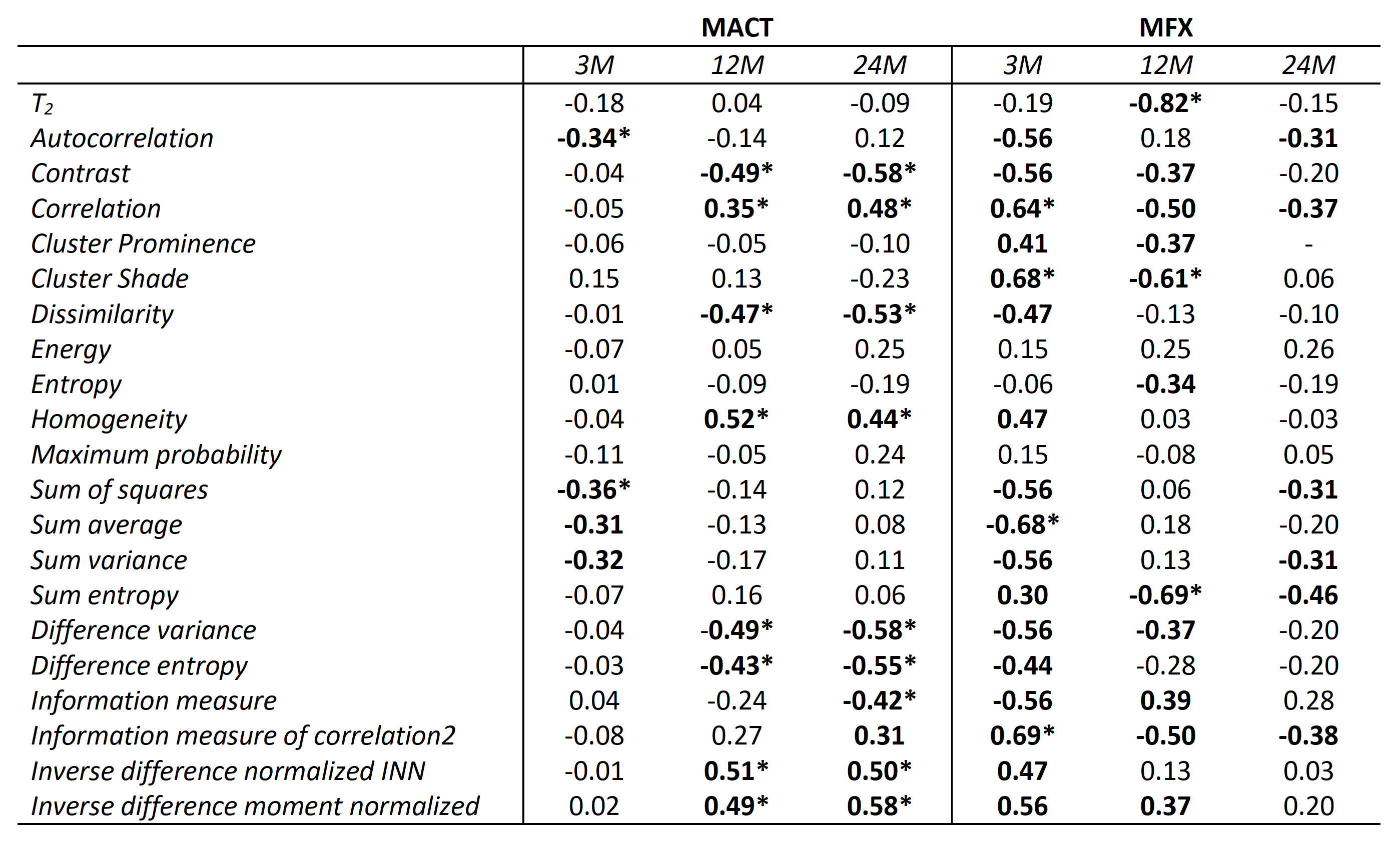
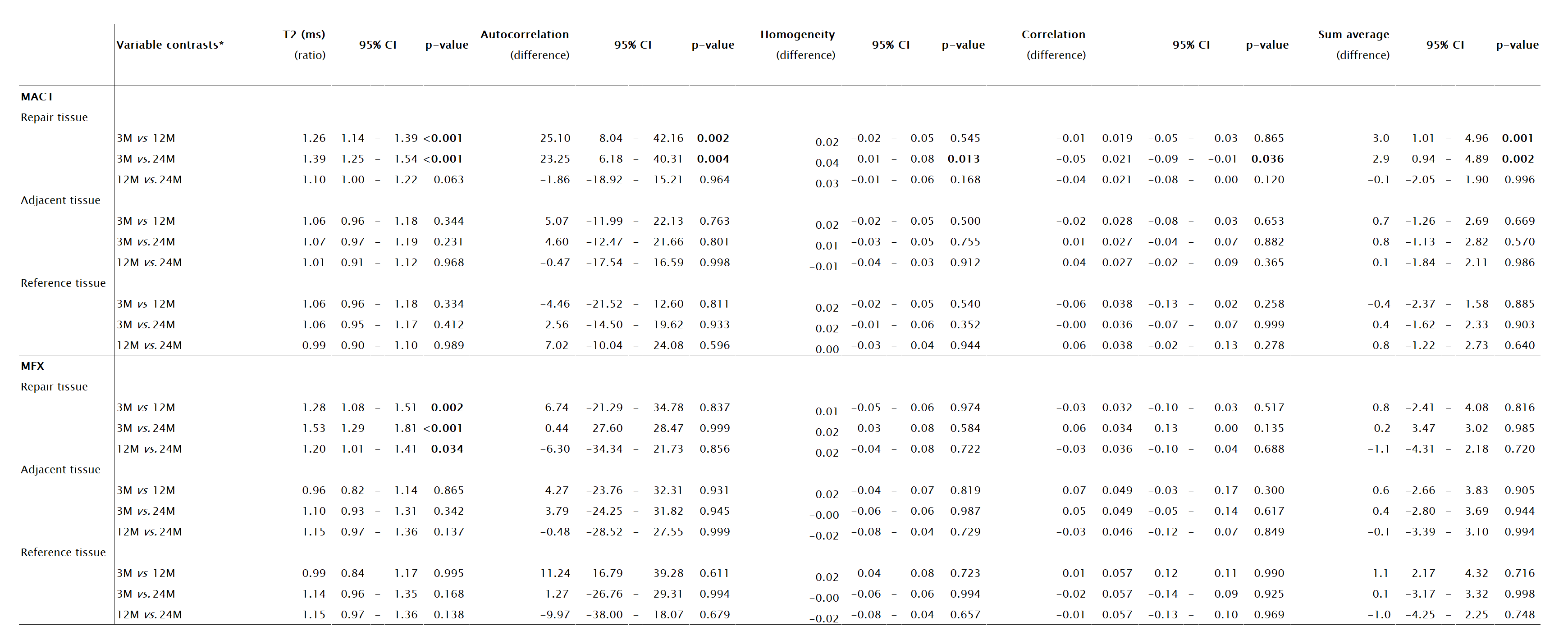
Correlation analysis showed weak to strong correlation between GLCM features and MOCART 2.0 score at different time-points. Models did not show significant differences between the procedures (p > 0.05 in all models). However, when comparing MACT and MFX separately, we found significant decrease in T2 of repair tissue in the MACT group (3Mvs12M: p<0.001; 3Mvs24M: p<0.001) and MFX group (3Mvs12M: p=0.001; 3Mvs24M: p<0.001; 12Mvs24M: p=0.034). Moreover significant change of autocorrelation (3Mvs12M: p=0.002; 3Mvs24M: p=0.004), homogeneity (3Mvs24M: p=0.013), correlation (3Mvs24M: p=0.036) and sum average (3Mvs12M: p=0.001; 3Mvs24M: p=0.002) in the MACT repair tissue was found.Discussion
This is the first study to analyse the correlation of textural features with the MOCART 2.0 score. Both surgical procedures show significant correlations with the MOCART score at 3M and 12M. However, only textural features of MACT showed significant correlations with the MOCART score at 24M. This suggests, that texture of the repair tissue is tied to the morphological outcome and this relationship is more prominent in the MACT group mainly at the later follow-ups. Repair tissue in both procedures is highly hydrated after surgery and the swelling goes down during maturation. This is reflected by the T2 values, as they are significantly reduced between 3M and 12M follow-up. We haven’t found significant differences in T2 and GLCM features between the two surgical procedures in this sub-cohort. However, when analysing the two groups separately, repair tissue texture develops significantly over time only in the MACT group, MFX repair tissue texture stays the same during the 24 months period. This suggest not only reduction of increased water content and swelling, but also development of the tissue structure of the MACT, in particular the collagen fiber network. Based on our previous work5, we incorporated the adjacent and reference tissue into analysis. Although these tissues did not change significantly, they assured better model fits and more complex understanding of the maturation process.Conclusion
GLCM features are correlated to morphological MOCART 2.0 score in both types of investigated cartilage repair procedures. Even though there was not significant difference in parameters between the procedures, significant tissue development in time was uncovered by the GLCM analysis in the case of MACT. This is potentially clinically useful information for normal maturation in the MACT repair technique.Acknowledgements
No acknowledgement found.References
1. Seo, S.-S., C.-W. Kim, and D.-W. Jung, Management of Focal Chondral Lesion in the Knee Joint. Knee Surgery & Related Research. Vol. 23. 2011: Korean Knee Society. 185.
2. Schreiner, M.M., et al., The MOCART (Magnetic Resonance Observation of Cartilage Repair Tissue) 2.0 Knee Score and Atlas. Cartilage, 2021. 13(1_suppl): p. 571s-587s.
3. Haralick, R.M., K. Shanmugam, and I. Dinstein, Textural Features for Image Classification. IEEE Transactions on Systems, Man, and Cybernetics, 1973. SMC-3(6): p. 610-621.
4. Uppuluri, A. GLCM_Features4.m: Vectorized version of GLCM_Features1.m [With code changes]. 2023 October 24, 2023.]; Available from: https://www.mathworks.com/matlabcentral/fileexchange/22354-glcm_features4-m-vectorized-version-of-glcm_features1-m-with-code-changes.
5. Janacova, V., et al., Adjacent cartilage tissue structure after successful transplantation: a quantitative MRI study using T2 mapping and texture analysis. European Radiology, 2022. 32(12): p. 8364-8375.
Figures