0509
MRI-Based Machine Learning Fusion Models to Distinguish Encephalitis and Gliomas1Peking University people' hospital, Beijing, China, 2Beijing Tiantan Hospital, Beijing, China
Synopsis
Keywords: Neuroinflammation, Brain, Encephalitis · Gliomas · Magnetic resonance imaging
Motivation: Encephalitis and glioma can appear very similar in atypical cases. However, their treatment protocols differ significantly. As such, distinguishing between these two diseases is crucial.
Goal(s): Our objective is to assess and compare the performance of various machine learning (ML) techniques in discriminating between encephalitis and glioma in atypical cases.
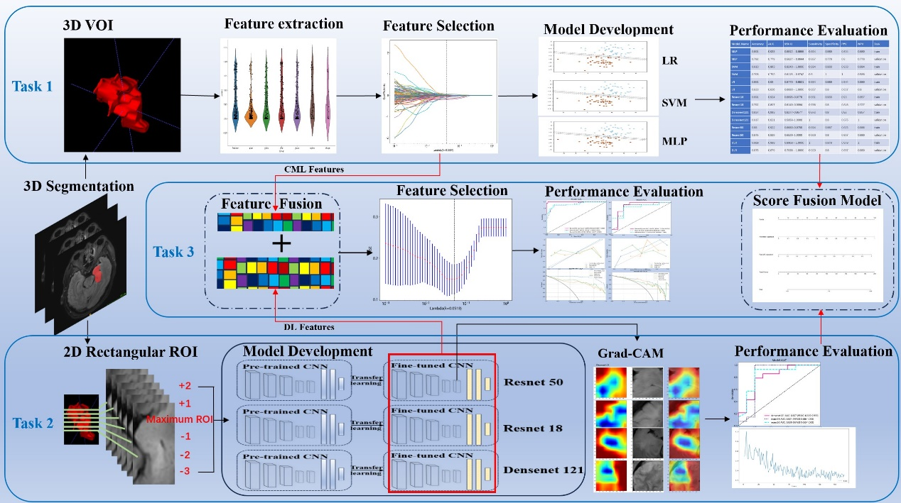
Approach: We compare the performance of the classical machine learning (CML) model and the deep learning (DL) model, and assess the effectiveness of utilizing radiomics features extracted from both CML and DL in distinguishing encephalitis from glioma in atypical cases.
Results: ML models can distinguish between encephalitis and glioma in atypical cases.
Impact: Surgery is commonly considered as the initial treatment for glioma, while non-operative therapy is the primary approach for managing encephalitis. Precise identification of glioma and encephalitis facilitates physicians in avoiding misdiagnosis and delays in treatment.
Introduction
In atypical cases where encephalitis and glioma exhibit very similar manifestations, the laboratory tests are atypical, and the clinical symptoms and signs of these conditions often coincide [1-6]. This diagnostic dilemma can result in unintentional surgery or delayed treatment [7]. Therefore, it is paramount to explore alternative noninvasive diagnostic tools to guide appropriate treatment.Presently, machine learning (ML) is extensively employed in the field of neurological diseases to enhance clinical decision-making. Several studies have demonstrated that ML can distinguish the various pathological subtypes of gliomas [8] and assess the status of molecular and genetic markers associated with the brain tumor [9]. It has been employed to distinguish between glioblastoma and tumefactive demyelinating lesions [10]. These studies suggest that ML proves to be a potent analytical tool in evaluating radiological data related to glioma and encephalitis.
The objective of this study was to compare the performance of the classical machine learning (CML) model and the deep learning (DL) model, and to assess the effectiveness of utilizing radiomics features extracted from both CML and DL in distinguishing encephalitis from glioma in atypical cases.
Methods
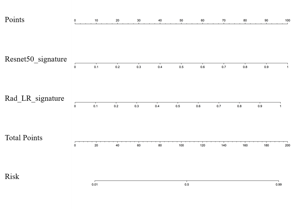
In this study, 116 patients (mean age ± standard deviation, 42.3 ± 17.2 years old; 63 men and 53 women) pathologically confirmed as gliomas and clinically diagnosed with encephalitis in our medical institute between January 1, 2019 and March 31, 2023 were recruited.In the current study, we aimed to establish 3 ML models: (1) Task 1 consisted of establishing 3 CML models (Logistic Regression, LR; Support Vector Machine, SVM; and Multi-Layer Perceptron, MLP) using the FLAIR sequence; (2) Task 2 involved constructing 3 DL models (DenseNet 121, ResNet 50, and ResNet 18) based on FLAIR sequence; and (3) Task 3 focused on building 2 fusion models, which are feature fusion model and predictive score fusion model. The feature fusion model was based on selecting FLAIR-based CML features and DL features. The features were then combined to create a deep learning radiomics (DLR) model. The predictive score fusion model, a deep learning radiomics nomogram (DLRN), was constructed by combining CML and DL scores using multivariate LR. An online web calculator embedding a dynamic nomogram with binary logistic regression model was also developed.
Results
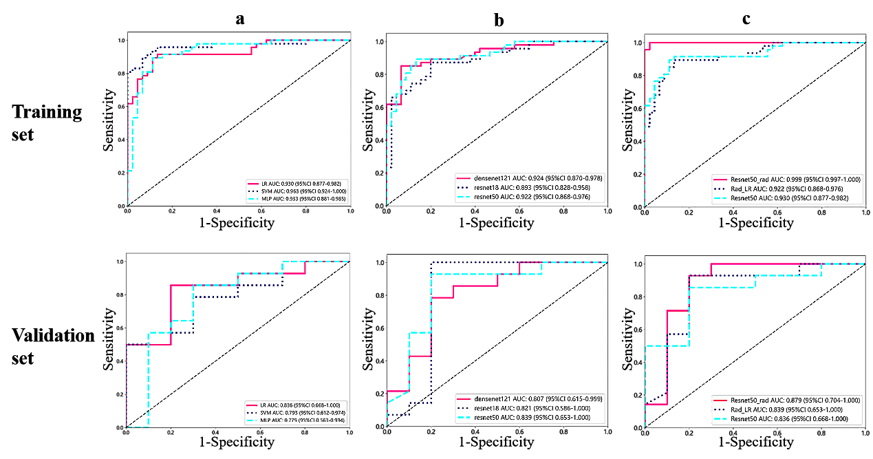
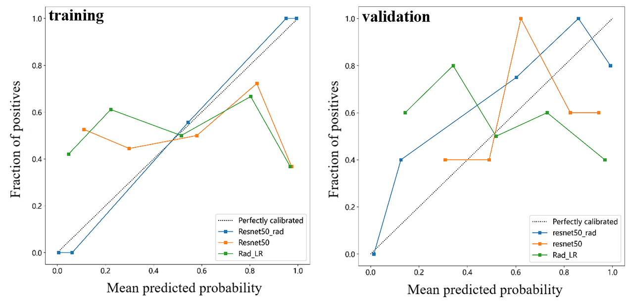
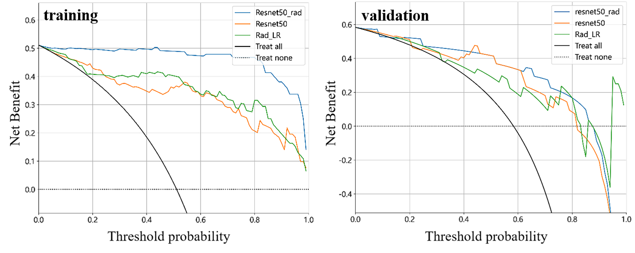
In the validation set, the best DL model (ResNet50) consistently outperformed the best CML model(LR), achieving an AUC of 0.839, accuracy of 0.875, sensitivity of 0.929, specificity of 0.800, PPV of 0.867, and NPV of 0.889. The DLR model exhibited the highest performance. The DLR model, which is considered the optimal model, demonstrated the highest AUC values on both the training and validation cohorts, reaching 0.999 and 0.879 respectively. In addition, a deep learning radiomics nomogram (DLRN) and a web calculator were designed as a tool to aid clinical decision-making.Conclusion
In conclusion, our findings demonstrate the potential utility of ML based on FLAIR for distinguishing atypical cases of encephalitis and glioma which suggests its potential application in assisting clinical decision-making is noteworthy.Acknowledgements
NoneReferences
[1] Lu J, Zhang JH, Miao AL, et al. Brain astrocytoma misdiagnosed as anti-NMDAR encephalitis: a case report. BMC Neurol. 2019;19(1):210.
[2] Nagata R, Ikeda K, Nakamura Y, et al. A case of gliomatosis cerebri mimicking limbic encephalitis: malignant transformation to glioblastoma. Intern Med. 2010;49(13):1307-1310.
[3] Panagopoulos D, Themistocleous M, Apostolopoulou K, Sfakianos G. Herpes Simplex Encephalitis Initially Erroneously Diagnosed as Glioma of the Cerebellum: Case Report and Literature Review. World Neurosurg. 2019;129:421-427.
[4] Piper K, Foster H, Gabel B, Nabors B, Cobbs C. Glioblastoma Mimicking Viral Encephalitis Responds to Acyclovir: A Case Series and Literature Review. Front Oncol. 2019;9:8.
[5] Talathi S, Gupta N, Reddivalla N, Prokhorov S, Gold M. Anaplastic astrocytoma mimicking herpes simplex encephalitis in 13-year old girl. Eur J Paediatr Neurol. 2015;19(6):722-725.
[6] Vogrig A, Joubert B, Ducray F, et al. Glioblastoma as differential diagnosis of autoimmune encephalitis. J Neurol. 2018;265(3):669-677.
[7] Goodfellow JA, Mackay GA. Autoimmune encephalitis. J R Coll Physicians Edinb. 2019;49(4):287-294.
[8] Qian Z, Zhang L, Hu J, et al. Corrigendum: Machine Learning-Based Analysis of Magnetic Resonance Radiomics for the Classification of Gliosarcoma and Glioblastoma. Front Oncol. 2021;11:774369.
[9] Zheng F, Chen B, Zhang L, et al. Radiogenomic Analysis of Vascular Endothelial Growth Factor in Patients With Glioblastoma. J Comput Assist Tomogr. 2023.
[10] Zhang Y, Liang K, He J, et al. Deep Learning With Data Enhancement for the Differentiation of Solitary and Multiple Cerebral Glioblastoma, Lymphoma, and Tumefactive Demyelinating Lesion. Front Oncol. 2021;11:665891.
Figures