0504
High-Resolution Sodium MRI of Human Gliomas at 3T Using Physics-Based Generative AI1Radiological Sciences, UCLA Brain Tumor Imaging Laboratory, Los Angeles, CA, United States, 2Siemens Healthcare GmbH, Erlangen, Germany, 3Siemens Medical Solutions USA, Boston, MA, United States, 4Radiological Sciences, Magnetic Resonance Research Laboratories, David Geffen School of Medicine, University of California Los Angeles, Los Angeles, CA, United States, 5Radiological Sciences, David Geffen School of Medicine, University of California, Los Angeles, CA, United States, 6Neurosurgery, David Geffen School of Medicine, University of California, Los Angeles, CA, United States, 7Neurology, David Geffen School of Medicine, University of California, Los Angeles, CA, United States
Synopsis
Keywords: Tumors (Post-Treatment), Non-Proton, Sodium
Motivation: Sodium MRI is a promising technique for understanding the brain tumor microenvironment. However, sodium MRI at 3T suffers from extremely low SNR, resulting in compromised resolution and long acquisition times.
Goal(s): Our goal is to create a high-resolution sodium MRI at 3T using generative AI to improve biological characterization, treatment monitoring, and surgical planning for brain tumor patients.
Approach: We developed a physics-informed synthetic dataset to train an anatomically-constrained GAN for high-resolution neuroimaging of brain tumors.
Results: When applied to brain tumor patients' images, the synthetic-sodium MRI improved resolution, SNR, and correlated with expression of sodium-proton exchanger (NHE1) on image-guided biopsy.
Impact: High-resolution sodium neuroimaging at 3T using physics-informed anatomically-constrained GAN has the potential to make multinuclear MRI feasible in the clinical environment, leading to conceivable improvements in diagnosis, monitoring, treatment, and our understanding of the biology of brain tumors.
Introduction
Sodium neuroimaging is a promising technique for the diagnosis and monitoring brain tumors, as it can provide information about the brain microenvironment, cellular composition, and metabolic activity1-4. However, sodium neuroimaging at 3T is limited by low signal-to-noise ratio (SNR) and resolution. This results in long acquisition times and low-quality images that prevent the routine clinical use of sodium neuroimaging. To address these limitations, we developed and evaluated a physics-informed generative adversarial network (GAN) training approach for high-resolution sodium neuroimaging of brain tumors at 3T.Methods
5,078 artifact-free anatomical acquisitions from 1,330 suspected brain tumor patients undergoing routine proton MRI using the standard brain tumor protocol5 acquired at 3T were used to create a synthetic dataset with physics-based simulated artifacts. By including multiple proton MRI contrasts in our training dataset, we hypothesized the network would be able to learn to correct for artifacts while preserving the inherent information of all modalities, including sodium.Artifact dataset synthesis
Synthetic data was generated by simulating:
- B0 magnetic susceptibility artifacts (Δχ) manifested as signal drop-out/pile-up and geometric distortion. To accomplish this, a ΔB0 field map was created using a template CT image in MNI space to identify air, tissue, and bone using a Fourier-based calculation method proposed by Bouwman et al.6. Off-resonance artifact simulation was then performed using a simulated field map and the FORECAST algorithm7.
- Chemical shift artifact simulated as a fractional pixel shift using the fat-water frequency difference at 3T (430Hz) and the phase encode pixel bandwidth applied to skull bone marrow estimated using skull stripping algorithms8.
- Nyquist aliasing artifact added along the phase-encoding direction (ky) using an alternating linear phase ramp simulated using a constant term b and a first-order term a: ϕ(kx)=(akx+b)π . The orientation of frequency- and phase-encoding were randomized with 80% probability of A-P and 20% L-R phase encoding.
- Gibbs artifact and low-resolution images simulated by sampling the center of k-space using a rectangular window of random size between 60% and 30% of the original size.
- Rician noise was randomly added to the images during training to augment the dataset.
Model and Training
We used a transfer learning approach, in which we trained a series of models on increasingly more complex artifacts and used the weights of each model to initialize the next (Fig.1). The networks were built with a modified Pix2Pix9GAN architecture with an Attention10R2UNet11 generator. Fig.2 shows an example image with artifacts incrementally added to create the synthetic datasets. Additionally, to anatomically constrain the reconstruction, we generated an edge image used as the second input to the GAN to ensure realistic and accurate reconstruction.
In-Vivo Data
Twenty glioma patients classified based on the WHO classification12 consented to participate in this IRB-approved prospective study. The study population involved mostly previously-treated tumors (75%) IDH-wildtype glioblastoma (80%). Image-guided biopsies from 8 patients were available for sodium-proton exchanger (NHE1) expression evaluation on immunohistochemistry. Imaging was performed on a 3T Siemens scanner. Proton and sodium scans were conducted during the same session using a dual-tuned head coil (16-channel 1H/1-channel 23Na; RAPID MR International). Anatomical pre-/post-contrast high-resolution T1-weighted, T2-weighted, T2/FLAIR, and DWI images were obtained5. Sodium MRI was performed using a 3D spoiled gradient-echo sequence optimized for short TE with parameters: TE/TR=2.39/10.52ms, 5.5mm isotropic resolution, 264×264×264mm3 FOV, 39.8o flip-angle, 80 Hz/pixel bandwidth, 26 averages, and 10.5min scan time. Sodium images were normalized to the mean intensity of a VOI in the vitreous humor. High-resolution synthetic-sodium MR images were generated using the previously-trained GAN model on this cohort. Statistical analyses were performed using Pearson and Spearman’s correlation and t-test for group differences.
Results
The proposed method was able to reconstruct high-resolution synthetic-sodium MR images with appreciably improved SNR compared to traditional sodium images (Fig.3) for both high and lower-grade gliomas. The synthetic values were consistent with the native measurements (Fig.4A; P=0.9066) and there was a strong linear correlation between the two measurements (Fig.4B; R2=0.8539, P<0.0001). When comparing synthetic-sodium MR measurements to the relative NHE1 expression obtained from image-guided biopsies, there was a significant correlation (Fig.5A; ρ=0.5817, P=0.0036), with a notable difference in relative sodium MR signal within samples expressing little to no NHE1 compared with tumor regions with elevated expression (Fig.5B; P<0.0001). Together, these data suggest high-resolution synthetic-sodium MRI retains much of the inherent information from the native sodium MR while providing accurate biological representation of tumor tissue as verified by image-guided biopsies.Conclusion
High-resolution sodium neuroimaging at 3T using physics-informed anatomically-constrained GAN makes multinuclear MRI feasible in the clinical environment, leading to conceivable improvements in diagnosis, monitoring, treatment, and our understanding of the biology of brain tumors.Acknowledgements
This research was partially supported by grants from Siemens Healthcare and the Department of Defense (DOD) grant CA20029. We also express our sincere gratitude to the patients who participated in this study.References
1. Hagiwara, A. et al. Sodium MR Neuroimaging. AJNR Am J Neuroradiol 42, 1920-1926 (2021). https://doi.org:10.3174/ajnr.A7261
2. Thulborn, K. R., Lu, A., Atkinson, I. C., Damen, F. & Villano, J. L. Quantitative sodium MR imaging and sodium bioscales for the management of brain tumors. Neuroimaging Clin N Am 19, 615-624 (2009). https://doi.org:10.1016/j.nic.2009.09.001
3. Cho, N. S. et al. Multi-nuclear sodium, diffusion, and perfusion MRI in human gliomas. J Neurooncol 163, 417-427 (2023). https://doi.org:10.1007/s11060-023-04363-x4. Madelin, G. & Regatte, R. R. Biomedical applications of sodium MRI in vivo. J Magn Reson Imaging 38, 511-529 (2013). https://doi.org:10.1002/jmri.24168
5. Ellingson, B. M. et al. Consensus recommendations for a standardized Brain Tumor Imaging Protocol in clinical trials. Neuro Oncol 17, 1188-1198 (2015). https://doi.org:10.1093/neuonc/nov095
6. Bouwman, J. G. & Bakker, C. J. Alias subtraction more efficient than conventional zero-padding in the Fourier-based calculation of the susceptibility induced perturbation of the magnetic field in MR. Magn Reson Med 68, 621-630 (2012). https://doi.org:10.1002/mrm.24343
7. Zijlstra, F., Bouwman, J. G., Braškutė, I., Viergever, M. A. & Seevinck, P. R. Fast Fourier-based simulation of off-resonance artifacts in steady-state gradient echo MRI applied to metal object localization. Magn Reson Med 78, 2035-2041 (2017). https://doi.org:10.1002/mrm.26556
8. Smith, S. M. Fast robust automated brain extraction. Hum Brain Mapp 17, 143-155 (2002). https://doi.org:10.1002/hbm.10062
9. Isola, P., Zhu, J. Y., Zhou, T. & Efros, A. A. in 2017 IEEE Conference on Computer Vision and Pattern Recognition (CVPR). 5967-5976.
10. Oktay, O. et al. Attention u-net: Learning where to look for the pancreas. arXiv preprint arXiv:1804.03999 (2018).
11. Alom, M. Z., Hasan, M., Yakopcic, C., Taha, T. M. & Asari, V. K. Recurrent residual convolutional neural network based on u-net (r2u-net) for medical image segmentation. arXiv preprint arXiv:1802.06955 (2018).
12. Louis, D. N. et al. The 2021 WHO Classification of Tumors of the Central Nervous System: a summary. Neuro Oncol 23, 1231-1251 (2021). https://doi.org:10.1093/neuonc/noab106
Figures
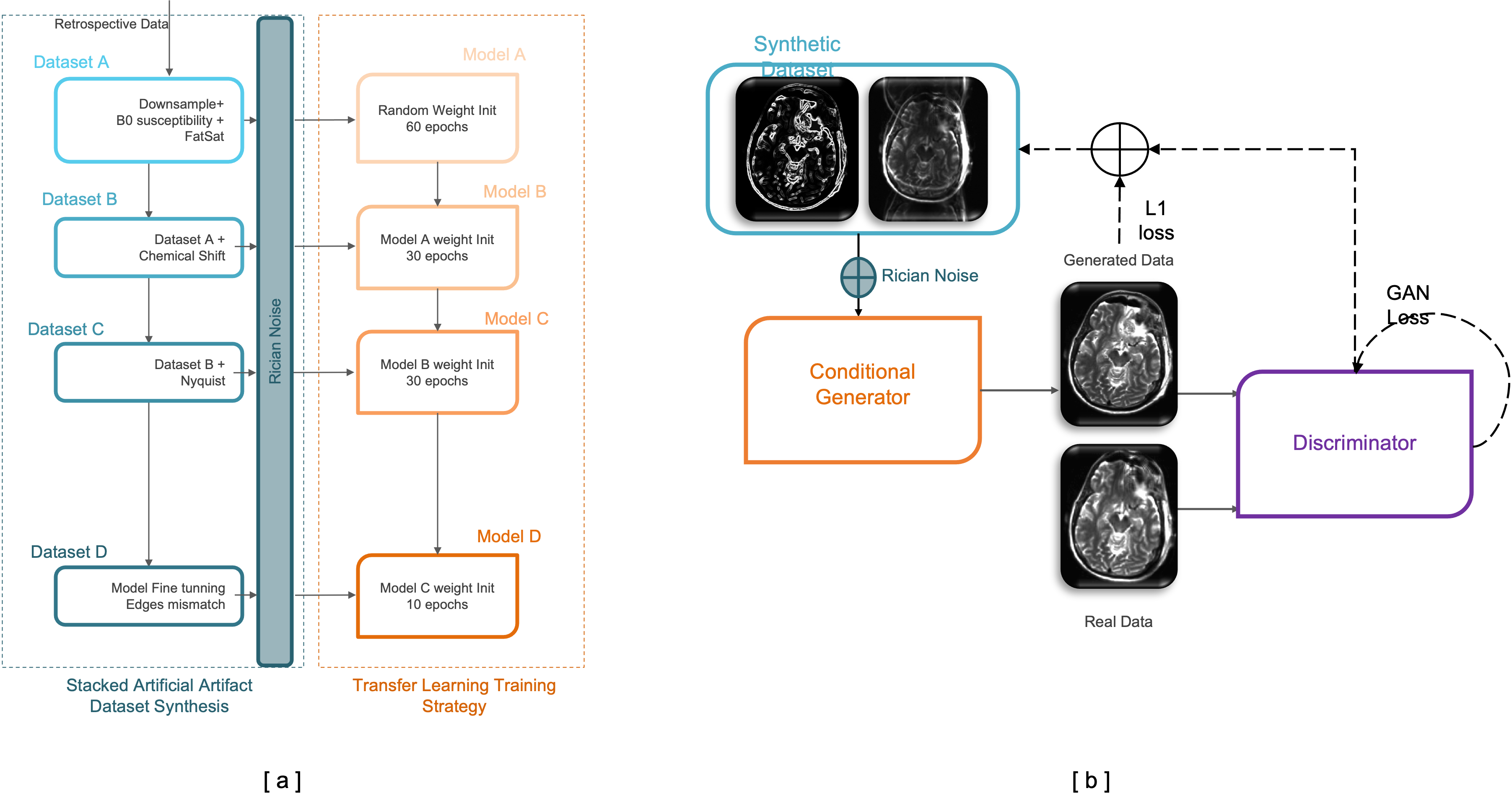
Fig. 1. (a) Network training schema. The network was trained with a stacked approach, starting with simple artifacts and progressively increasing the complexity of the artifacts. The trained weights from the previous model were used to initialize the next model. (b) GAN architecture used for all models.
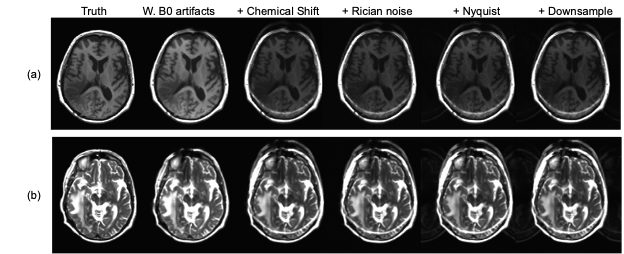
Fig. 2. Example case of simulated artifacts sequentially added on (a) T1-weighted MPRAGE images and (b) T2-weighted TSE images
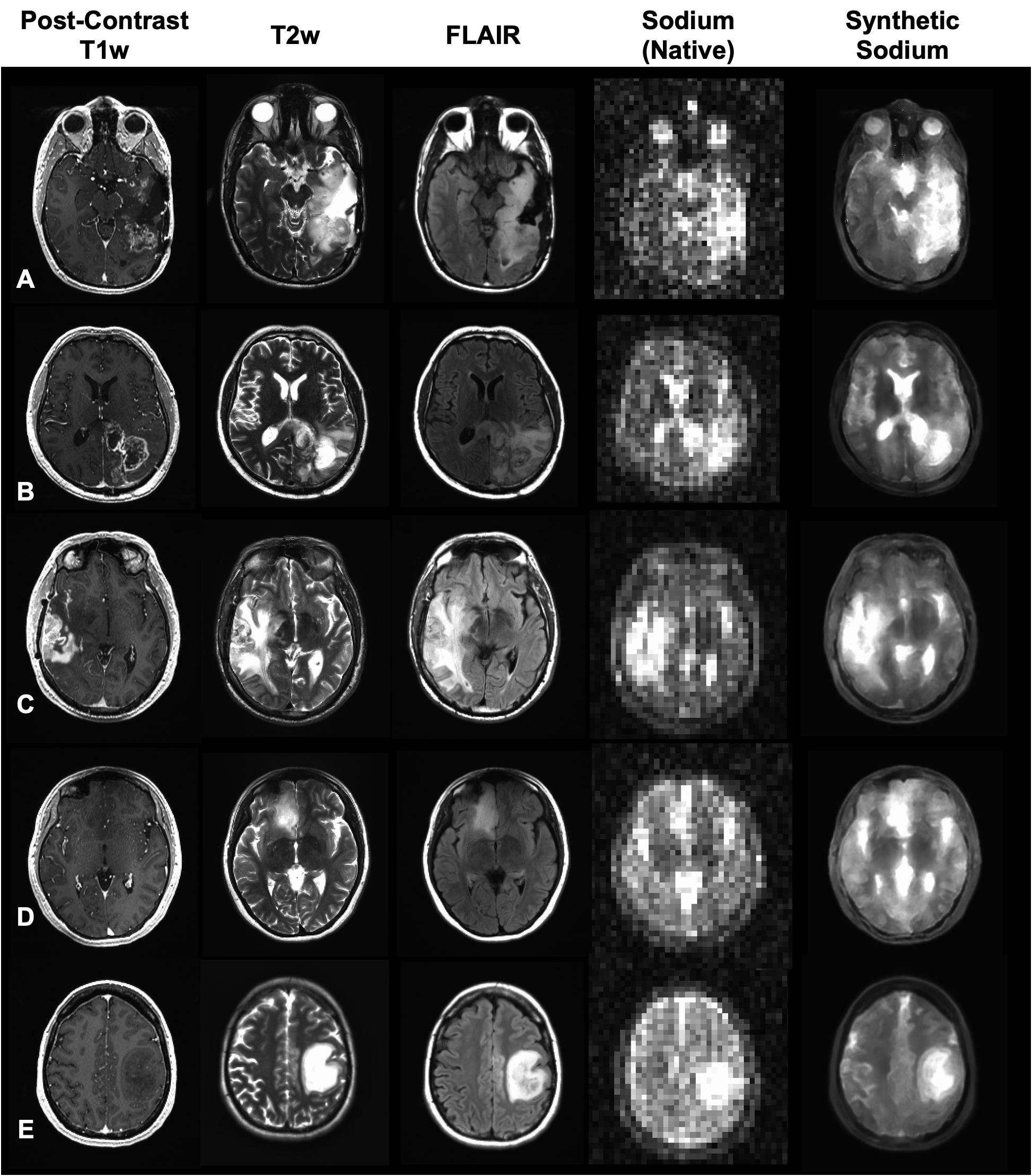
Fig. 3. (Left-to-right) Post-contrast T1-weighted, T2-weighted, T2-weighted FLAIR, native 3T sodium MR images, and synthetic high-resolution sodium MR images for (A-C) patients with recurrent glioblastoma and (D-E) IDH mutant lower grade gliomas.

Fig. 4. (A) Paired t-test comparison of synthetic and native sodium MRI signal (normalized to the eye) within contrast-enhancing tumor regions showing no significant difference between the measurements. (B) Pearson’s Linear correlation between synthetic and native sodium MR measurements of enhancing tumors shows a strong association.

Fig. 5. (A) Spearman’s Correlation between synthetic sodium MR signal and NHE1 IHC expression from image-guided biopsies (relative expression). (B) t-test comparison of synthetic sodium MR measurements of image-guided biopsy samples with no NHE1 expression compared with those having notable expression.